

Kidney nontumor / medical renal
General
Anatomy & histology

Copyright: 2002-2024, PathologyOutlines.com, Inc.
PubMed Search: Kidney anatomy
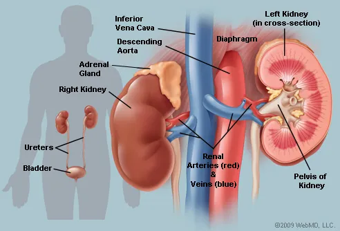
- Posterior abdomen on either side of vertebral column in retroperitoneum
- Surrounded by fat and loose areolar tissue
- Superior border is at T12, inferior border is at L3
- 11 cm long x 5 - 8 cm x 3 cm
- Weighs 125 - 170 g in males, 115 - 155 g in females
- Capsule: covers kidney is surrounded by perirenal fat
- Cortex: outer 1.2 cm of kidney surrounds inner medulla containing pyramids and lacking glomeruli
- Renal sinus: fatty compartment within confines of kidney not delineated from renal cortex by a fibrous capsule
- Gerota fascia: fibromembranous tissue surrounding the kidney that separates it from adjacent musculature
- Ureter ascends into renal pelvis and divides into calyces (2 - 3 major, 12 minor total)
- Related to a calyx are renal pyramids with apices called papillae
- Vasculature: receives 25% of cardiac output, 90% goes to cortex, via interlobar, arcuate, interlobular, afferent arterioles, then into glomeruli, efferent arterioles and peritubular vascular network
- Deeper juxtamedullary glomeruli give rise to vasa recta, which supply outer and inner medulla
- Since arteries are end vessels, their occlusion causes infarction
- Regional lymph nodes: renal hilar, paracaval, aortic and retroperitoneal
- References: Wikipedia: Kidney, eMedicine: Kidney Anatomy, WebMD: Picture of the Kidneys
- Metanephric blastema: forms glomerulus, proximal convoluted tubules, loops of Henle, distal convoluted tubules and connective tissue of renal interstitium
- Ureteric bud: forms cortical and medullary collecting tubules and medullary collecting ducts
- Stages of kidney development:
- Pronephros (based on Wolffian duct, kidneys non-functional)
- Mesonephros (appear at week 4, form nephron-like tubules but degenerate)
- Metanephros (form at week 5, function by week 11)
- References: UNSW Embryology, Wikpedia
- Age related changes occur; are affected by hypertension and heart failure (Kidney Int 1997;51:1196)
- Aldosterone:
- Causes increased reabsorption of NaCl to increase blood volume
- Antidiuretic hormone (vasopressin):
- Stimulates water reabsorption by stimulating insertion of "water channels" or aquaporins (Google: Aquaporins - Water Channels) into the membranes of kidney tubules
- These channels transport solute free water through tubular cells and back into blood, leading to a decrease in plasma osmolarity and an increased osmolarity of urine
- In diabetes insipidus (without ADH), kidney tubules are virtually impermeable to water, which flows out as urine (up to 10 liters of dilute urine/day)
- Erythropoietin:
- Secreted in response to low serum pO2, promotes red blood cell production
- Natriuretic hormones:
- Cause increase in glomerular filtration rate after nephron destruction
- Renin:
- Produced by juxtaglomerular apparatus in response to hypotension, converts angiotensinogen to angiotensin I, which is converted to angiotensin II in the lung by angiotensin converting enzyme (ACE)
- Angiotensin II increases aldosterone production and promotes vasoconstriction
Images hosted on other servers:


Renin pathway


- Glomerular filtration: glomeruli are highly permeable to water and solutes through fenestrated endothelium; they are impermeable to large proteins like albumin (proteins are more permeable if smaller and more cationic)
- Glomerular disease causes tubular disease, since efferent arterioles supply tubules
Images hosted on other servers:

Vertical section


Normal glomerulus

Vessels surrounding glomeruli and tubules


Relation to other organs

Juxtaglomerular apparatus

Renal column

Vessels supplying tubule

Vessels surrounding glomeruli and tubules
Images hosted on other servers:

Fetal kidneys at 25 weeks gestation

Infant kidney with fetal lobulations


Normal adult kidney
- Glomeruli:
- Tuft-like vascular structure composed of lobules of specialized capillaries that arise from an afferent arteriole and eventually coalesce to drain into an efferent arteriole
- 200 microns in diameter, 20% larger in juxtamedullary area
- Hypercellularity: the presence of more than 3 cells in an individual glomerular mesangial region away from the vascular pole
- References: Mod Pathol 2002;15:988, Wikipedia: Glomerulus
- Juxtaglomerular apparatus:
- Close to glomerulus where afferent arteriole enters it; consists of juxtaglomerular cells (modified smooth muscle cells) plus macula densa (region of distal tubule as it returns to vascular pole of parent glomeruli) plus lacis or Goormaghtigh cells (nongranular cells that reside near afferent arteriole, macula densa and glomerulus and resemble mesangial cells); produces renin; cells of macula densa show reverse polarity (with abluminal nuclei and basal cytoplasmic clearing due to Golgi apparatus) to direct the synthesized molecules towards the glomerulus
- Interstitium: contains fibroblast-like cells and peritubular capillaries; expands due to edema and inflammation
- Medullary rays: in cortex, contain cortical collecting tubules and loops of Henle of superficial nephrons
- Proximal tubules: long microvilli, numerous mitochondria and extensive intercellular interdigitations assist in reabsorption of sodium, water, proteins, glucose, potassium, phosphate and amino acids; vulnerable to toxins and ischemic damage
- Renal columns of Bertin: cortical tissue extending into spaces between pyramids
Contributed by Grigory Demyashkin, M.D., Ph.D.
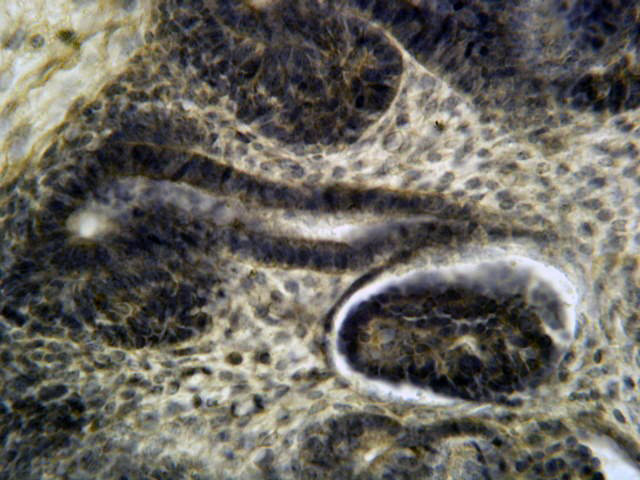
6 - 8 week embryo:
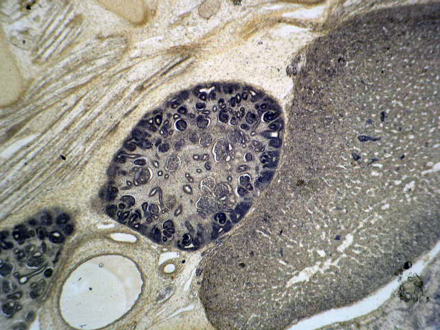
Metanephros; adrenal gland medulla
Metanephros; colon; pancreas

Small intestine; stomach;
gonad and kidney primary
(mesonephros); symphysis
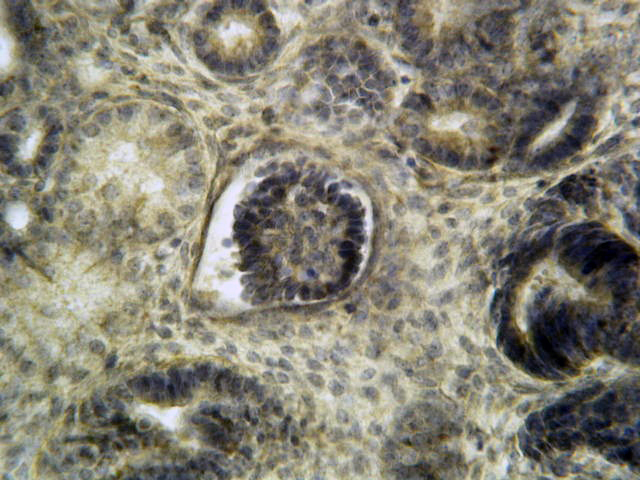
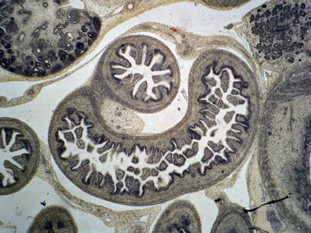
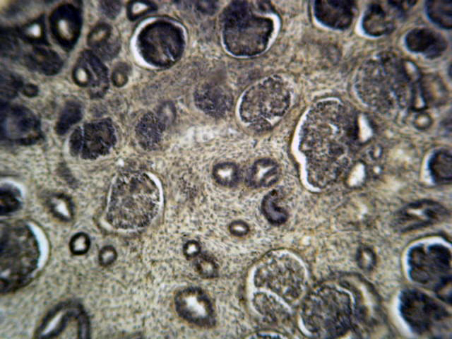
Final kidney (metanephros), renal corpuscle
Final kidney (metanephros), renal corpuscle, collecting ducts
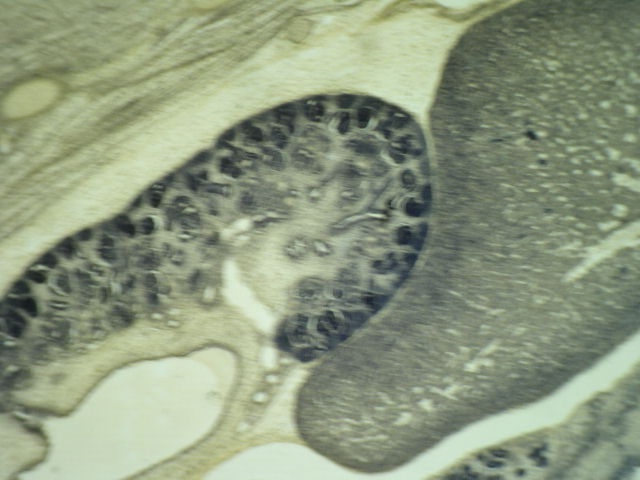
Metanephros; adrenal gland medulla
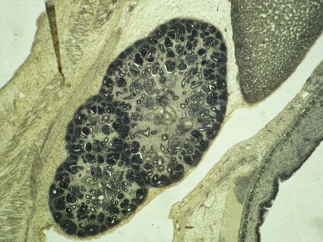
Final kidney (metanephros)

Final kidney (metanephros);
liver; gonad & kidney
primary (mesonephros);
symphysis
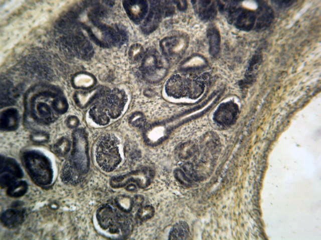
Final kidney (metanephros)
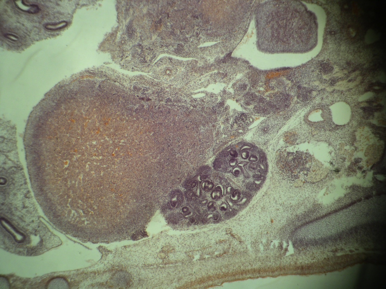
Adrenal gland, medulla & final kidney (metanephros)
Images hosted on other servers:

Glomerular capillary loops are thin and delicate

Basement membranes
of glomerular
capillary loops and
tubular epithelium
Collecting duct: brush border

Juxtaglomerular apparatus and macula densa
Medullary rays
- Layers (inner to outer) are: fenestrated endothelium, then glomerular basement membrane (lamina rare interna, lamina densa and lamina rare externa), then podocytes (visceral epithelium with foot processes); also parietal epithelium which lines Bowman space (which contains the ultrafiltrate of plasma)
- Glomerular basement membrane (GBM): normally 250 - 380 nm, composed of type IV collagen, laminin, polyanionic proteoglycans (mostly heparan sulfate), fibronectin and entactin
- Type IV collagen forms suprastructure to which other glycoproteins attach; composed of 3 alpha chains
- Each alpha chain has amino 7S domain, middle triple helical domain and a carboxyl noncollagenous (NC1) domain; NC1 domain is site of anti-GBM nephritis and dimer formation
- Mesangial cells: type of myofibroblast that supports glomerular tuft, regulates capillary width and blood flow; are phagocytic and can proliferate; the mesangium on the capillary side is covered by endothelial cell; the capillary basement membrane extends to form the paramesangial basement membrane; at ultrastructural level, the mesangium shows cell membrane dense bodies (attachment plaques), which anchors the mesangial cell to the cell membrane
- Podocytes (visceral epithelium): their foot processes embed in lamina rare externa of glomerular basement membrane; the distal diffusion barrier to filtration of proteins is a filtration slit diaphragm between foot processes
- PAS stain allows assessment of glomerular basement membranes, mesangial matrix and tubular basement membranes
- Jones methenamine silver (JMS) highlights these better than PAS
- Masson trichrome stain highlights hyalinosis, scarring, immune deposits and fibrinoid deposits
