
Larynx, hypopharynx & trachea
General
Anatomy & histology
Last author update: 1 November 2013
Last staff update: 5 March 2024 (update in progress)
Copyright: 2002-2024, PathologyOutlines.com, Inc.
PubMed Search: Larynx anatomy histology


Table of Contents
Definition / general | Staging anatomy | Structures | Diagrams / tables | Gross images | Microscopic (histologic) description | Microscopic (histologic) imagesCite this page: Pernick N, Handra-Luca A. Anatomy & histology. PathologyOutlines.com website. https://www.pathologyoutlines.com/topic/larynxanatomy.html. Accessed April 20th, 2024.
Definition / general
Larynx and hypopharynx
Trachea
- Tubular structure between pharynx / root of tongue and trachea at level of cervical vertebrae C4 - C6 in males, somewhat higher in females and during childhood
- Composed of cartilaginous tissue that undergoes ossification and may completely replace cartilage by age 20
- At puberty, increases in size in males due to enlargement of cartilages
- Cartilages are connected by ligaments and moved by numerous muscles
Trachea
- Composed of imperfect rings of hyaline cartilage, fibrous tissue, muscular fibers, mucous membranes and glands
Staging anatomy
Larynx and hypopharynx
- Anterior limit is anterior or lingual surface of suprahyoid epiglottis, thyrohyoid membrane, anterior commissure and anterior wall of subglottic region (composed of thyroid cartilage, cricothyroid membrane and anterior arch of cricoid cartilage)
- Posterior and lateral limits include laryngeal aspect of aryepiglottic folds, arytenoids region, interarytenoid space and posterior surface of subglottic space (mucous membrane covering surface of cricoid cartilage)
- Superolateral limits are tip and lateral borders of epiglottis
- Inferior limit is plane passing through inferior edge of cricoid cartilage
Structures
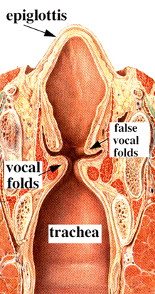
- Supraglottic portion:
- Epiglottis (lingual and laryngeal aspects), false vocal cords (ventricular bands), aryepiglottic folds (laryngeal aspect), arytenoid cartilages and ventricles
- Derived from third and fourth branchial pouches
- Inferior boundary is horizontal plane passing through lateral margin of ventricle at its junction with superior surface of vocal cord
- Glottic portion:
- True vocal cords (superior and inferior surfaces) and anterior and posterior commissures
- Derived from sixth branchial pouch
- Subglottic portion:
- Between lower border of true vocal cords and first tracheal cartilage (or lower margin of cricoid cartilage)
- Derived from sixth branchial pouch
- Anterior commissure:
- Convergence of thyroepiglottic, vestibular and vocal ligaments and conus elasticus
- Tendon provides anterior attachment for true vocal cords
- Tendon also separates glottic and supraglottic parts of larynx
- Arytenoid cartilage:
- 2 hyaline cartilages at upper border of cricoid cartilage at back of larynx that support the vocal cords
- Each is pyramidal
- Apex is surmounted by small, conical, corniculate cartilage
- Conus elasticus:
- Extends from superior border of cricoid cartilage to free edge of vocal cord, then thickens to form vocal ligament, which runs length of true vocal cord close to mucosal surface, then continues along floor of ventricle as thyroglottic ligament
- Cricoid cartilage:
- Hyaline cartilage that is smaller but thicker and stronger than thyroid cartilage
- Upper edge is 1 cm below true vocal cords at mid larynx
- Forms the only complete trachiobronchial ring with posterior quadrate lamina (deep and broad, 2 - 3 cm high) and anterior arch that is narrow and convex
- Articulates with inferior horns of thyroid cartilage
- Cuneiform cartilages: 2 small, elongated pieces of cartilage on either side of aryepiglottic fold
- Epiglottis: thin, bicycle saddle-like and elastic fibrocartilage
- Apex is attached to inner thyroid cartilage just above anterior commissure by thyroepiglottic ligament
- Projects up behind tongue and body of hyoid bone, partly covers laryngeal entrance
- Sides are attached to arytenoid cartilages by aryepiglottic folds
- Upper and anterior surface is free, covered by mucous membrane reflected onto pharyngeal tongue and lateral wall of pharynx to form median and lateral glossoepiglottic folds
- Median glossoepiglottic fold divides area between base of tongue and epiglottis into 2 valleculae
- Not essential for respiration, phonation or deglutition
- Hypopharynx:
- Comprises posterolateral pharyngeal wall (from level of floor of valleculae to level of inferior border of cricoid cartilage), postcricoid esophagus (has anterior wall and extends from level of arytenoids cartilages superiorly to inferior border of cricoid cartilage) and pyriform sinuses (lie lateral to and below opening of larynx, each bounded laterally by medial aspect of thyroid lamina and medially by aryepiglottic fold)
- Trachea:
- Cartilaginous and membranous tube (also called windpipe), extending from lower larynx at C6 to upper border of T5 vertebrae, where it divides into right and left mainstem bronchi
- Flattened posteriorly
- 11 cm long, 2 - 2.5 cm in diameter, diameter greater in men than women, greater in adults than children
- Anteriorly: contacts thyroid isthmus, inferior thyroid veins, neck muscles, cervical fascia, anterior jugular veins, manubrium sterni, thymus, left innominate vein, aortic arch, innominate and left common carotid arteries, deep cardiac plexus
- Posteriorly: contacts esophagus
- Right bronchus appears to be a more direct continuation of trachea and so is the site of most foreign bodies
- Supplied by inferior thyroid arteries
- Pre-epiglottic space: bounded posteriorly by epiglottic cartilage and thyroepiglottic ligament, anteriorly by thyroid cartilage and thyrohyoid membrane and superiorly by hypoepiglottic ligament; space communicates laterally with paraglottic space above ventricle
- Pyriform sinus: not strictly part of larynx; inverted 3 sided pyramid with apex inferiorly at level of cricopharyngeus muscle, bounded superiorly by glossoepiglottic folds, medially by aryepiglottic folds, laterally by pharyngeal wall
- Thyroid cartilage:
- Largest cartilage of larynx
- Shield shaped forming anterior surface of larynx and acute angle in midline of neck (laryngeal prominence, Adam's apple)
- Originally only hyaline cartilage but ossifies at age 25 at sites of muscle origin or insertion
- Lower edge is 1 cm below anterior commissure
- Lymphatics:
- Separate systems exist above and below ventricle
- Are also subdivided as superficial (mucosal) and deep
- Few lymphatics are present in true vocal cord
- Pyriform sinus drains laterally into deep cervical nodes, occasionally to paratracheal nodes
- Supraglottic lymphatics drain through thyrohyoid membrane into upper cervical and anterosuperior nodes
- Subglottic lymphatics drain into prelaryngeal (Delphian) node on cricothyroid membrane, then into pretracheal, paratracheal and supraclavicular nodes
Diagrams / tables
Images hosted on other servers:

Sagittal section

Coronal section

Entrance viewed from above

Laryngoscopic view

Laryngeal cartilages

Vocal cords and muscles

Muscles


Ligaments

Trachea and bronchi

Tracheal bifurcation

Transverse section
Gross images
Images hosted on other servers:
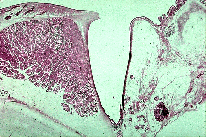
True and false vocal cords
Microscopic (histologic) description
- Junction between epithelial types may be abrupt or separated by transitional area; patches of squamous epithelium in respiratory epithelium are common, particularly in smokers
- Dendritic melanocytes may be present in basal layer, particularly in blacks
- Epiglottis: stratified squamous epithelium similar to oral cavity with modified salivary glands that secrete thick mucous; laryngeal surface also has pits containing mucous glands
- Cartilage has full thickness fenestrae that communicate with pre-epiglottic space, which contains fat and areolar tissue
- False vocal cords and other supraglottic larynx: ciliated, columnar epithelium extending into ventricle of Morgagni with submucosal modified salivary gland epithelium
- Glottis: space between 2 vocal cords
- Hypopharynx: covered by nonkeratinizing stratified squamous epithelium; contains mucosal glands, scattered lymphoid aggregates and rich lymphatic plexus
- Reinke space: lamina propria of true vocal cord, between base of squamous epithelium and vocal ligament
- True vocal cords: stratified squamous epithelium with no / rare submucosal glands
- Subglottic larynx: epithelium resembles trachea / major bronchi - ciliated columnar epithelium with submucosal glands
- Trachea:
- Cartilage:
- 16 - 20 imperfect rings, with circular cartilaginous defect posterior and replaced by fibrous tissue and muscular fibers
- Each cartilage is 4 mm in depth, 1 mm in thickness
- Are elastic but may be calcified later in life
- Fibrous tissue:
- Thick layer covers outer surface of cartilaginous ring, thin layer covers inner surface
- Both layers merge at upper and lower margins of cartilaginous rings
- Muscular tissue:
- Longitudinal and transverse layers of smooth muscle
- Mucus membrane:
- Continuous with laryngeal and bronchial membranes
- Ciliated columnar epithelium overlying areolar and lymphoid tissue with elastic fibers, blood vessels, nerves, mucous glands
- Cartilage:
Microscopic (histologic) images
Images hosted on other servers:
True and false vocal cords

Transverse section of trachea
