
Table of Contents
Definition / general | Clinical features | Case reports | Microscopic (histologic) description | Microscopic (histologic) images | Peripheral smear images | Positive stains | Negative stains | Flow cytometry description | Electron microscopy description | Electron microscopy images | Molecular / cytogenetics description | Differential diagnosisCite this page: Mihova D. AML without maturation (FAB AML M1). PathologyOutlines.com website. https://www.pathologyoutlines.com/topic/leukemiaM1.html. Accessed April 23rd, 2024.
Definition / general
- 10 - 20% of AML cases, 44% in one Brazil hospital (Sao Paulo Med J 2006;124:45)
- Usually adults (median age 46 years), present with anemia, thrombocytopenia and neutropenia; may have leukocytosis with markedly increased blasts
- 4% of childhood AML
Clinical features
- Criteria for diagnosis: 90%+ of nonerythroid cells in marrow are myeloblasts; < 10% granulocytic elements; 3%+ of blasts must be positive for myeloperoxidase or Sudan Black B and / or Auer rods by enzyme cytochemistry
- Enzyme cytochemistry: 3%+ of blasts are positive for myeloperoxidase or Sudan Black B (confirm by immunohistochemistry if only 3 - 10% positive for MPO by enzyme cytochemistry); chloroacetate esterase positive
- Poor prognosis
Case reports
- 4 year old child with t(6;9) and basophilia (Ann Biol Clin (Paris) 2003;61:352)
- 44 year old man with large and small blasts (Arch Pathol Lab Med 2004;128:448)
- Presenting with arterial thromboembolism (Leuk Res 2007;31:869)
Microscopic (histologic) description
- Typically markedly hypercellular marrow, but normocellular and hypocellular cases occur
- Very immature cells, usually round with few azurophilic cytoplasmic granules or Auer rods
- Nuclei are round or indented; little maturation beyond myeloblast stage
- Cells may may resemble lymphoblasts and not appear myeloid
Microscopic (histologic) images
AFIP images
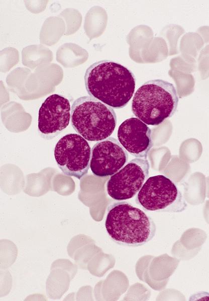
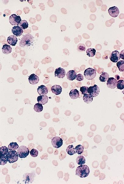
Blasts show mild size variation
Blasts show more variation in size and number of nucleoli
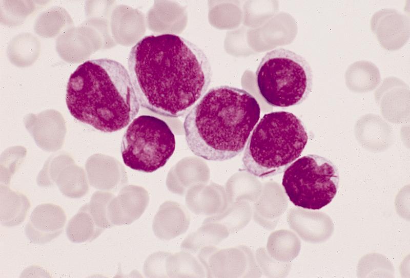
Myeloblasts have irregular nuclei
Myeloblasts have marked size variation
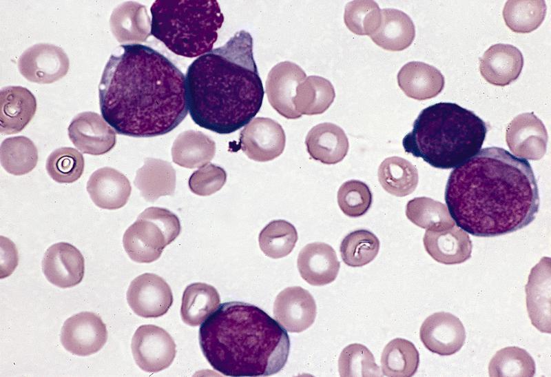
Some variation in size
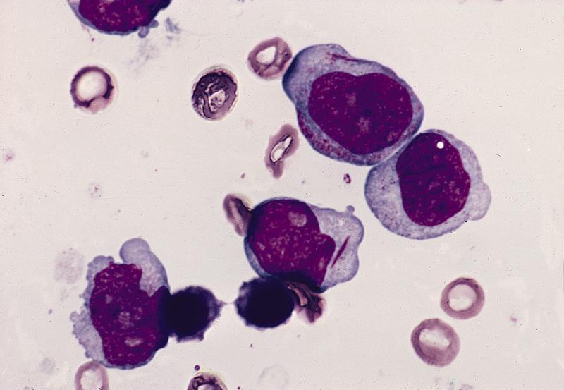
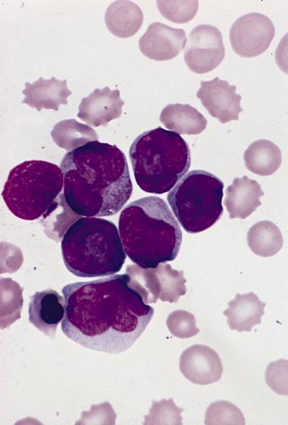
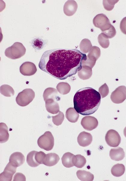
Large myeloblasts
with abundant
eosinophilic
cytoplasm
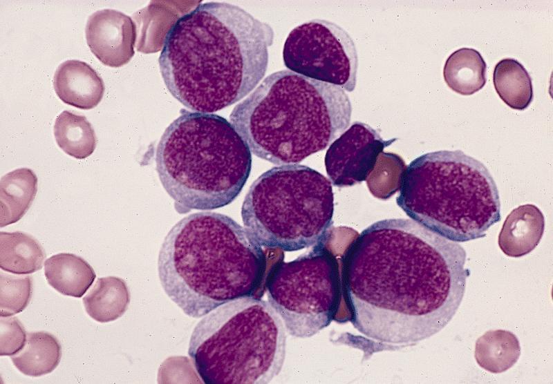
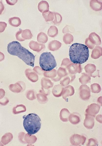
Agranular myeloblasts
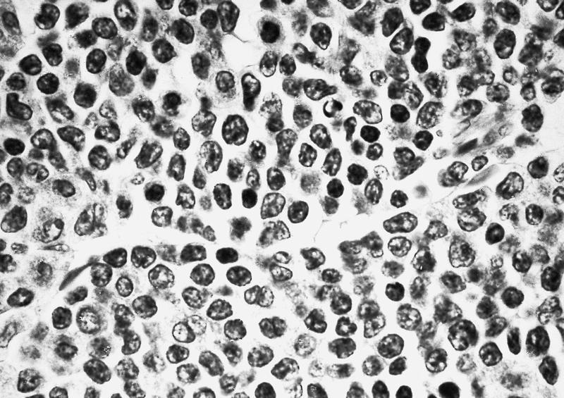
Bone marrow biopsy
Myeloblasts are myeloperoxidase+
Numerous granules are Sudan black B+
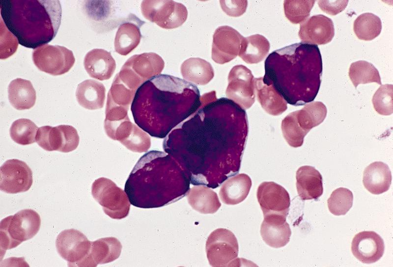
Peripheral smear images
AFIP images
Peripheral blood smear
Positive stains
Negative stains
Flow cytometry description
Electron microscopy description
- May have heterogeneous features (Ultrastruct Pathol 1995;19:9)
Electron microscopy images
AFIP images
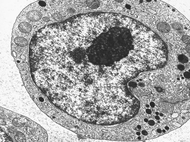
Numerous electron dense granules in Golgi region
Molecular / cytogenetics description
- Associated with t(8;21)
- FLT3 ITD in 22% (Ai Zheng 2007;26:58)
- FLT3 mutations associated with HLA-DR negative patients (Leuk Res 2007;31:921)
