
Table of Contents
Definition / general | Case reports | Microscopic (histologic) description | Microscopic (histologic) images | Positive stains | Negative stains | Flow cytometry description | Electron microscopy images | Molecular / cytogenetics description | Differential diagnosisCite this page: Mihova D. AML with maturation (FAB AML M2). PathologyOutlines.com website. https://www.pathologyoutlines.com/topic/leukemiaM2.html. Accessed April 24th, 2024.
Definition / general
- 10% of AML cases; 5% of childhood leukemias
- Any age, 20% are < 25 years and 40% are 60 years+
- Anemia, thrombocytopenia, neutropenia; variable number of blasts in peripheral blood
- Variable prognosis
- Criteria for diagnosis: 20%+ nonerythroid cells in peripheral blood or bone marrow are myeloblasts; monocytic precursors are < 20% in bone marrow and granulocytes are 10%+ of cells
- Enzyme cytochemistry: most blasts are positive for myeloperoxidase or Sudan Black B, and chloroacetate esterase
Case reports
- 7 year old boy with myeloid sarcoma with acute myeloblastic leukemia (J Coll Physicians Surg Pak 2011;21:369)
- 30 year old man with t(5;11) (Cancer Genet Cytogenet 2007;172:154)
- 35 year old pregnant woman (Rinsho Ketsueki 2011;52:18)
- Three patients with variant t(8;21) (Cancer Genet Cytogenet 2010;199:31)
Microscopic (histologic) description
- Usually hypercellular marrow
- Full range of myeloid maturation through maturing neutrophils, often with abnormal segmentation and 10%+ bone marrow cells with variable degree of dysplasia
- Auer rods in 70% of blasts; myeloblasts with or without azurophilic granules
- Erythroid and megakaryocyte precursors may have dysplastic changes
- Often increased eosinophilic precursors without cytological and cytochemical abnormalities of inv(16)(p13.1q22)
- Basophils may be increased, rarely mast cell hyperplasia (Indian J Pathol Microbiol 2007;50:655)
Microscopic (histologic) images
AFIP images
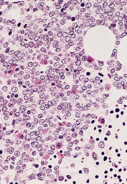
Bone marrow
biopsy: markedly
hypercellular
marrow
Diagnosed as AML with maturation because no t(15;17) and no DIC but FISH not done, so may actually be acute promyelocytic leukemia
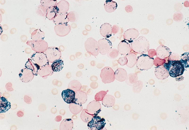
Myeloperoxidase positive blasts

Erythroid cells are negative
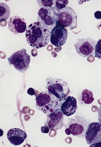
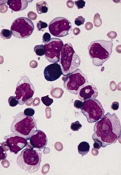
Bone marrow smears (Wright-Giemsa):
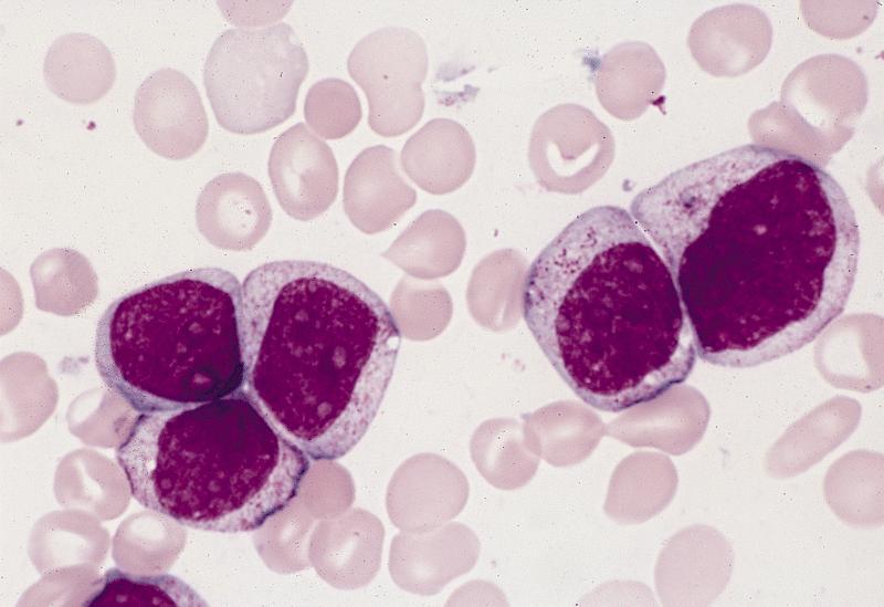
Type III myeloblasts
Myeloblasts,
promyelocytes,
myelocytes and
neutrophils

Several myeloblasts and maturing forms
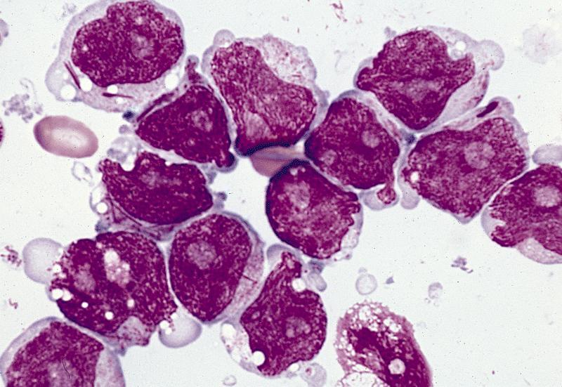
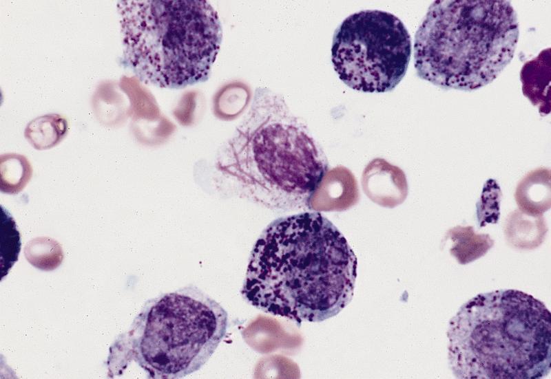
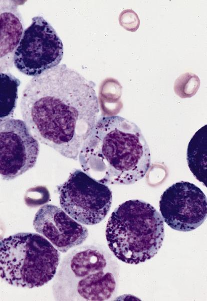
Pseudopods, cytoplasm and prominent Auer rods
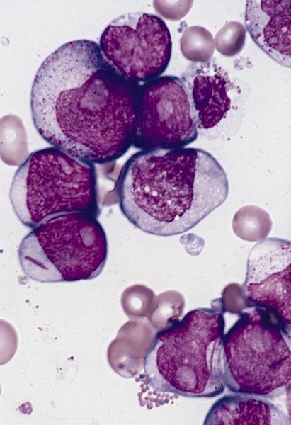
Several blasts have prominent nucleoli and Auer rods
Positive stains
Flow cytometry description
Electron microscopy images
AFIP images
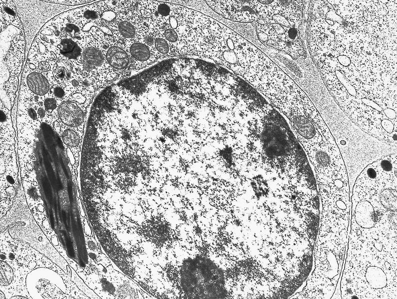
Numerous primary granules and fusion of Auer rods
Molecular / cytogenetics description
- t(8;21) cases classified as AML with t(8;21); RUNX1-RUNX1T1; may be only genetic abnormality or part of more complex abnormalities (Cytometry B Clin Cytom 2008;74:25)
- FLT3 mutations associated with HLA-DR negative patients (Leuk Res 2007;31:921)
Differential diagnosis
