
Liver & intrahepatic bile ducts
Infectious nonviral
Entamoeba histolytica abscess
Author: Komal Arora, M.D.

Last author update: 1 May 2012
Last staff update: 21 July 2022
Copyright: 2002-2024, PathologyOutlines.com, Inc.
PubMed Search: Entamoeba histolytica liver abscess
Table of Contents
Definition / general | Diagrams / tables | Radiology images | Treatment | Gross description | Microscopic (histologic) description | Microscopic (histologic) images | Cytology description | Positive stains | Differential diagnosisCite this page: Arora K. Entamoeba histolytica abscess. PathologyOutlines.com website. https://www.pathologyoutlines.com/topic/liveramebicabscess.html. Accessed April 25th, 2024.
Definition / general
- Also called amebic abscess; more common than pyogenic abscess worldwide but not in US (Med Clin North Am 1989;73:847)
- Common in developing world (Exp Parasitol 2010;126:366)
- Usually fecal oral spread; may also be sexually transmitted (Clin Infect Dis 2009;49:346)
- Complication of 3 - 9% of amebiasis cases
- Trophozoite:
- Active stage that exists only in host and in fresh loose feces; 10 - 60 microns with small, single, round nucleus with distinctive karyosome
- Thick, beaded nuclear membrane
- Bubbly cytoplasm with ingested red blood cells
- Complications: bacterial superinfection, extension or perforation into other structures; death in < 1% of cases
- Diagnosis: serology is 90% sensitive
Diagrams / tables
Images hosted on other servers:

Diagram of life cycle
Radiology images
Images hosted on other servers:

CT scan
Treatment
- Metronidazole, surgery only if complications
Gross description
- Well circumscribed, firm cavities, usually right sided near liver dome
- Initially yellow, later odorless, orange brown, pasty, necrotic material resembles anchovy sauce
Microscopic (histologic) description
- Shaggy necrotic fibrinous zone, granulation tissue, no / rare neutrophils (not actually an "abscess"), peripheral trophozoites up to 60 microns with small eccentric nucleus and cytoplasmic vacuoles that may contain red blood cells
- Resemble histiocytes
- Adjacent liver has fibrosis, chronic inflammation and reactive hepatocytes
Microscopic (histologic) images
Images hosted on other servers:
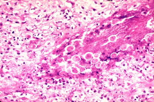
Trophozoites

Cysts
Cytology description
- Use saline wet mounts and stained material
- 50% positive if material obtained from periphery; very rarely positive if from center of cyst
Differential diagnosis
- Macrophages:
- Bean shaped nuclei, fine chromatin, delicate nuclear membrane, small nucleoli
