
Liver & intrahepatic bile ducts
Dysplasia
Liver cell dysplasia
Last author update: 27 January 2021
Last staff update: 28 January 2021
Copyright: 2002-2024, PathologyOutlines.com, Inc.
PubMed search: Liver cell dysplasia[TI]


Table of Contents
Definition / general | Essential features | Terminology | Gross description | Microscopic (histologic) description | Microscopic (histologic) images | Sample pathology report | Board review style question #1 | Board review style answer #1Cite this page: Assarzadegan N, Gonzalez RS. Liver cell dysplasia. PathologyOutlines.com website. https://www.pathologyoutlines.com/topic/livertumorlivercelldysplasia.html. Accessed April 23rd, 2024.
Definition / general
- Premalignant foci in liver characterized by morphologic alterations of hepatocytes and sometimes by architectural changes
- Challenging category that encompasses macroscopic and microscopic patterns of change (see also low grade dysplastic nodule and high grade dysplastic nodule) (Hepatology 2009;49:658)
Essential features
- Dysplastic features in hepatocytes that can progress to malignancy as part of a multistep sequence
- May be incidentally discovered or seen in a discrete nodule
Terminology
- Dysplastic foci are incidental submillimeter microscopic lesions in cirrhotic livers containing dysplastic hepatocytes (Arch Pathol Lab Med 2011;135:704)
- Large cell change and small cell change refer to morphologic changes in hepatocytes; these were formerly called large cell dysplasia and small cell dysplasia
- Iron free foci are areas of hepatocytes lacking iron in a liver otherwise suffering significant iron overload; they appear to be preneoplastic (Hepatology 1993;18:1363)
- Terms adenomatous hyperplasia and atypical adenomatous hyperplasia are no longer in use
Gross description
- Dysplastic foci cannot be identified grossly
Microscopic (histologic) description
- Large cell change is defined as an increase in both nuclear and cytoplasmic size, preserving nuclear to cytoplasmic ratio; nuclei are hyperchromatic, pleomorphic and frequently multinucleated
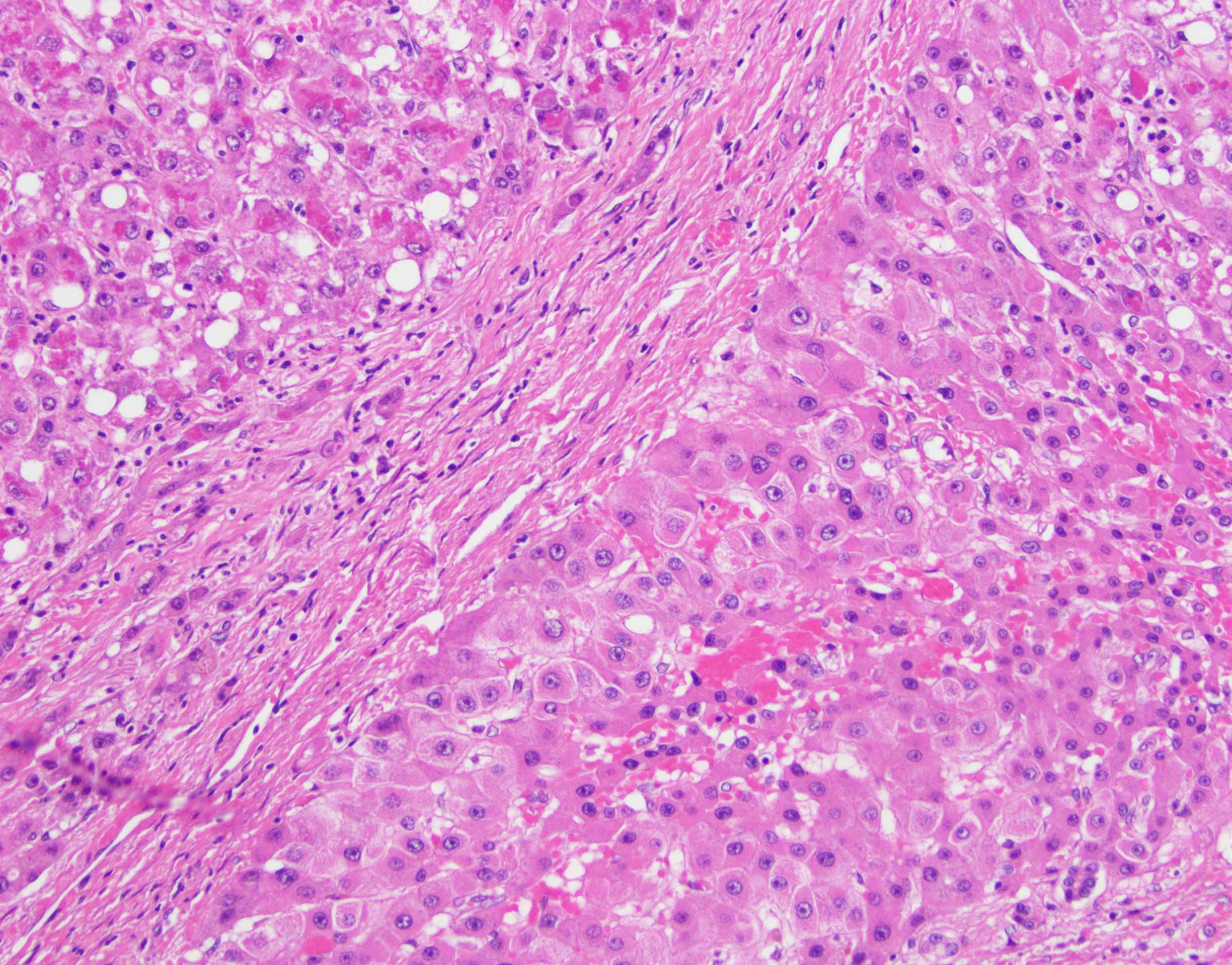
- Small cell change is defined as hepatocytes showing decreased cell volume, increased nuclear to cytoplasmic ratio, mild nuclear pleomorphism, hyperchromasia and cytoplasmic basophilia, giving the impression of nuclear crowding; this pattern is typical of high grade dysplastic nodules
Microscopic (histologic) images
Contributed by Naziheh Assarzadegan, M.D.
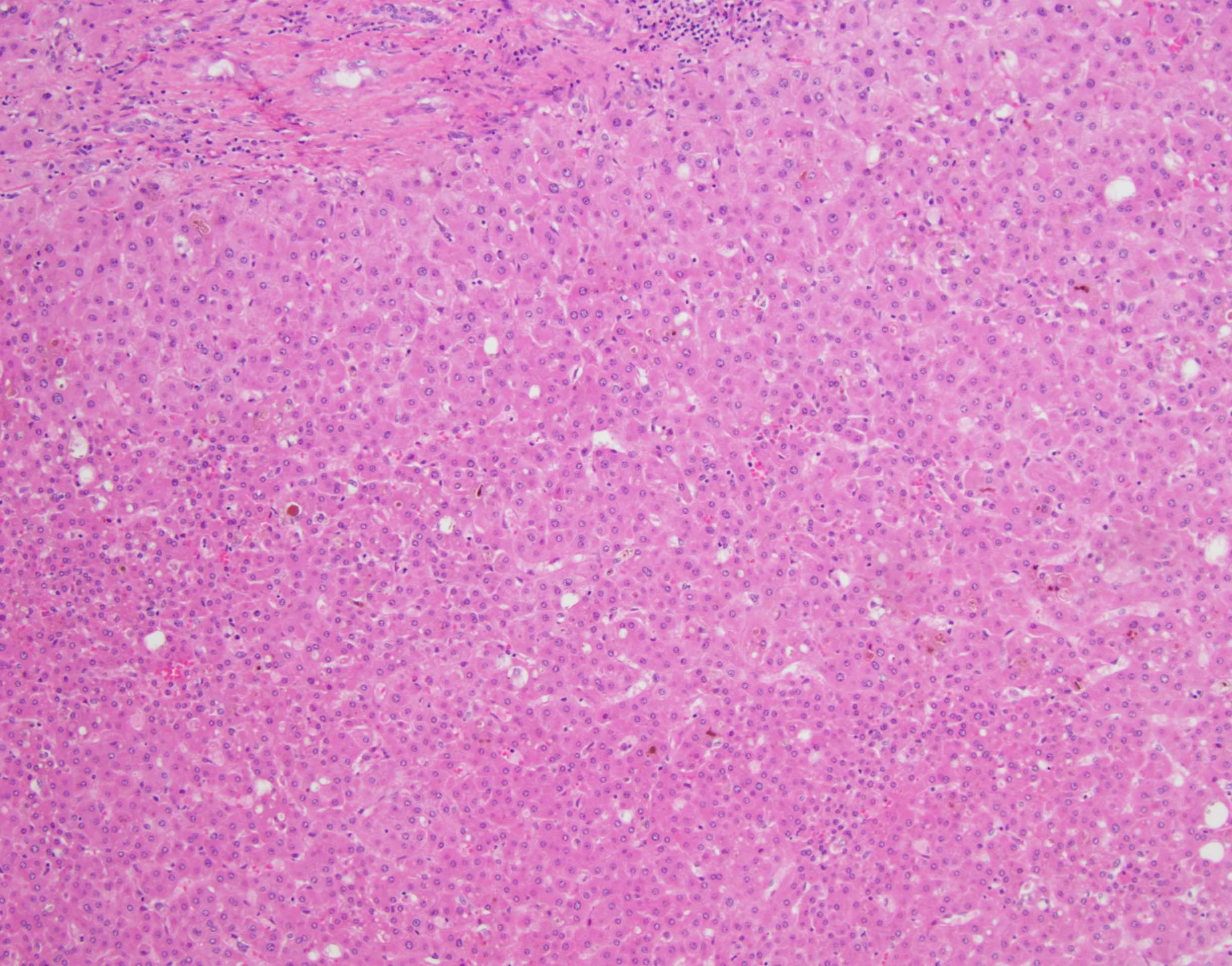
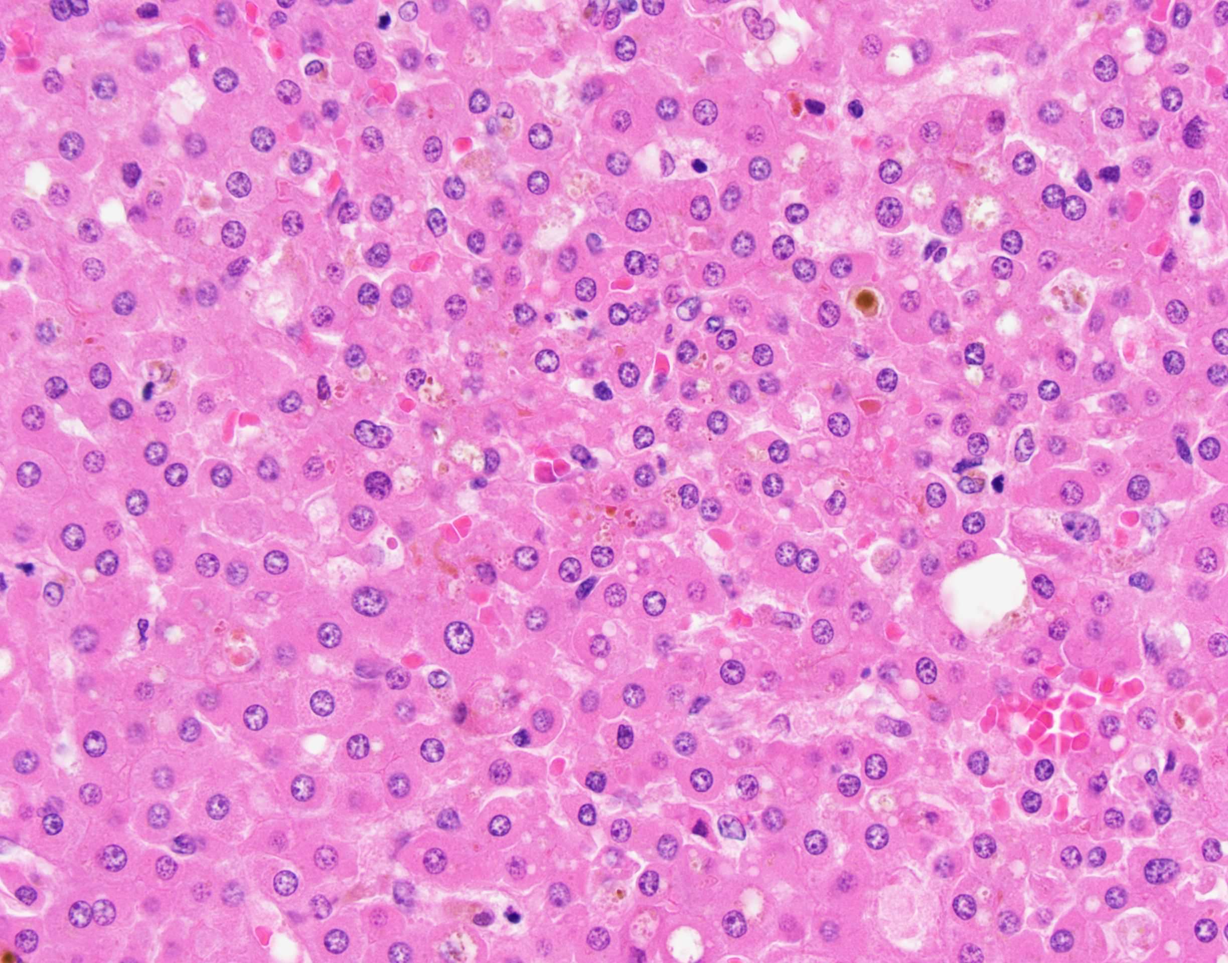
Small cell change
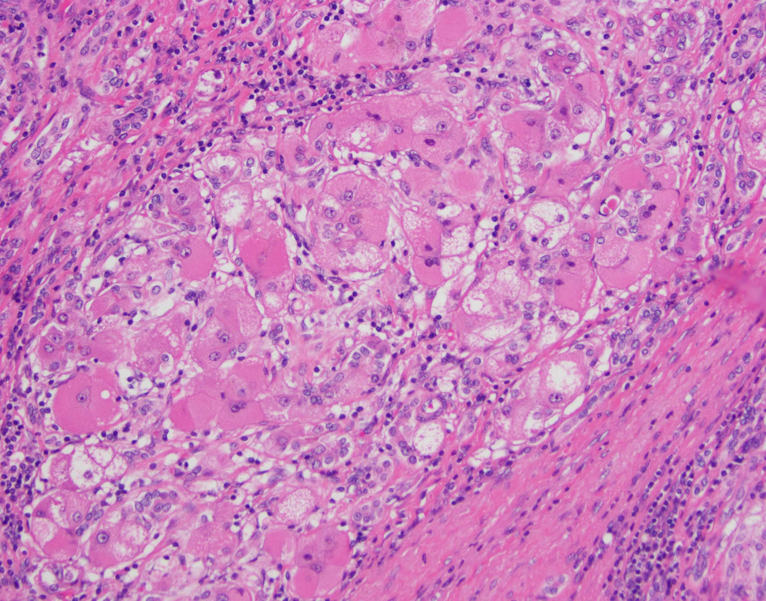
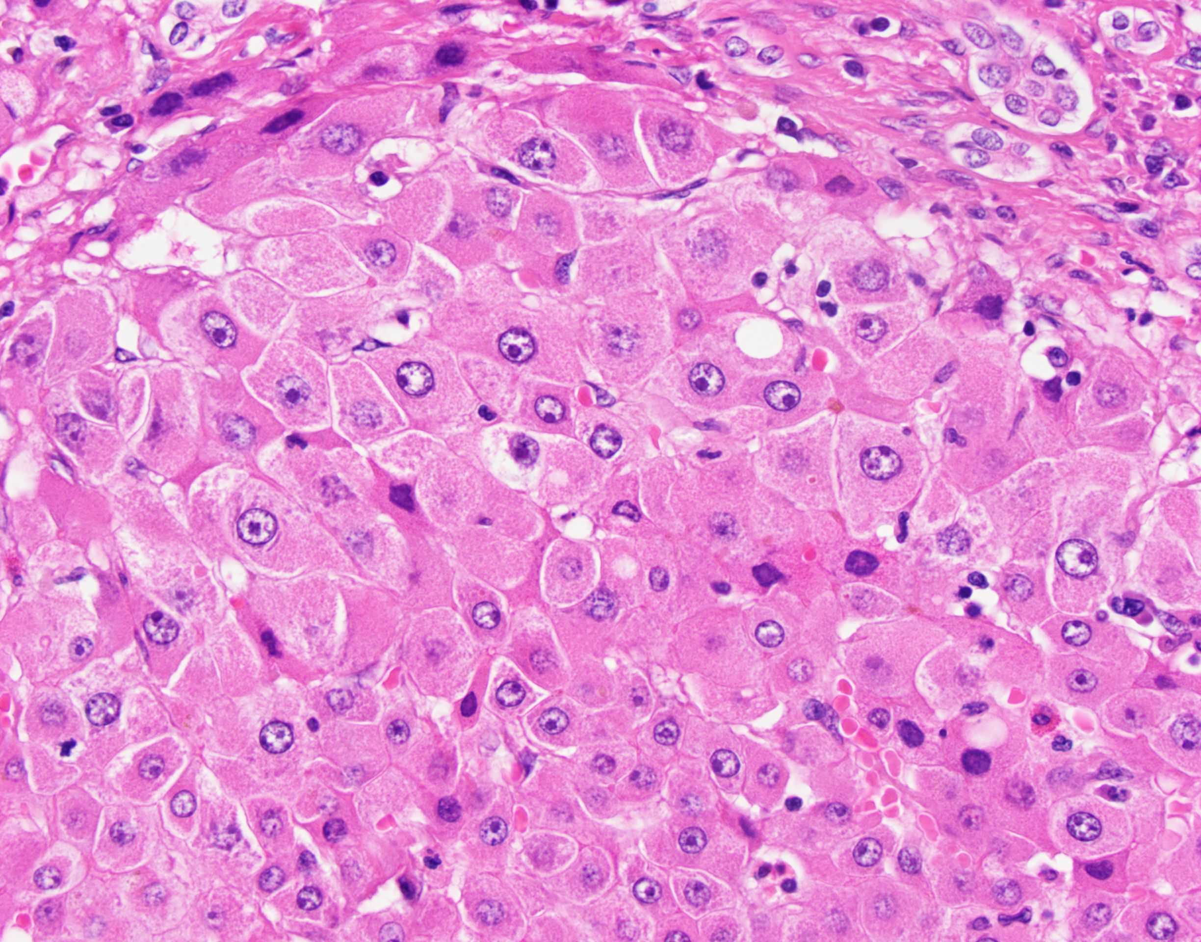
Large cell change
Large cell change with adjacent cirrhotic nodule
Sample pathology report
- Liver, native, orthotopic transplantation:
- Cirrhosis with mild chronic inflammation and patchy low grade dysplasia (see comment)
- Negative for high grade dysplasia malignancy.
- Margins of resection unremarkable.
- Comment: The findings are consistent with the patient’s reported history of nonalcoholic hepatitis. A trichrome stain confirms cirrhosis. An iron stain is unremarkable.
Board review style question #1
Which of the following is true about hepatocellular dysplasia?
- Dysplastic foci can be identified grossly
- It acts as the precursor to cholangiocarcinoma
- Large cell change has an increased nuclear to cytoplasmic ratio
- Small cell change has mild nuclear pleomorphism and cytoplasmic basophilia
Board review style answer #1
D. Small cell change has mild nuclear pleomorphism and cytoplasmic basophilia
Comment Here
Reference: Liver cell dysplasia
Comment Here
Reference: Liver cell dysplasia
