
Table of Contents
Asplenia | Hepatolienal fusion | Hyposplenism | Polysplenia | Splenic gonadal fusion | Splenorenal fusion | Wandering spleen | Case reports | Gross description | Microscopic (histologic) description | Microscopic (histologic) imagesCite this page: Mansouri J. Splenic malformations. PathologyOutlines.com website. https://www.pathologyoutlines.com/topic/lymphnodessplenicmalformations.html. Accessed April 18th, 2024.
Asplenia
- Rare, associated with cardiac malformations (80%, usually involving atrioventricular endocardial cushion and ventricular outflow tract), situs inversus, anomalies of blood vessels, lung, abdominal viscera
- Can present with pneumococcal sepsis (OPSI)
Hepatolienal fusion
- Fusion of liver and spleen (Hum Pathol 1978;9:234)
- Very rare
Hyposplenism
- Atrophy of spleen with concomitant compromised splenic function (Lancet 2011;378:86)
- Usually acquired but rare congenital forms exist (isolated congenital hyposplenia, Ivemark syndrome, hypoparathyroidism syndrome, autoimmune polyendocrinopathy candidiasis ectodermal dystrophy [APECED] syndrome, Stormorken syndrome)
Polysplenia
- Multiple spleens, found as part of heterotaxy syndromes (malpositioning of organs on opposite side of body, e.g. situs inversus)
- Associated with extrahepatic biliary atresia (Arch Pathol Lab Med 1980;104:212), cardiac abnormalities and developmental anomalies of pancreas, portal vein, inferior vena cava
Splenic gonadal fusion
- Rare congenital anomaly in which ectopic splenic tissue unites with a gonad (< 200 cases reported)
- Continuous: spleen connected to ectopic splenic mass by cord of splenic and fibrous tissue
- Discontinuous: no connection between spleen and ectopic splenic mass
- 20% of continuous types associated with other congenital defects, including peromelus (fetus with malformed limbs) and micrognathia; also testicular ectopia, inguinal hernia
- 90% in males; usually left sided; usually less than 20 years old
- Diagnosis: Technetium Tc 99m sulfur colloid scan
- Treatment: Surgical excision of ectopic splenic tissue to prevent testicular atrophy, torsion or infarction and preserve fertility
Splenorenal fusion
- May be due to splenosis after splenic trauma or splenectomy; less commonly is developmental anomaly resulting in fusion of splenic and renal tissue
- May present as a renal mass or with symptoms of hypersplenism
Wandering spleen
- Due to congenital loss, weakness of splenic ligaments or abnormal length of splenic vessels
- Susceptible to torsion
- May rarely be associated with polysplenia
Case reports
- 16 year old boy with chronic, painless testicular mass (Case #309)
- 22 year old woman with intermittent hypochondrial pain for over a year (Int J Surg Case Rep 2012;3:151)
- 27 year old man with bilateral cryptorchidism and nonseminomatous germ cell tumor in intra-abdominal splenic gonadal mass (Arch Pathol Lab Med 2002;126:1222)
- 51 year old woman with renal mass (Arch Pathol Lab Med 2003;127:e1)
- 56 year old man with asymptomatic testicular mass (Arch Pathol Lab Med 2003;127:e277)
- 62 year old woman with polysplenia with agenesis of dorsal pancreas and preduodenal portal vein presenting with obstructive jaundice (Br J Radiol 2011;84:e217)
- Familial congenital asplenia (Eur J Pediatr 2010;169:315)
- Woman with discontinuous type (Hum Pathol 1989;20:486)
Gross description
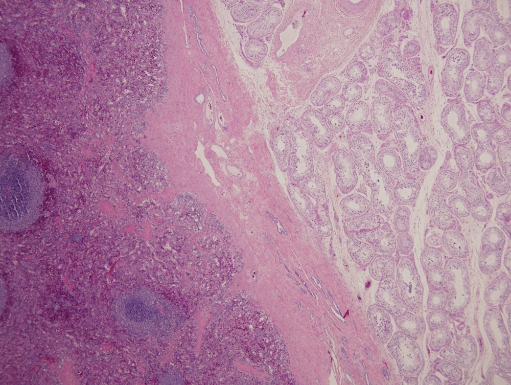
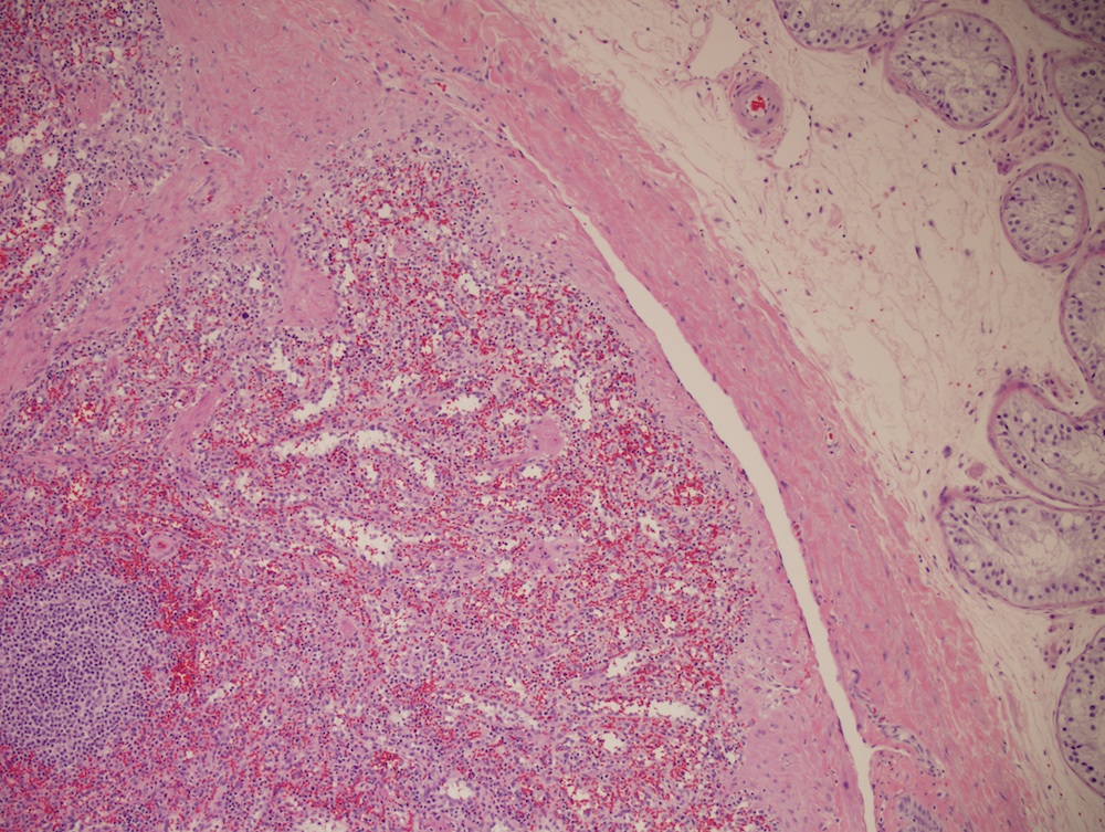
- Splenic gonadal fusion: ectopic splenic tissue is well demarcated from gonad, only rarely is intermingling of tissue
Microscopic (histologic) description
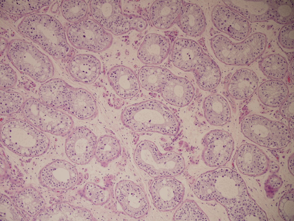
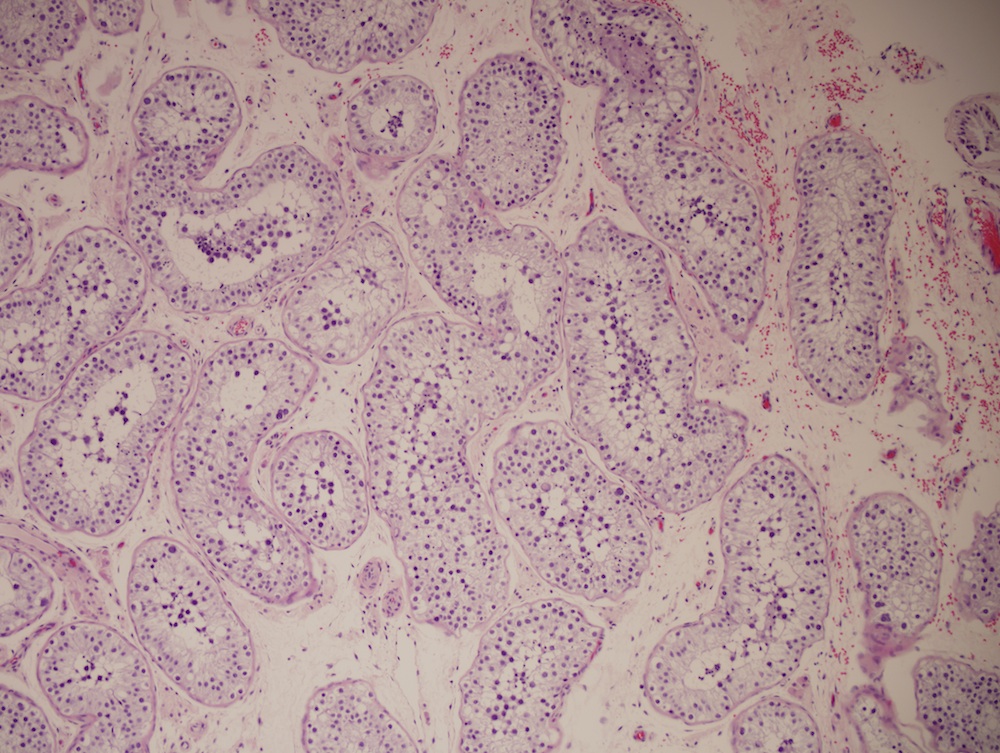
- Splenic gonadal fusion:
- Normal splenic parenchyma but may have thrombosis, calcification, fibrosis, fat degeneration, hemosiderin deposits
- Testicular tissue may be normal but may have atrophy or fibrosis of seminiferous tubules, increased Leydig cells, thrombosis of spermatic vessels or testicular tumors
Microscopic (histologic) images
Case #309

Splenic gonadal fusion
