Staging criteria for mycosis fungoides / Sézary syndrome (MF / SS) is different from non mycosis fungoides / Sézary syndrome lymphomas (including primary cutaneous B cell lymphomas [CBCL], other primary cutaneous T cell lymphomas [CTCL] and NK cell lymphomas) (Blood 2007;110:1713, Blood 2007;110:479)
pN0: no clinically or pathologically abnormal lymph nodes
pN1: involvement of 1 peripheral lymph node region that drains an area of current or prior skin involvement
pN2: involvement of 2 or more peripheral lymph node regions or involvement of any lymph node region that does not drain an area of current or prior skin involvement
pN3: involvement of central lymph nodes
Notes:
Mycosis fungoides and Sézary syndrome
Abnormal peripheral lymph node(s) indicates any palpable peripheral node that on physical examination is firm, irregular, clustered, fixed or 1.5 cm or larger in diameter
Node groups examined on physical examination include cervical, supraclavicular, epitrochlear, axillary and inguinal
Central nodes, which are not generally amenable to pathologic assessment, are not currently considered in the nodal classification unless used to establish N3 histopathologically
T cell clone is defined by PCR or Southern blot analysis of the T cell receptor gene
Non mycosis fungoides / Sézary syndrome primary cutaneous lymphomas
Definition of lymph node regions is consistent with the Ann Arbor system
Peripheral sites: antecubital, cervical, supraclavicular, axillary, inguinal femoral and popliteal
Central sites: mediastinal, pulmonary hilar, para-aortic, iliac
pB1: > 5% of peripheral blood lymphocytes are atypical (Sézary) cells but does not meet the criteria of B2
pB1a: clone negative
pB1b: clone positive
pB2: > 1,000/µL Sézary cells with positive clone
Notes:
Sézary cells are defined as lymphocytes with hyperconvoluted cerebriform nuclei
If Sézary cells are not able to be used to determine tumor burden for B2, then 1 of the following modified ISCL criteria along with a positive clonal rearrangement of the TCR may be used instead:
Expanded CD4+ or CD3+ cells with CD4/CD8 ratio of 10 or more
Expanded CD4+ cells with abnormal immunophenotype including loss of CD7 or CD26
T cell clone is defined by PCR or Southern blot analysis of the T cell receptor gene
Prefixes
Not standardized for primary cutaneous lymphomas, therefore not used currently
Primary cutaneous CD8+ aggressive epidermotropic cytotoxic T cell lymphoma
Primary cutaneous acral CD8+ T cell lymphoma
Primary cutaneous CD4+ small / medium T cell lymphoproliferative disorder
Primary cutaneous peripheral T cell lymphoma, NOS
Cutaneous B cell lymphomas
Primary cutaneous marginal zone lymphoma
Primary cutaneous follicle center lymphoma
Primary cutaneous diffuse large B cell lymphoma, leg type
Clinical images
Contributed by Henry K. Wong, M.D., Ph.D.
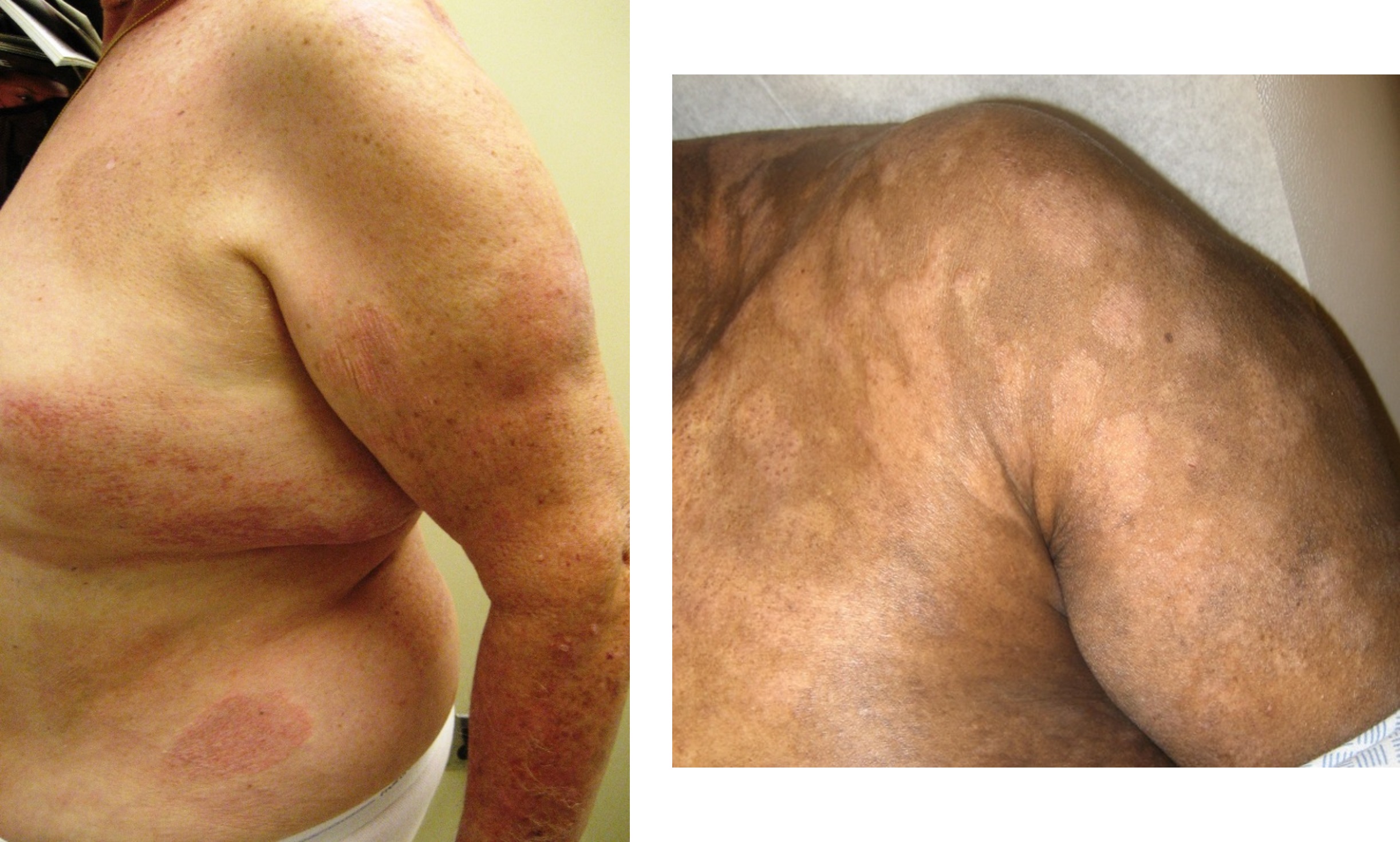
Mycosis fungoides, patch stage
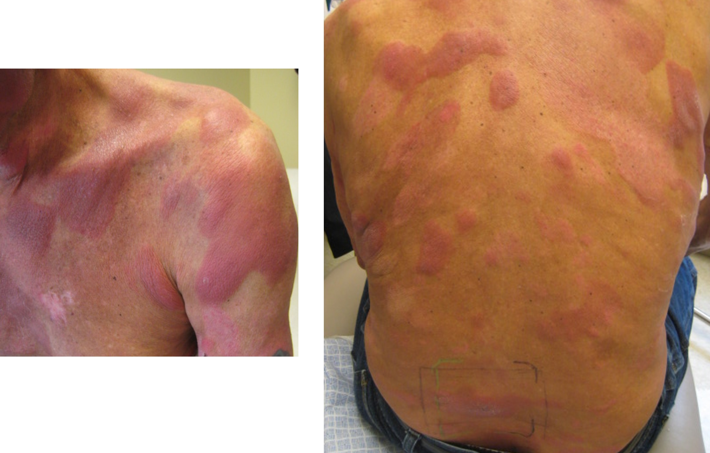
Mycosis fungoides, plaque stage
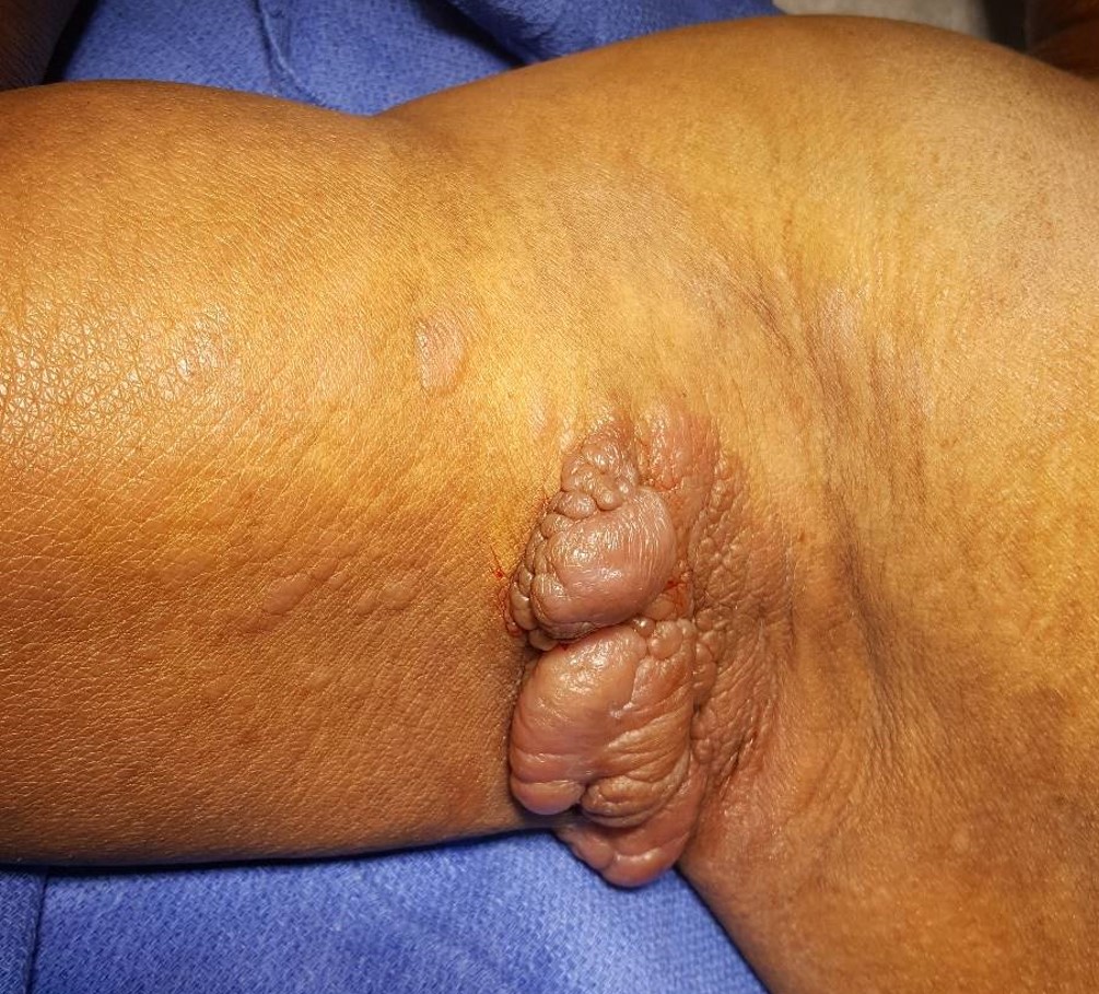
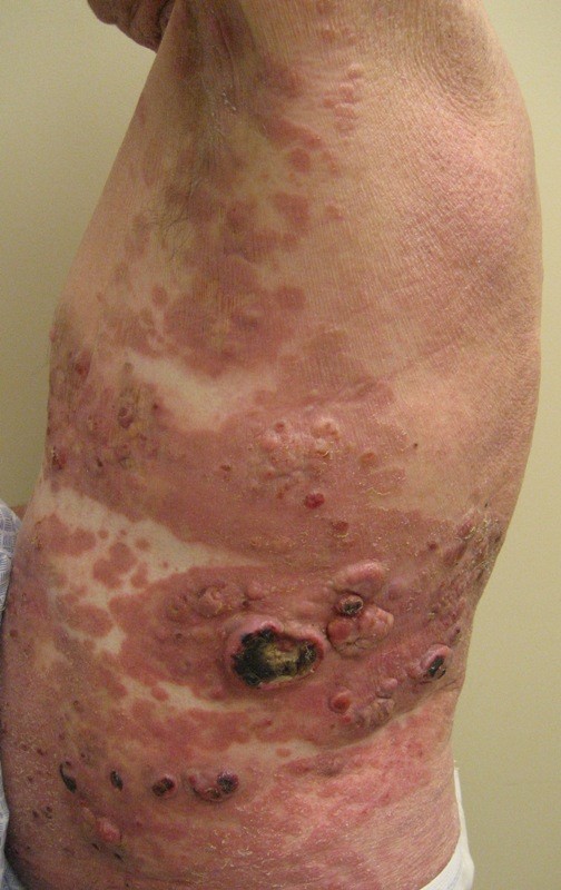
Mycosis fungoides, tumor stage
Peripheral smear images
Images hosted on other servers:
Sézary cells, peripheral smear
Videos
Diagnosis and staging of cutaneous lymphoma
Board review style question #1
A 45 year old woman presents with new onset violaceous plaques and tumors on the lower leg and foot with rapid progression (see picture above). The lesions measure 3.2 cm and 4.9 cm in greatest dimension, respectively. No other abnormalities are found on physical exam. Biopsy of the lesions shows sheet of large, atypical cells that stain positively with CD20, MUM1 and BCL2. A diagnosis of diffuse large B cell lymphoma, leg type is made. What is the correct T stage of the disease?
T1a
T2a
T3a
T3b
Board review style answer #1
B. T2a. Multiple lesions of diffuse large B cell lymphoma, leg type are present on 2 contiguous regions of the body, hence, the T stage is T2a using the non mycosis fungoides / Sézary syndrome primary cutaneous lymphoma staging criteria.
A 68 year old man presents with erythema of the entire back, chest, abdomen and bilateral extremities. On further inspection, there is a 1.8 cm raised ulcerated lesion present on the left upper thigh and multiple enlarged palpable lymph nodes in the inguinal region. Biopsy of the thigh lesion shows tagging of CD4 + T cells along the dermal epidermal junction and epidermotropism. The cells show loss of CD7 and CD26. What is the most likely diagnosis and correct T stage of the disease?
Mycosis fungoides; stage T1a
Mycosis fungoides; stage T2b
Mycosis fungoides; stage T3
Mycosis fungoides; stage T4
Board review style answer #2
D. Mycosis fungoides; stage T4. The erythema covers > 80% of body surface which is stage T4 according to the mycosis fungoides / Sézary syndrome primary cutaneous lymphoma staging criteria.