
Lymph nodes & spleen, nonlymphoma
Lymph nodes-general
B & T cells
Author: Nikhil Sangle, M.D.

Last author update: 1 January 2011
Last staff update: 18 July 2022
Copyright: 2001-2024, PathologyOutlines.com, Inc.
PubMed Search: b cells lymphocyte development lymphoma
Table of Contents
Definition / general | Pathophysiology | Diagrams / tables | Clinical features | Positive stains | Negative stains | Additional referencesCite this page: Sangle N. B & T cells. PathologyOutlines.com website. https://www.pathologyoutlines.com/topic/lymphomanormalbcells.html. Accessed April 24th, 2024.
Definition / general
-
B cells:
- Progressive maturation of hematopoietic stem cells through several stages to ultimately give rise to mature B cell pool that has been selected for reactivity against non-self antigens (Expert Rev Clin Immunol 2010;6:765)
- Develop from stem cells of yolk sac, fetal liver, spleen and bone marrow
- B cells complete most of their development within the bone marrow, in contrast to T cells that mature within the thymus
- In intersinusoidal bone marrow, hematopoietic stem cells mature to early lymphoid progenitors, the pro B, pre B stages
- Developmental stages are in close contact with slender CD10+ stromal cells or their extensions, which allows tightly regulated signaling (J Pathol 2005;205:311)
- Earliest stem cells are in subendosteum, adjacent to inner bone surface; with maturation, B lineage cells move towards central axis of marrow; final stages of development of immature B cells occur in peripheral lymphoid organs (spleen, lymph nodes)
- Develop from bone marrow, become prothymocytes, then migrate to thymus gland, where self recognizing T cells are eliminated
T cells:
Pathophysiology
-
B cells:
- B cells express surface immunoglobulin (Ig), composed of 2 heavy (H) and 2 light (L) chains (either kappa or lambda)
- B cell antigen receptor loci may have 4 types of modification: (a) recombination of variable, diversity and joining regions (VDJ); (b) somatic hypermutation of V segments; (c) immunoglobulin heavy chain gene class switching; and (d) receptor editing
- Early B cell precursor is TdT+, CD34+, HLA-DR+, then undergoes heavy (H) chain rearrangement and adds CD19, then adds CD10, then adds IgM heavy chain; then adds light (L) chain rearrangement and adds cytoplasmic IgM with heavy and light chains, then B cells express IgM and IgD with the same binding site, then adds CD20 (now called pre B cell); then adds surface Ig, then adds CD21 and CD22 and drops TdT (now called B cell)
- If B cell encounters an antigen that interacts with its variable region, it becomes a plasma cell
- Precursor B cells contain immunoglobulin related components but not immunoglobulin; express CD179a and CD179b (precursor to light chains) as part of their pre B cell receptor, which disappears when replaced with conventional light chains
- B cells express surface immunoglobulin, consisting of 2 heavy chains and 2 light chains (kappa or lambda); immunoglobulin is associated with CD79a / CD79b complex to form a B cell antigen receptor complex
- IgH gene (heavy chain of immunoglobulin) is at 14q32; variable portion is coded by VDJ regions
- IgL gene (light chain of immunoglobulin): kappa is at 2p11, lambda is at 22q11; no diversity region is present
- Heavy chain isotype switch: determines if immunoglobulin is IgM, IgD, IgG1-4, IgA1-2 or IgE (9 constant regions); mediated by switch genes
- T cell receptors (TCR) are either alpha / beta (95%) or gamma / delta (5%) heterodimers
- T cell progenitors migrate to the thymus and undertake a highly ordered developmental program, regulated by signals derived from microenvironment
- Most immature thymocytes are CD4- / CD8- ("double negative" / DN); DN is divided into 4 stages based on CD44 / CD25 expression: DN1 is CD44+, CD25-; DN2 is CD44+, CD25+; DN3 is CD44-, CD25+; DN4 is CD44-, CD25- (Immunol Res 2010;47:45)
- Precursor cell is TdT+, CD34+, HLA-DR+, then drops HLA-DR, then adds CD2, CD5, CD7 (early thymocyte) while undergoing gamma / beta chain rearrangement, then adds CD1 and drops CD34, now a common thymocyte (CD4-, CD8-, "double negative"), then undergoes beta / alpha chain rearrangement (DN3 stage) and adds CD4 and CD8, then splits into helper (CD4) or cytotoxic (CD8) T cell ("single positive") with CD2 and CD3, and without TdT, CD1, CD5 and CD7
- T alpha and delta genes are on 14q11; T beta gene is on 7q34; T gamma gene is on 7p15
- Note: T cells and NK cells arise from common progenitor that expresses CD3 epsilon and cannot develop into B cells
T cells:
Diagrams / tables
AFIP images
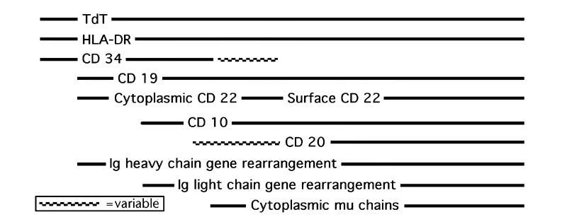
B cell biomarker expression during development
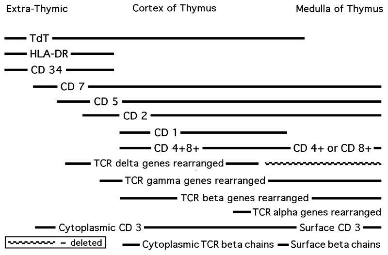
T cell biomarker expression during development
Images hosted on other servers:
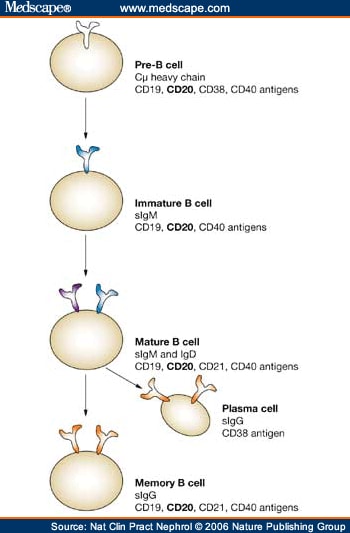
B cell development

B cell response to antigen

B cell biomarker expression during development

niches in the bone
marrow required for
B cell development

T cell activation

T cell development
Clinical features
-
B cells:
- B cell lymphomas: a clonal light chain rearrangement is usually specific for the presence of a B cell neoplasm
- X linked agammaglobulinemia: no B cell development
- Defects in receiving T cell signals can cause hyper IgM syndrome (J Allergy Clin Immunol 2010;125:778)
- 90% of peripheral T cell lymphomas have rearrangements of T alpha, beta and gamma, including all cases of mycosis fungoides and Sezary syndrome
- T cell lymphomas have no distinct marker of clonality, but cells may express an abnormal immunophenotype or TCR gene rearrangement
- As there are only 10 V (variable) regions, a polyclonal population of cells can appear oligoclonal
- T cell clonality is seen in AIDS and congenital immunodeficiency syndromes, but does NOT indicate malignancy
- Rarely a clonal band may comigrate with the germline band; solution: use 2 - 3 restriction enzymes (HindIII, EcoRI, BamHI)
T cells:
Positive stains
Additional references
