
Nasal cavity, paranasal sinuses, nasopharynx
Infectious lesions
Fungal ball
Last author update: 1 February 2016
Last staff update: 17 February 2023
Copyright: 2003-2024, PathologyOutlines.com, Inc.
PubMed Search: Fungal ball nasal

Table of Contents
Definition / general | Essential features | Terminology | Sites | Pathophysiology / etiology | Clinical features | Radiology description | Radiology images | Case reports | Treatment | Gross description | Gross images | Microscopic (histologic) description | Microscopic (histologic) images | Differential diagnosis | Additional referencesCite this page: Sun J, Brandwein-Weber MS. Fungal ball. PathologyOutlines.com website. https://www.pathologyoutlines.com/topic/nasalfungalball.html. Accessed April 18th, 2024.
Definition / general
- Noninvasive accumulation of fungal hyphae that branch at 45 degrees
- Aspergillus causes fungus balls in nasal antrum of immunocompetent patients with minimal inflammatory response, microabscesses or multinucleated giant cells
- Also causes invasive aspergillosis, regardless of immune status, with extension into retroorbital region, cranium or parapharyngeal space; often fatal
- Also causes allergic fungal sinusitis
Essential features
- Dense fungal growth with no tissue invasion
- Fruiting heads (sexual reproduction) may be seen
Terminology
- Fungal ball, mycetoma, chronic noninvasive fungal sinusitis
Sites
- Maxilla is most commonly affected
Pathophysiology / etiology
- A. fumigatus and A. flavus are the most common isolates
- Usually immunocompetent patients, often prior history of sinus disease, trauma or foreign body
Clinical features
- Nasal congestion / obstruction
- Sinus pain
Radiology description
- Expansile massive process with bony remodeling
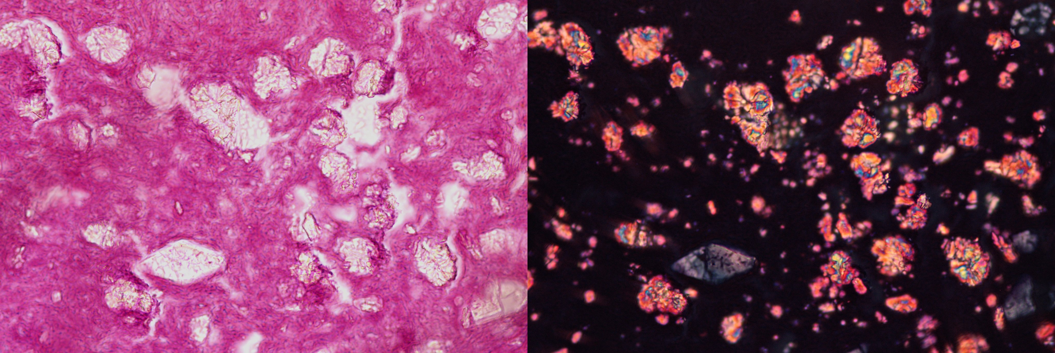
- MR / CT signaling reflects the iron, manganese and calcium content of fungal hyphae ("iron-like signalling")
Radiology images
Images hosted on other servers:

CT of face

MR of face

Likely calcific in nature

Normal pneumatization

Opacification
Case reports
- 70 and 78 year old women with fungus ball of the paranasal sinuses (Int Arch Otorhinolaryngol 2012;16:286)
Treatment
- Conservative curettage, irrigation with saline or iodine solution, surgery
Gross description
- May present as a large, expansile mass, without involvement of the underlying mucous membrane
- Grumous, friable, gray-brown-black mass, often with clotted blood
Gross images
Contributed by @Andrew_Fltv on Twitter
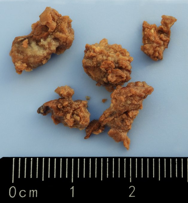
Fungal ball
Images hosted on other servers:

Maxillary sinusotomy
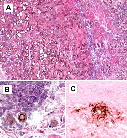
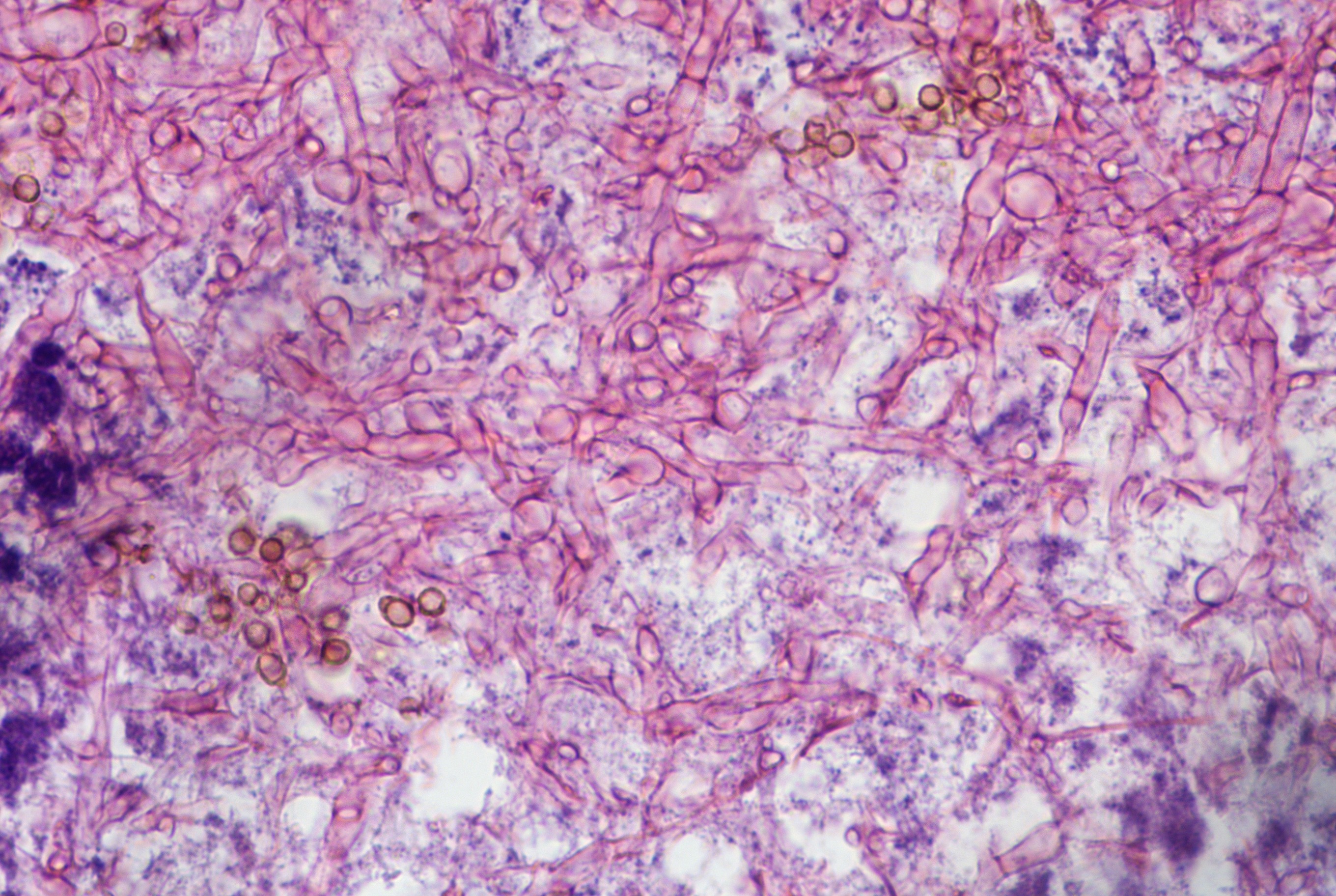
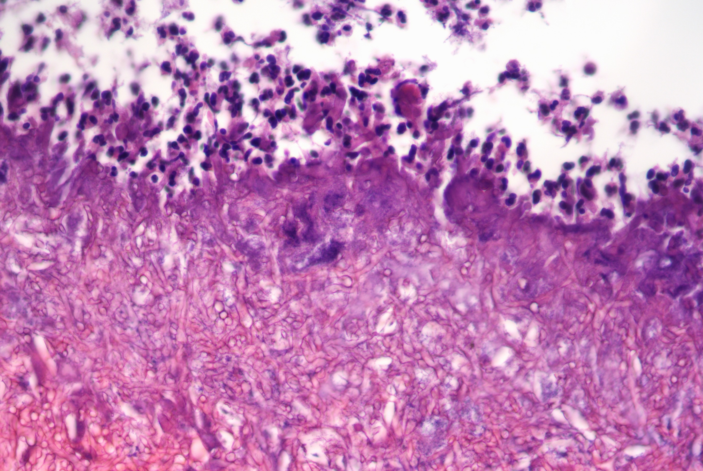
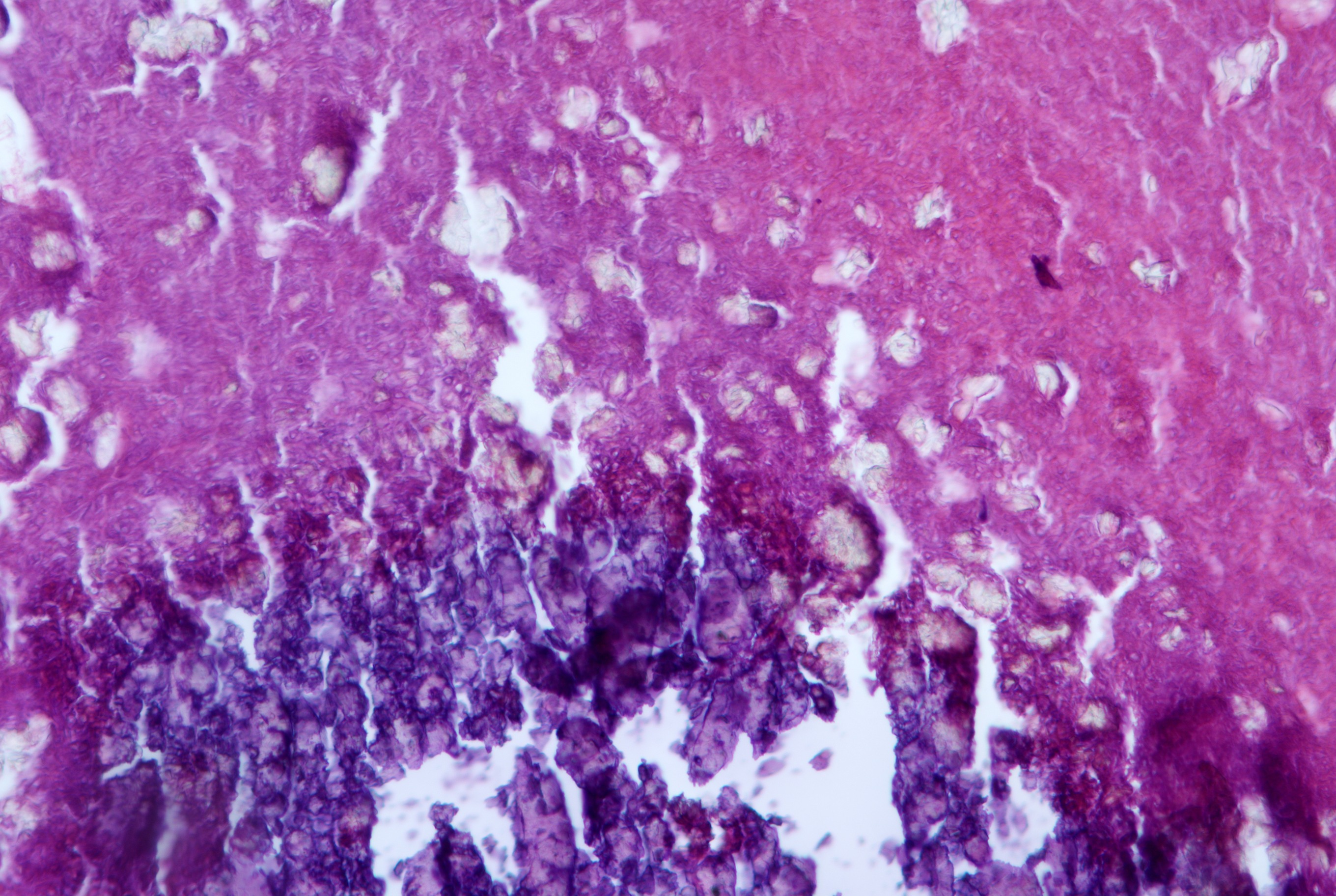
Microscopic (histologic) description
- Tightly packed laminated hyphae with inflammatory exudates and cell debris
- Pigmented hyphae may be dematiaceous group of fungi
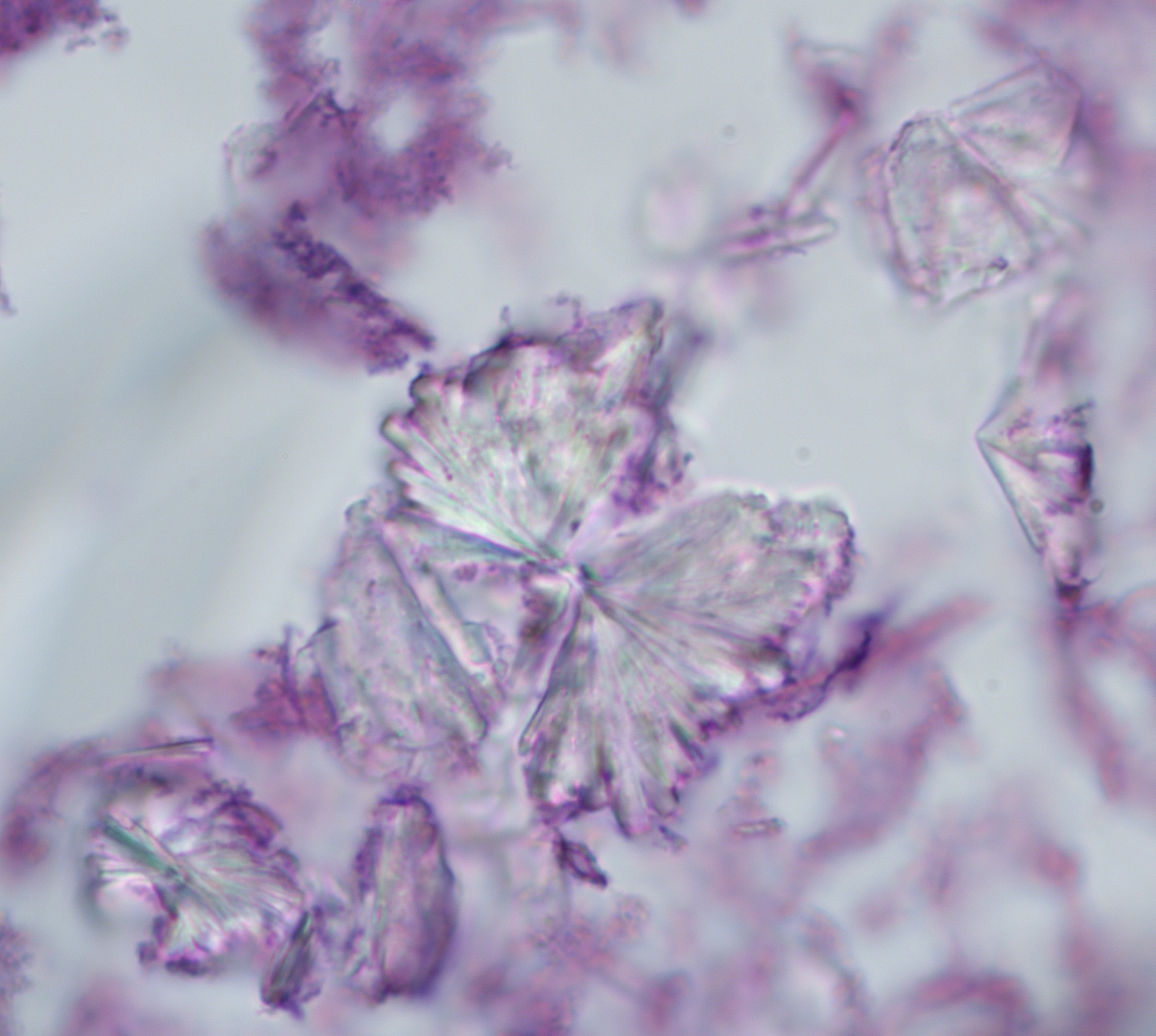
- Presence of characteristic fruiting heads is diagnostic for Aspergillus sp
- Black conidia specifically indicate Aspergillus niger
- Fungal invasion of tissue is usually not seen, although it has been reported
- No / minimal host response in mucosa
Microscopic (histologic) images
Contributed by Margie Brandwein-Gensler, M.D. and @Andrew_Fltv on Twitter
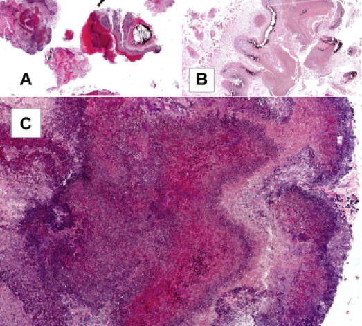
Large fungus ball
Fungal hyphae, fungal sexual reproduction (fruiting heads)
Fungal ball
Fungal ball
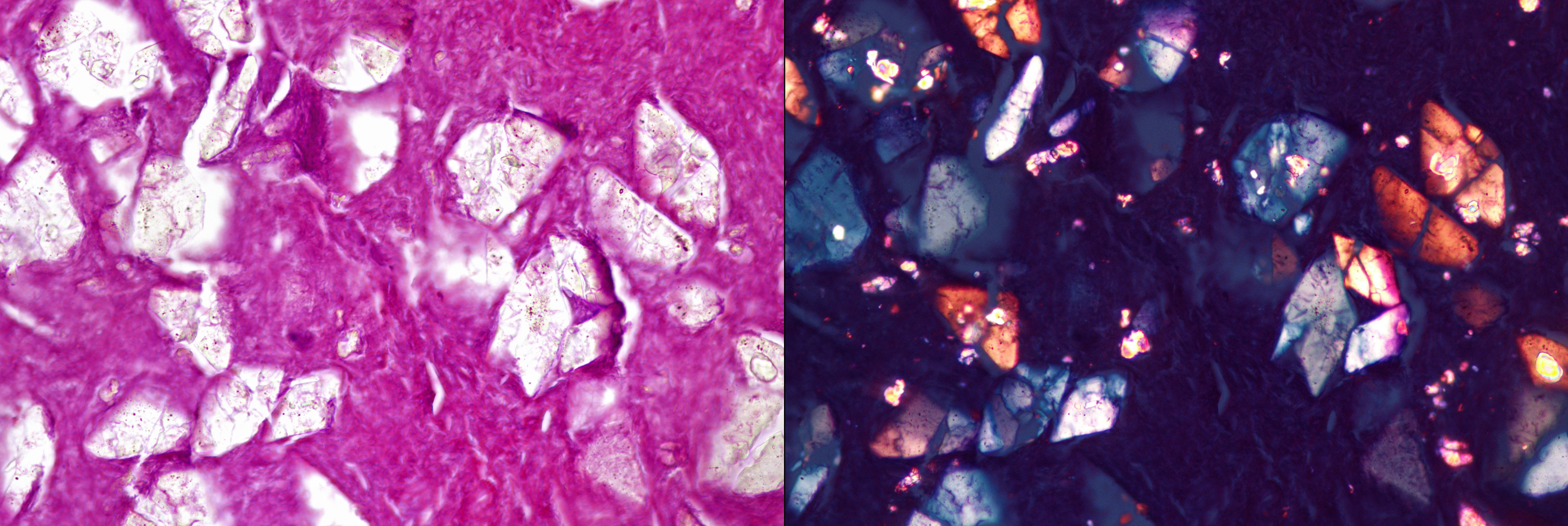
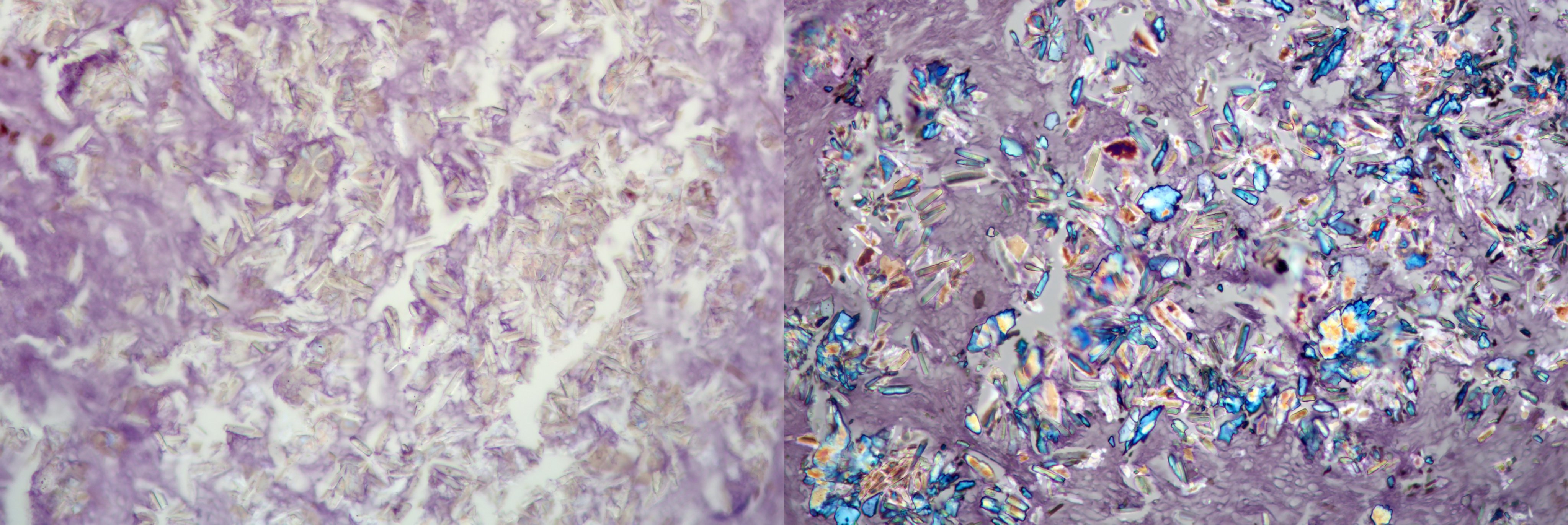
Aspergillus lesions of the sinuses
Images hosted on other servers:

Aspergilloma

Hyphae of mycetes

Foot
Differential diagnosis
- Chronic sinusitis
- Sinonasal neoplasm
- Of fungal infections:
- Alternaria
- Cladosporium trichoides
- Fusarium
- Paecilomyces
- Pseudallescheria boydii
- Zygomycetes
