
Oral cavity & oropharynx
Soft tissue tumors & proliferations
Choristoma
Author: Nella Cristina Fernandez, M.D.

Last author update: 1 August 2013
Last staff update: 12 December 2023
Copyright: 2004-2024, PathologyOutlines.com, Inc.
PubMed Search: Choristoma minor salivary glands
Table of Contents
Definition / general | Epidemiology | Sites | Clinical features | Radiology images | Case reports | Treatment | Clinical images | Gross description | Gross images | Microscopic (histologic) description | Microscopic (histologic) images | Differential diagnosisCite this page: Fernandez NC. Choristoma. PathologyOutlines.com website. https://www.pathologyoutlines.com/topic/oralcavitychoristoma.html. Accessed April 18th, 2024.
Definition / general
- Choristoma is a tumor-like mass consisting of normal cells in an abnormal location (J Oral Maxillofac Surg 2012;24:110)
- Hamartoma is a tumor-like malformation composed of mature normal cells in usual location but as a disorganized mass
Epidemiology
- Most cases occur in adults but may occur at all ages
- > 70% of lingual osseous and cartilaginous choristomas occur in females (Barnes: Surgical Pathology of the Head and Neck, 3rd Edition, 2008 [p822])
Sites
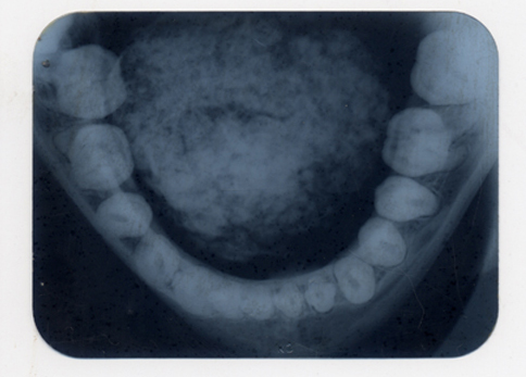
- 85% of osseous and cartilaginous choristomas occur on tongue (dorsal posterior third near foramen cecum)
Clinical features
- Firm, exophytic, asymptomatic nodule covered by intact oral mucosa
- Base may be sessile or pedunculated
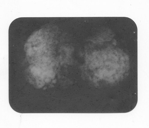
Radiology images
Case reports
- Glial choristomas of palate in newborn / one month old (J Oral Pathol Med 1997;26:147)
- 5 year old boy with buccal osseous choristoma (Oral Oncology 2005;41:198)
- 73 year old woman with asymptomatic ventral tongue lesion (Gerodontology 2009;26:78)
Treatment
- Surgical excision
Clinical images
Images hosted on other servers:

Osseous choristoma
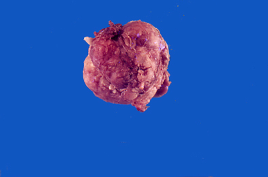
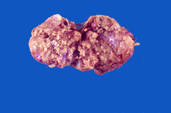
Gross description
- Firm mass covered by oral mucosa, with smooth contour
- Usually < 1.0 cm; lesions > 2.0 cm warrant careful microscopic examination (Barnes: Surgical Pathology of the Head and Neck, 3rd Edition, 2008 [p822])
Gross images

Microscopic (histologic) description
- Oral choristomas can be classified according to types of tissues they constitute (J Oral Maxillofac Surg 2012;24:110):
- Salivary gland choristoma
- Central
- Gingival
- Both have ectopic salivary gland tissue appearing as a raised tumor-like mass; must not have any connection with normal minor or major salivary glands
- Cartilagenous choristoma: composed of mature hyaline cartilage in fibrous tissue that resembles perichondrium; usually multilobulated; chondrocytes vary from small to large but lack atypia
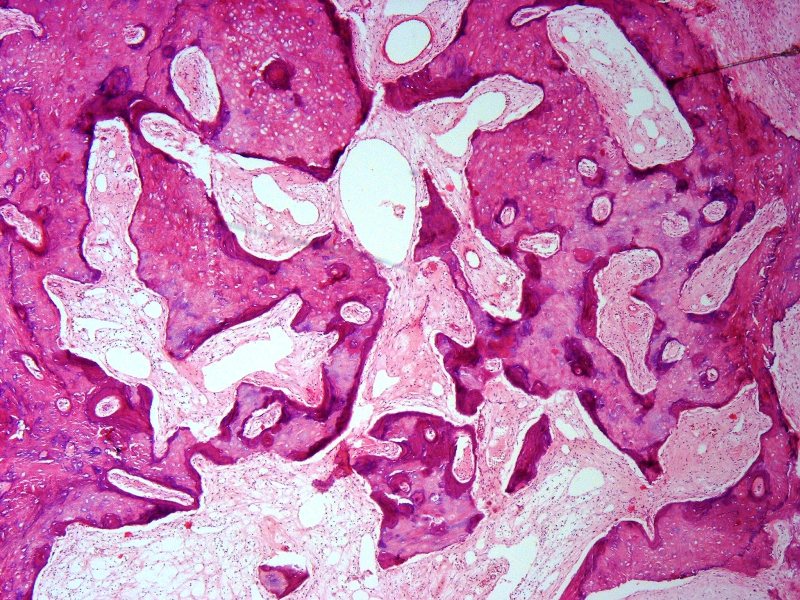
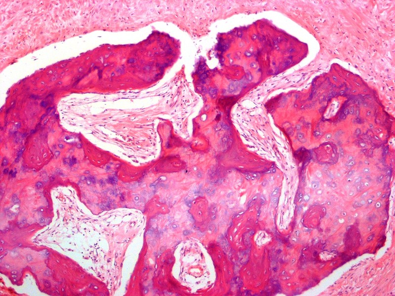
- Osseous choristoma: composed of dense mature bone; osteocytes are compact and unremarkable; no prominent osteoblastic rimming; occasionally bone and cartilage are present in same lesion
- Lingual thyroid choristoma
- Lingual sebaceous choristoma
- Glial choristoma
- Gastric / respiratory mucosal choristoma
- Solid
- Cystic
- Salivary gland choristoma
Microscopic (histologic) images


Differential diagnosis
- Cartilaginous metaplasia:
- Usually occurs in soft tissue beneath ill fitting dentures, has diffuse deposits of calcium, scattered cartilaginous cells arranged in various stages of maturation in single or clustered foci
- Pleomorphic adenoma:
- May have osteocartilaginous foci
- Salivary gland tissue:
- Choristomas lack salivary gland ductal or myoepithelial components
