
Oral cavity & oropharynx
Soft tissue tumors & proliferations
Varix / varicosities / varices
Author: Molly Housley Smith, D.M.D.
Editorial Board Member: Kelly Magliocca, D.D.S., M.P.H.
Editor-in-Chief: Debra L. Zynger, M.D.

Last author update: 13 August 2020
Last staff update: 16 June 2021
Copyright: 2020-2024, PathologyOutlines.com, Inc.
PubMed Search: Oral[TIAB] (varix[TI] OR varicosities[TI] OR varices[TI])
Table of Contents
Definition / general | Essential features | Terminology | ICD coding | Epidemiology | Sites | Etiology | Clinical features | Diagnosis | Radiology description | Prognostic factors | Case reports | Treatment | Clinical images | Gross description | Gross images | Microscopic (histologic) description | Microscopic (histologic) images | Negative stains | Sample pathology report | Differential diagnosis | Additional references | Board review style question #1 | Board review style answer #1 | Board review style question #2 | Board review style answer #2Cite this page: Smith MH. Varix / varicosities / varices. PathologyOutlines.com website. https://www.pathologyoutlines.com/topic/oralcavityvarix.html. Accessed April 20th, 2024.
Definition / general
- Oral varicosities are tortuous and aberrantly dilated venous structures that most often present on the lips and ventral tongue of older individuals
- They represent a developmental anomaly of unknown etiology with minimal clinical implications
Essential features
- Abnormally dilated venous structures
- The most common oral mucosal lesion in older adults (J Dent Res Dent Clin Dent Prospects 2019;13:24)
- Most often found under the tongue and around the lips / labial mucosa
Terminology
- Varicosity / varicosities
- Varix
- Varices
- Caviar tongue
- Phlebectasia linguae
Epidemiology
- Older individuals (67% of patients are > 60 years) (Clin Dermatol 2016;34:458)
Sites
- Sublingual varices are most common
- May also be found on skin of lip, labial mucosa or buccal mucosa
Etiology
- Unknown etiology
- Loosely associated risk factors include smoking, cardiovascular disease (e.g. hypertension), female gender, denture wearing, chronic liver disease and old age (Clin Dermatol 2016;34:458, Folia Morphol (Warsz) 2019;78:325, Medicine (Baltimore) 2019;98:e16987, Saudi Med J 2015;36:310, BMC Oral Health 2015;15:78)
Clinical features
- Presents as a chain of compressible blue-purple nodules on either side of the lingual frenum or as a single elevated or flat submucosal nodule in other locations
- May be firm when thrombosed
- Ventral tongue is the most common site, although also found on labial mucosa, skin of lip, buccal mucosa or other rare oral sites
- Painless and often inconsequential (Gerodontology 2015;32:82)
Diagnosis
- Diagnosis often is made on clinical presentation, especially when in the sublingual location; biopsy often is not required
- Diascopy may prove useful in differential diagnosis, as varicosities demonstrate blanching (Gerodontology 2015;32:82)
- A single varicosity may be diagnosed upon histologic examination when the presentation is not clinically diagnostic
Radiology description
- Occasionally, oral varicosities may demonstrate small opacifications on radiographic imaging due to phlebolith formation within a thrombus (Gerodontology 2015;32:82)
Prognostic factors
- Varicosities are of little clinical consequence
- Patients have a good prognosis, although rare cases may be associated with bleeding (Medicine (Baltimore) 2019;98:e16987)
Case reports
- 42 year old woman with bleeding varicosity at base of tongue in association with liver cirrhosis (Medicine (Baltimore) 2019;98:e16987)
- 60 year old white woman with oral varicosity treated with sclerotherapy (Med Oral Patol Oral Cir Bucal 2006;11:E44)
- 62 year old woman with lingual varicosities visualized via dermoscopy / mucoscopy (Dermatol Pract Concept 2018;8:54)
Treatment
- Varicosities generally are not treated unless thrombosed or displeasing to the patient in form or function
- In such cases, conservative surgical excision or sclerotherapy is sufficient (Med Oral Patol Oral Cir Bucal 2018;23:e180)
- Safe and effective treatment has been performed with diode and Nd:YAG lasers (J Contemp Dent Pract 2015;16:723)
Clinical images
Contributed by Molly Housley Smith, D.M.D.
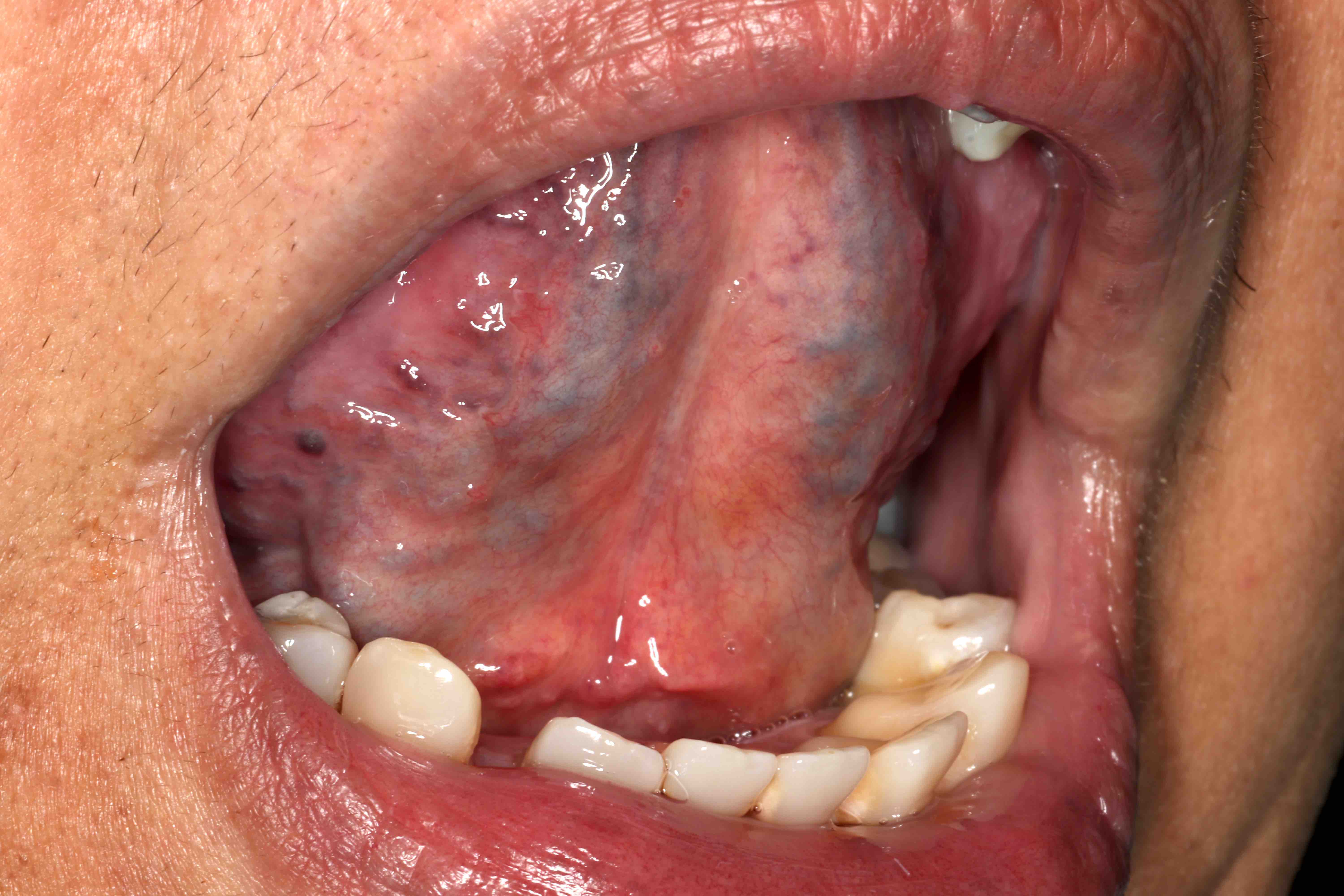
Sublingual varicosities
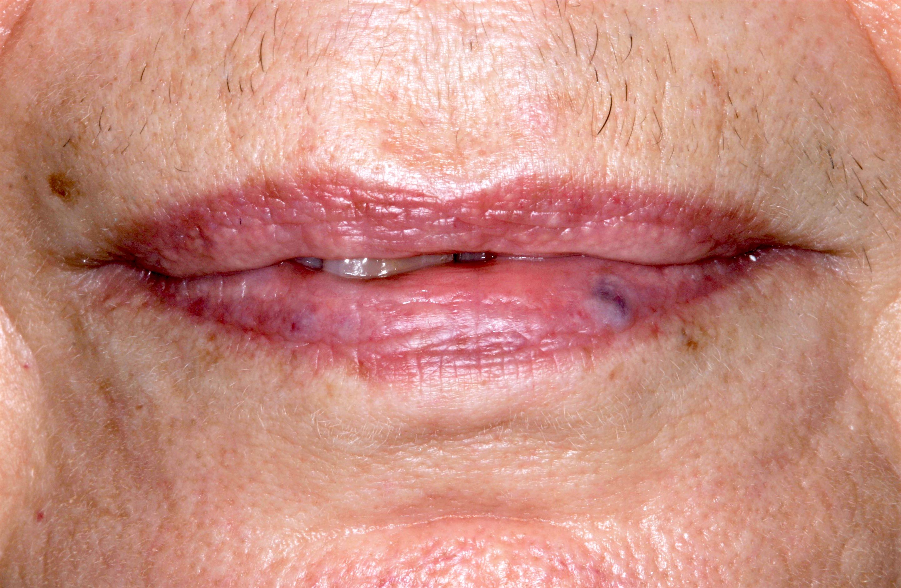
Varicosity on lower lip
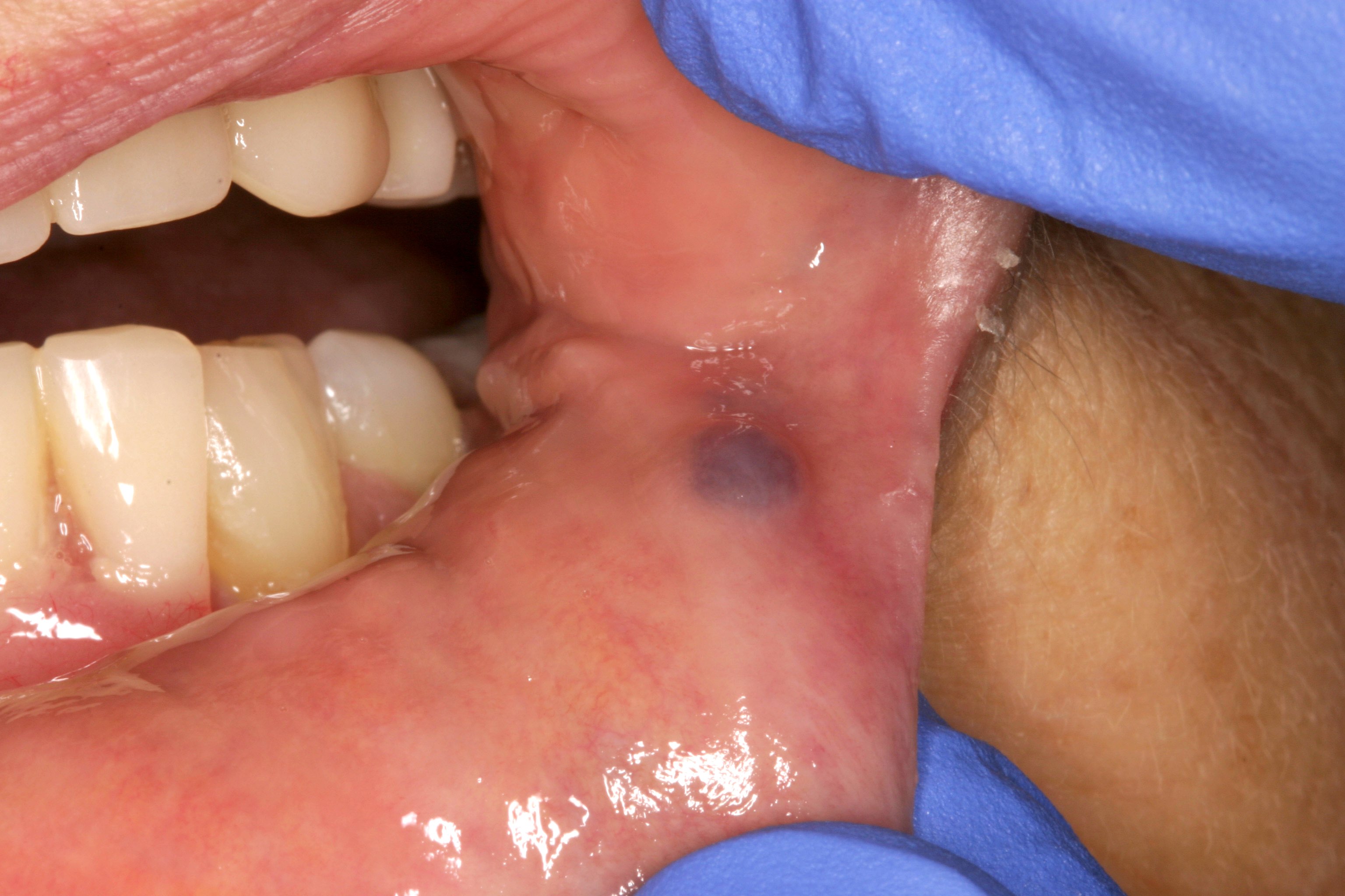
Thrombosed varicosity of labial mucosa
Gross description
- Tan surface with raised or flat blue-purple-black ovoid structure, which may be firm or depressible
- Cut surface shows dilated or collapsed vascular space that may demonstrate luminal accumulation of brown erythrocytes
Gross images
Contributed by Molly Housley Smith, D.M.D.
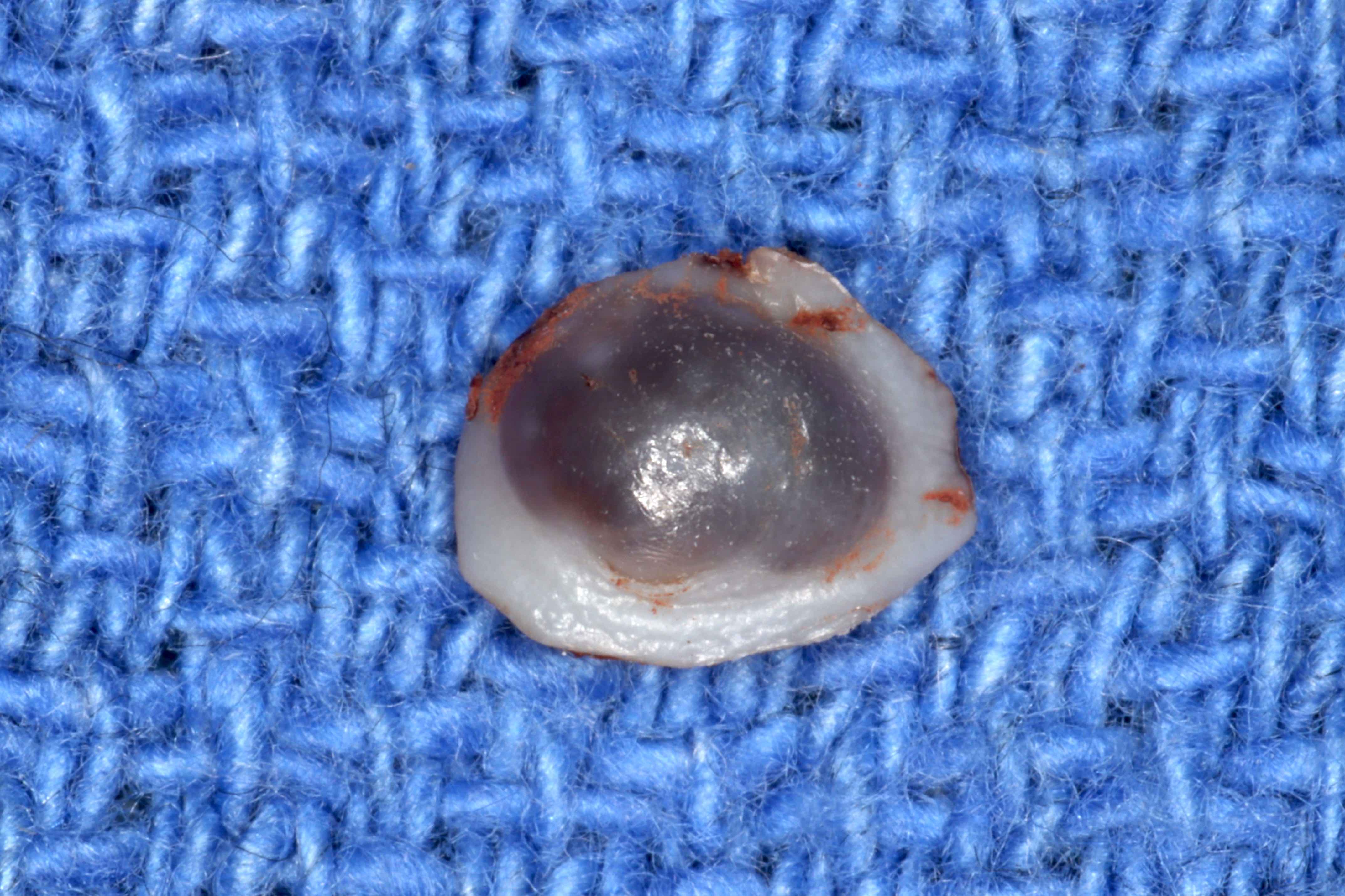
Oral varicosity
Microscopic (histologic) description
- 1 - 3 tortuous, enlarged venous structures lined by a thin layer of endothelium (Gerodontology 2015;32:82)
- Thrombosis or organizing thrombosis is often present, which is depicted by concentric layers of erythrocytes and platelets, referred to as lines of Zahn
- Aggregates of granulation tissue may also be present during formation
- Concentric rings of eosinophilic calcifications may be present within the lumen
Microscopic (histologic) images
Contributed by Molly Housley Smith, D.M.D.
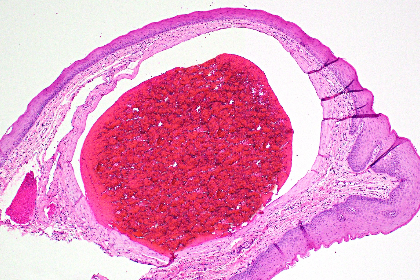
Oral varicosity
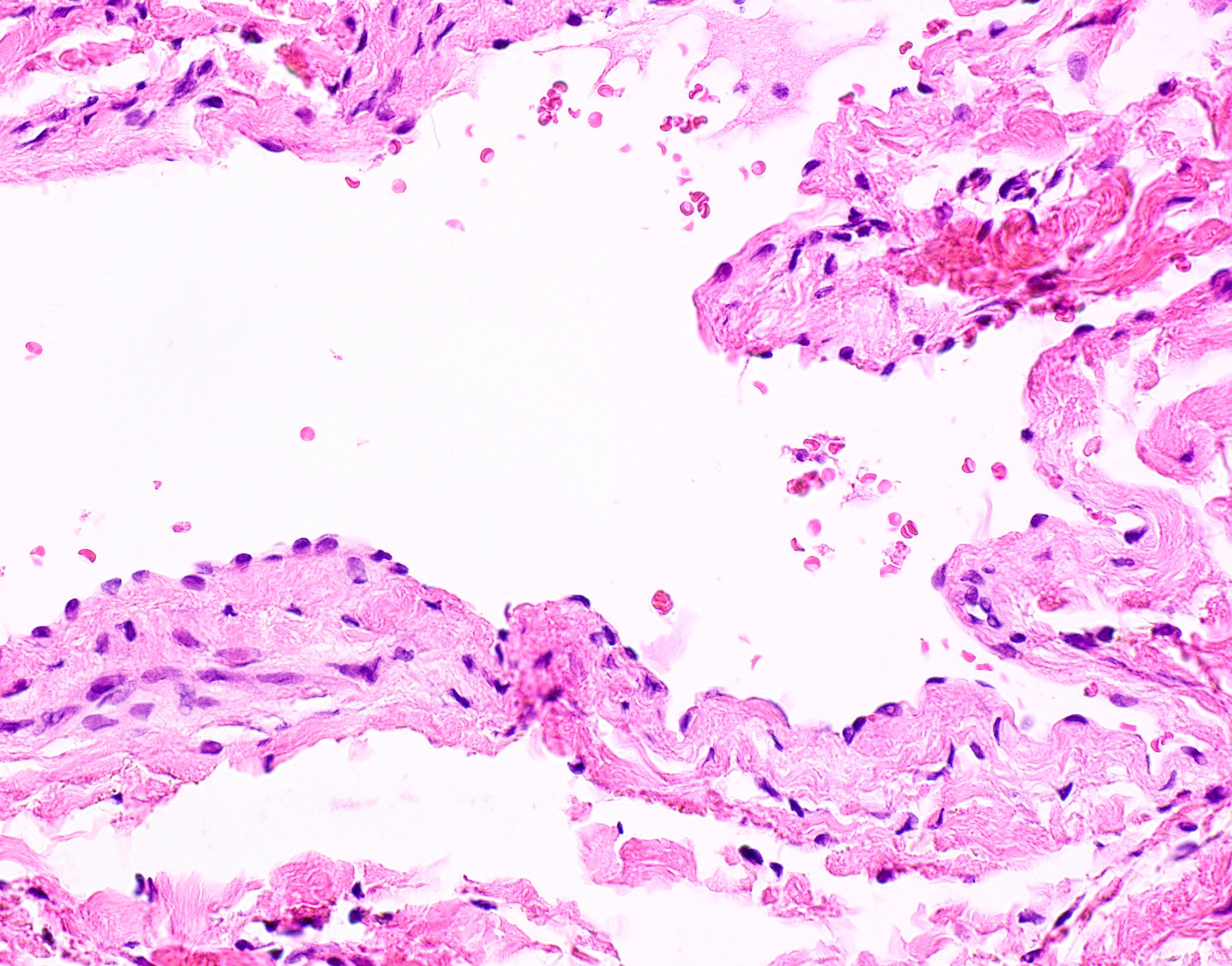
Compressed oral varicosity
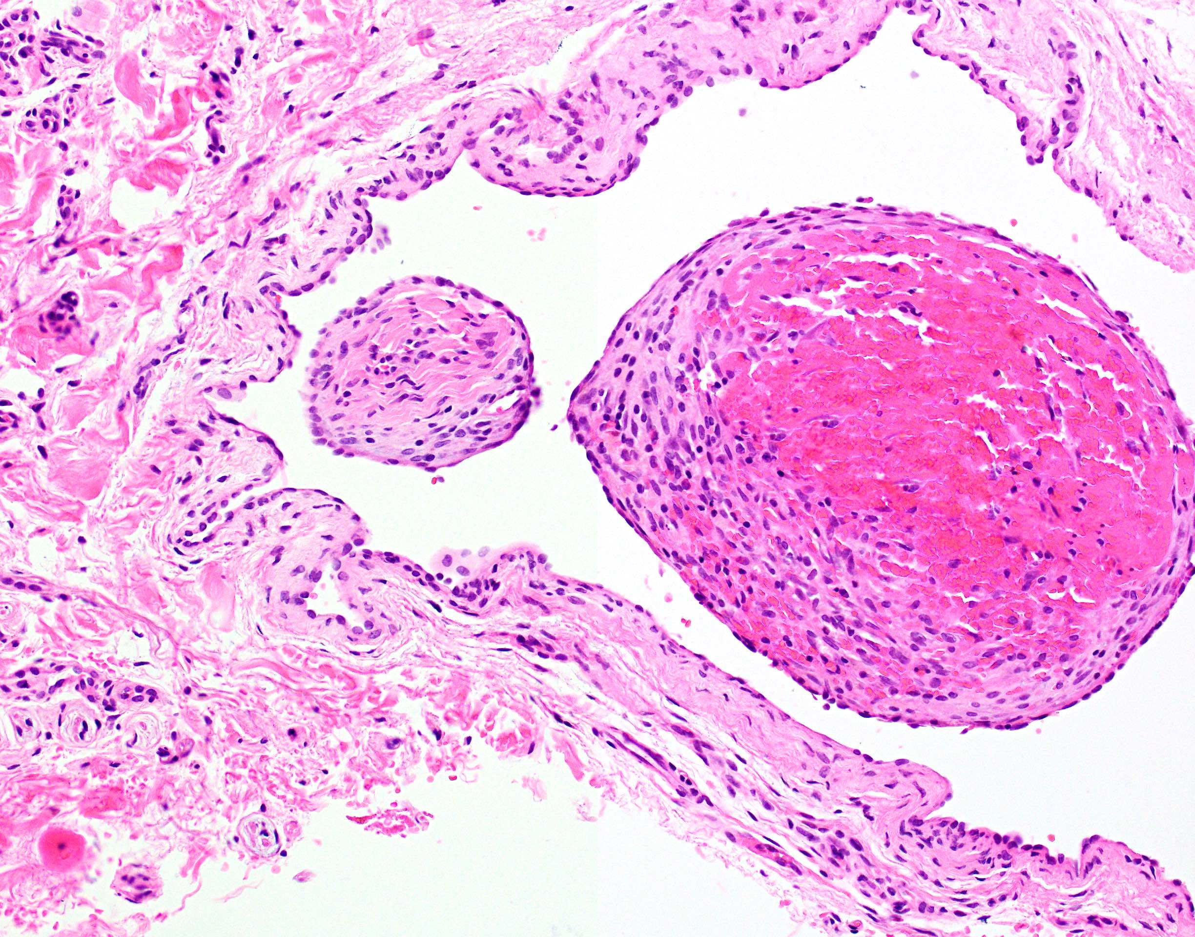
Oral varicosity with organizing thrombus
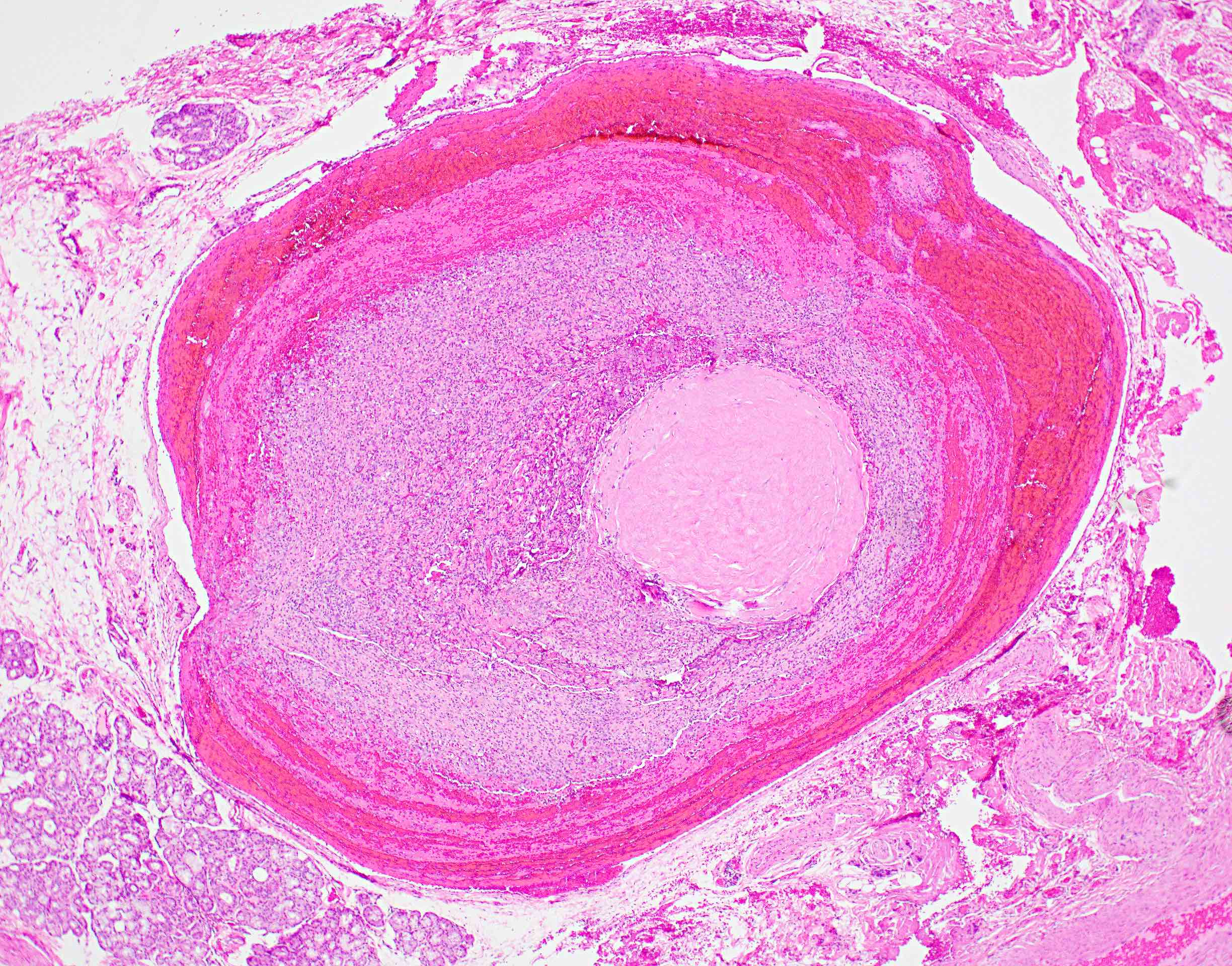
Phlebolith formation
Negative stains
Sample pathology report
- Lower lip, excision:
- Vascular anomaly consistent with varicosity
Differential diagnosis
- Sublingual varicosities are clinically diagnostic and do not require biopsy
- Clinical differential diagnosis for a single, blue oral varix may include the following:
- Hemangioma:
- An abnormal collection of more than 3 dilated vessels, seen on light microscopy
- Congenital
- Often GLUT1 positive (Oral Dis 2007;13:51)
- Vascular leiomyoma (angioleiomyoma):
- Histopathologic examination reveals a well defined proliferation of smooth muscle emanating from vascular walls
- Positive for alpha smooth muscle actin
- Salivary tumors / reactive processes, differentiated upon biopsy:
- Amalgam tattoo:
- May demonstrate opacification on radiograph
- Flat, blue-black pigmentation
- Histopathologic examination reveals foreign particles within connective tissue
- Melanocytic nevus (especially blue nevus):
- Histopathologic examination reveals focal collection of melanocytes
- Melanoma:
- Often irregular borders
- Histopathologic examination reveals abnormal increase in melanocytes
- Most common on the maxillary gingiva and hard palate
- Focal melanosis / melanocytic macule:
- Flat, never raised
- Focal accumulation of melanin without increase in melanocytes, seen on light microscopy
- Hemangioma:
Additional references
Board review style question #1
A biopsy of an elevated blue mass on the oral labial mucosa is shown above. What is the diagnosis?
- Amalgam tattoo
- Mucocele
- Mucoepidermoid carcinoma
- Varicosity
Board review style answer #1
Board review style question #2
A 68 year old woman with a history of hypertension presents to the clinic with concern about blue bumps under her tongue. What is the most likely diagnosis?
- Amalgam tattoo
- Medication related pigmentation
- Oral lichen planus
- Sublingual varicosities
Board review style answer #2
