

Ovary
Inflammatory
Xanthogranulomatous oophoritis
Authors: Jack Reid, M.D., Jian-Hua Qiao, M.D.
Editor-in-Chief: Debra L. Zynger, M.D.


Last author update: 25 October 2019
Last staff update: 23 August 2022
Copyright: 2019-2024, PathologyOutlines.com, Inc.
PubMed Search: Xanthogranulomatous oophoritis
Table of Contents
Definition / general | Essential features | Terminology | ICD coding | Epidemiology | Sites | Pathophysiology | Etiology | Clinical features | Diagnosis | Radiology description | Radiology images | Prognostic factors | Case reports | Treatment | Gross description | Gross images | Microscopic (histologic) description | Microscopic (histologic) images | Positive stains | Negative stains | Sample pathology report | Differential diagnosis | Additional references | Board review style question #1 | Board review style answer #1 | Board review style question #2 | Board review style answer #2Cite this page: Reid J, Qiao JH. Xanthogranulomatous oophoritis. PathologyOutlines.com website. https://www.pathologyoutlines.com/topic/ovarynontumorxanthogranoophoritis.html. Accessed April 24th, 2024.
Definition / general
- Xanthogranulomatous oophoritis is an uncommon, nonneoplastic, chronic process (Iran J Pathol 2018;13:372)
- Destruction by massive cellular infiltration of foamy histiocytes admixed with multinucleated giant cells, plasma cells, fibroblasts, neutrophils and foci of necrosis (Iran J Pathol 2018;13:372)
Essential features
- Xanthogranulomatous oophoritis is a rare form of chronic oophoritis (J Indian Med Assoc 2012;110:653)
- Affects fallopian tubes or ovaries focally or entirely, forming a mass-like lesion in the pelvic cavity that invades the surrounding tissues (J Cancer 2012;3:100)
Terminology
- Xanthogranulomatous oophoritis
- Xanthogranulomatous inflammation of the ovary
- Xanthogranulomatous inflammation of the female genital tract
- Ovarian fibroxanthoma
ICD coding
- ICD-10: N70.92 - oophoritis, unspecified
Epidemiology
- Most frequent in females of reproductive age (range: 23 - 72 years)
- Average age of patients with affected ovaries is 31 years (AJR Am J Roentgenol 2002;178:749)
- Only 13 related cases of xanthogranulomatous inflammation involving fallopian tube or ovary have been described through 2012 (J Cancer 2012;3:100)
Sites
- Xanthogranulomatous inflammation of the female genital tract is not common and usually involves the endometrium; however, xanthogranulomatous inflammation of the ovaries is a rare entity (J Ayub Med Coll Abbottabad 2017;29:162)
Pathophysiology
- Exact pathophysiology is unknown
- Associated with infection, ineffective antibiotic therapy
- Ineffective clearance of bacteria by phagocytes, endometriosis, intrauterine contraceptive device, pelvic inflammatory disease and drugs (antibiotics) is suggested (Iran J Pathol 2018;13:372)
Etiology
- Development can be of multiple predisposing factors, for example, pelvic inflammatory diseases, intrauterine device use, uterine leiomyoma, endometriosis and inappropriate antibiotic intake (BMJ Case Rep 2015 Jun 25;2015)
Clinical features
- Longstanding history of pelvic inflammatory disease
- Symptoms such as anorexia, fever, suprapubic pain, menorrhagia or vaginal bleeding, adnexal tenderness and a pelvic mass are the usual chief complaints (Iran J Pathol 2018;13:372)
Diagnosis
- Careful histopathology by experienced pathologists aided by immunohistochemistry can lead to a definitive diagnosis (J Ayub Med Coll Abbottabad 2017;29:162)
Radiology description
- Ultrasound of the abdomen reveals a thick walled, cystic hyperechoic ovarian mass (Iran J Pathol 2018;13:372)
- Pelvic CT scan shows a peripherally enhancing ovarian cystic lesion, which represents either an inflammatory process or ovarian neoplasm (Iran J Pathol 2018;13:372)
- Presence of nonenhancing intramural nodules in the thickened wall of an ovarian cystic mass may be a unique MRI indicator of xanthogranulomatous oophoritis (AJR Am J Roentgenol 2002;178:749)
Radiology images
Images hosted on other servers:

Pelvic sonography
shows a mixed
echogenicity mass

T2 MRI shows multiseptated cystic mass


Axial T2 weighted turbo spin echo MRI (TR / TE, 3900 / 99)

Axial T1 weighted spin echo MRI (800 / 12)
Prognostic factors
- Since xanthogranulomatous oophoritis is usually associated with pelvic inflammatory disease, endometriosis, intrauterine death, etc., these patients should be followed up closely (Indian J Pathol Microbiol 2010;53:197)
Case reports
- 20 year old woman presenting with a tubo-ovarian mass (Iran J Pathol 2018;13:372)
- 24 year old woman who was misdiagnosed with an ovarian neoplasm (J Midlife Health 2018;9:41)
- 25 year old woman presented with intermittent fever and lower abdominal pain (Indian J Pathol Microbiol 1999;42:89)
- 26 year old woman with a 10 year history of pelvic inflammatory disease (Int J Gynecol Pathol 1984;3:398)
- 31 year old Lebanese woman, 8 years following an open appendectomy as a reaction to talcum powder present on surgical gloves (J Med Liban 2012;60:169)
- 45 year old woman with heavy vaginal bleeding for 20 days (BMJ Case Rep 2015 Jun 25;2015)
- 47 year old woman with a several month history of intermittent abdominal pain localized to the left suprapubic region (Arch Pathol Lab Med 2001;125:260)
- 48 year old woman with a 3 day history of lower abdominal pain and fever (AJR Am J Roentgenol 2002;178:749)
Treatment
- Treatment of choice is oophorectomy (J Midlife Health 2018;9:41)
Gross description
- Ovarian lesion usually has a solid mass with rugged outer surface
- Serial sectioning mostly reveals tan, grayish white and necrotic cut surfaces (J Cancer 2012;3:100)
Gross images
Contributed by Jian-Hua Qiao, M.D.
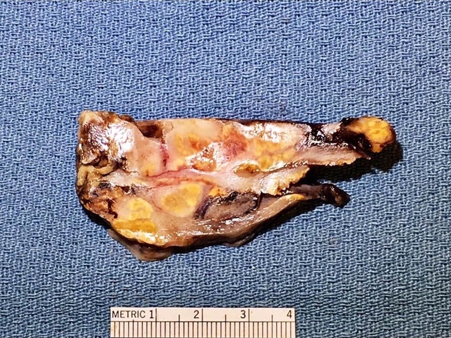
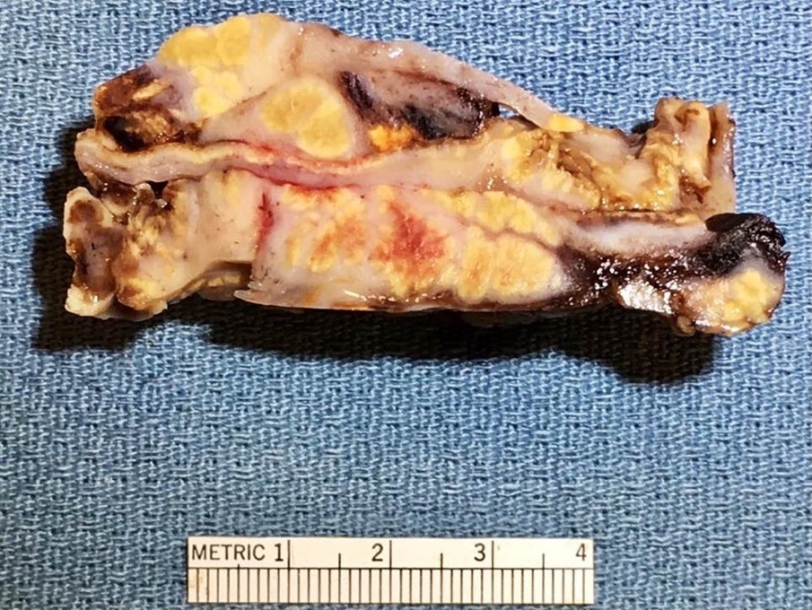
Yellow nodules
Microscopic (histologic) description
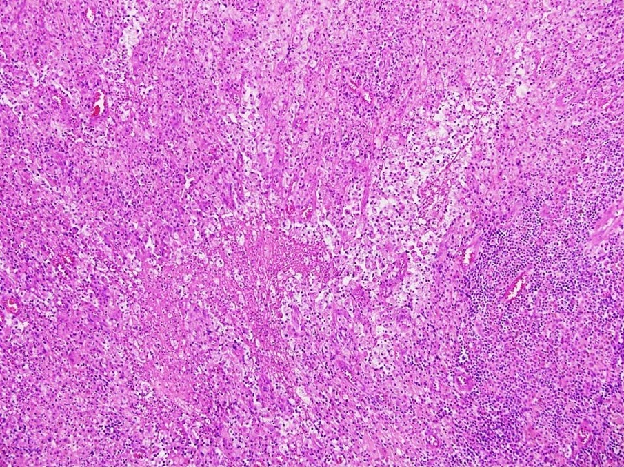
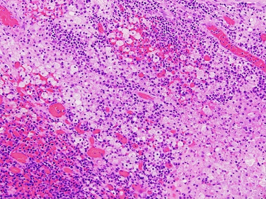
- Usually shows characteristic dense fibrosis with infiltration of the ovarian stroma by sheets and nodules of foamy macrophages, many histiocytes, lymphocytes and neutrophils (Iran J Pathol 2018;13:372)
Microscopic (histologic) images
Contributed by Jian-Hua Qiao, M.D.
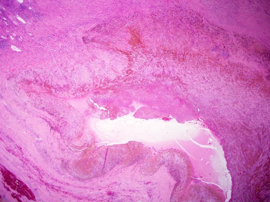

Foamy histiocytes in cystic wall
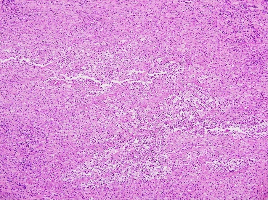
Foamy histiocytes mixed with inflammatory cells


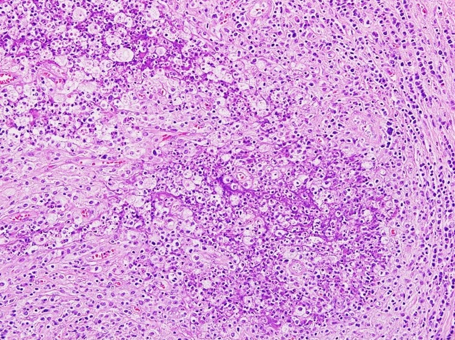
Foamy histiocytes
Positive stains
- CD68 in foamy histiocytes
- CD3 in T cells
- CD20 in B cells (J Cancer 2012;3:100)
Sample pathology report
- Left ovary and fallopian tube, left salpingo-oophorectomy:
- Cystic ovary with endometriosis
- Extensive xanthogranulomatous oophoritis
- Fallopian tube with no significant pathologic change
Differential diagnosis
- Ovarian carcinoma:
- May be difficult to differentiate with imaging
- Gross and microscopic pathologic findings will easily differentiate malignant tumors from inflammatory lesions
- Endometrioma:
- Usually filled with chocolate colored fluid
- Histologic sections of cystic wall show hemosiderin laden macrophages without abundant foamy histiocytes
- Tubo-ovarian mass:
- Benign tumors can occur in the tubo-ovarian area, i.e. leiomyoma
- Gross and histologic examinations reveal smooth muscle neoplasm rather than inflammatory process / lesion
Additional references
Board review style question #1
This gross photo of an ovary is most consistent with:

- Endometriotic cyst / endometrioma
- Leiomyoma
- Ovarian malignancy / carcinoma
- Ovarian simple cyst
- Xanthogranulomatous oophoritis
Board review style answer #1
E. Xanthogranulomatous oophoritis (the gross photo shows variably sized yellow nodules, which are
consistent with changes of xanthogranulomatous nodules of ovary).
Comment Here
Reference: Xanthogranulomatous oophoritis
Comment Here
Reference: Xanthogranulomatous oophoritis
Board review style question #2
This microscopic photo of an ovary includes changes of:

- Endometriosis and xanthogranulomatous oophoritis
- Hemorrhagic corpus luteum cyst
- Ovarian malignancy / carcinoma
- Ovarian simple cyst
Board review style answer #2
A. Endometriosis and xanthogranulomatous oophoritis. The microscopic photo shows endometriosis with increased foamy histiocytes in stroma and adjacent soft tissue, consistent with xanthomatous change.
Comment Here
Reference: Xanthogranulomatous oophoritis
Comment Here
Reference: Xanthogranulomatous oophoritis
