
Microbiology & infectious diseases
Parasites-cutaneous / subcutaneous / soft tissue
Toxocara
Author: Nat Pernick, M.D.

Last author update: 15 April 2019
Last staff update: 5 October 2020
Copyright: 2019-2024, PathologyOutlines.com, Inc.
PubMed Search: Toxocara pathology skin
Table of Contents
Clinical features | Case reports | Gross description | Gross images | Cytology images | Differential diagnosis | Additional referencesCite this page: Pernick N. Toxocara. PathologyOutlines.com website. https://www.pathologyoutlines.com/topic/parasitologytoxocara.html. Accessed April 20th, 2024.
Clinical features
- Member of ascarid family since adult worm has 3 lips
- Cats are infected by Toxocara cati and Toxascaris leonina
- Ingested eggs can hatch to release larva that cause larva migrans
- Adult worms are not a risk to humans
- T. leonina: little risk of clinical infection, although has been reported rarely in a human leg abscess and in children as visceral larval migrans
- T. cati: higher risk than T. leonina of visceral larval migrans (visceral, ocular and neural) (Pritt: Creepy Dreadful Wonderful Parasites Blog - Answer to Case 538 [Accessed 15 April 2019])
Case reports
- 50 year old woman with objects seen in a wet mount of a concentrated stool specimen (Pritt: Creepy Dreadful Wonderful Parasites Blog - Case of the Week 529 [Accessed 20 September 2019])
Gross description
- Cervical alae are short and wide in T. cati and long and narrow in T. leonina
- Both species have eggs about 70 microns
- T. cati has a pitted shell but T. leonina has a smooth shell (Pritt: Creepy Dreadful Wonderful Parasites Blog - Answer to Case 538 [Accessed 15 April 2019])
Gross images
Contributed by Bobbi Pritt, M.D.

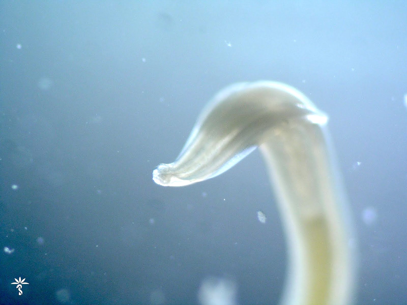
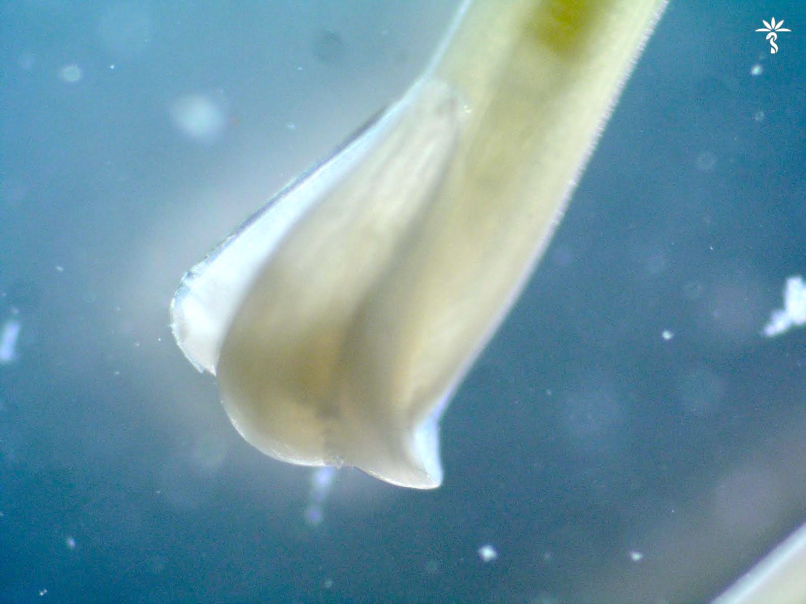

T. cati worms
Cytology images
Contributed by Bobbi Pritt, M.D.
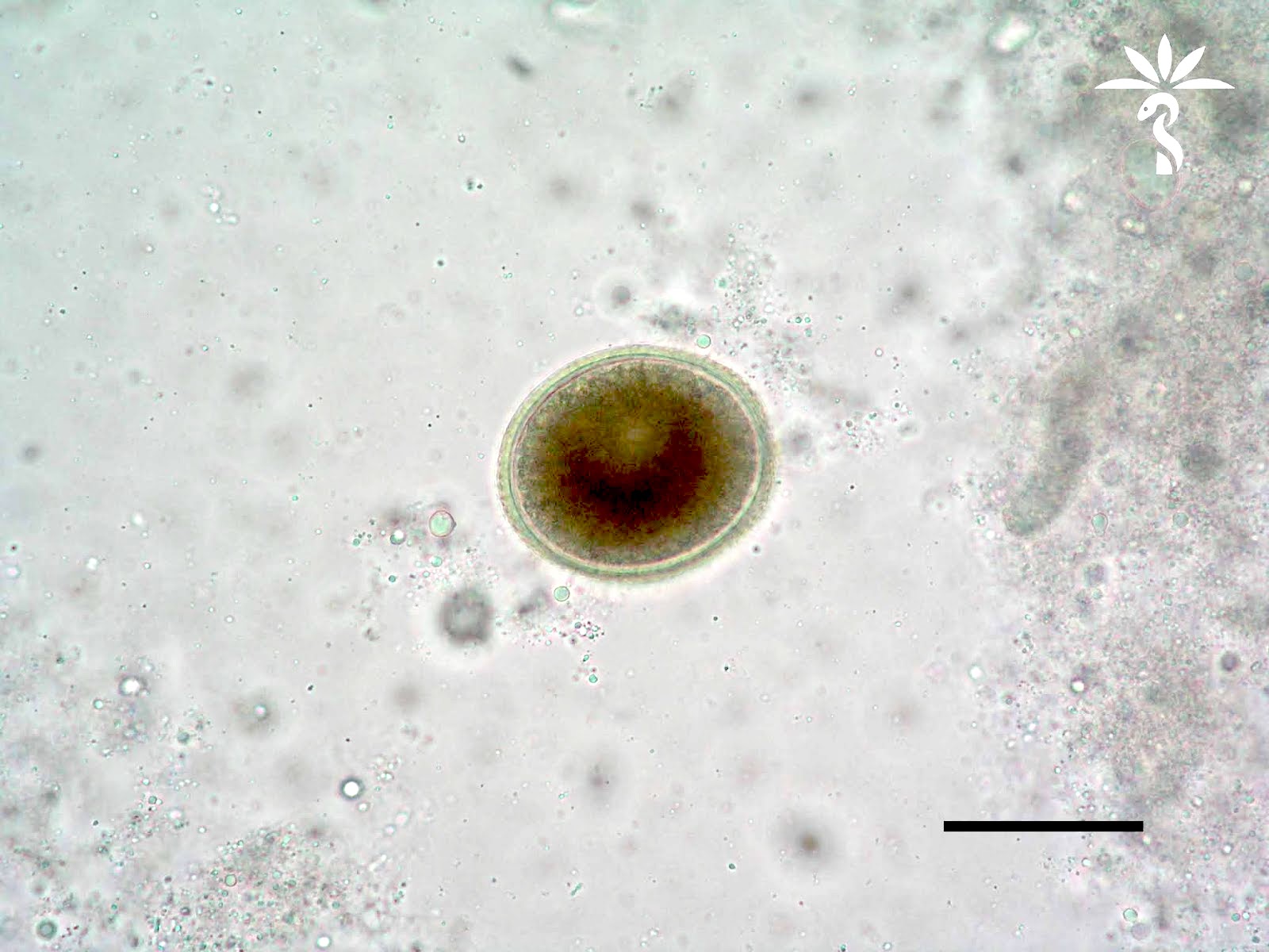
T. cati egg
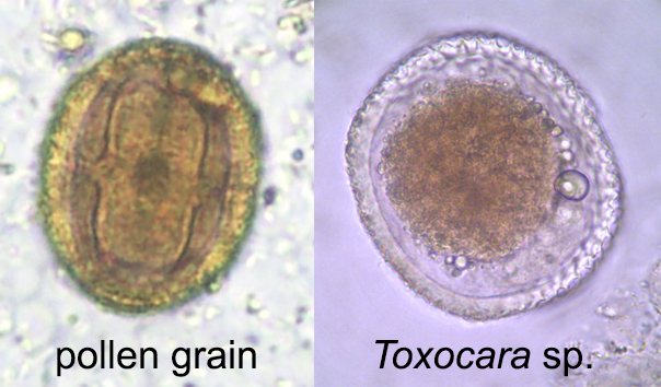
Pollen grain and Toxocara egg
Differential diagnosis
- Pollen grain (Pritt: Creepy Dreadful Wonderful Parasites Blog - Answer to Case 529 [Accessed 20 September 2019]):
- Superficially resembles ascarid eggs
- Size and surface texture of eggs help to differentiate
- Toxocara canis is 80 - 85 micrometers in greatest dimension with a golfball pitted surface texture
- T. cati is 65 - 70 micrometers and also has the golfball pitting but smaller and less distinct than T. canis
- Baylisascaris procyonis is 63 - 88 micrometers and has a granular surface texture
- Pores also help to differentiate the pollen grain from a true helminth egg
