

Penis & scrotum
Inflammatory
Scrotal calcinosis
Editorial Board Member: Debra L. Zynger, M.D.
Deputy Editor-in-Chief: Maria Tretiakova, M.D., Ph.D.


Last author update: 25 April 2023
Last staff update: 30 November 2023
Copyright: 2002-2024, PathologyOutlines.com, Inc.
PubMed Search: Scrotal calcinosis
Table of Contents
Definition / general | Essential features | Terminology | ICD coding | Epidemiology | Sites | Etiology | Clinical features | Diagnosis | Radiology description | Radiology images | Prognostic factors | Case reports | Treatment | Clinical images | Gross description | Gross images | Microscopic (histologic) description | Microscopic (histologic) images | Cytology description | Cytology images | Positive stains | Videos | Sample pathology report | Differential diagnosis | Board review style question #1 | Board review style answer #1 | Board review style question #2 | Board review style answer #2Cite this page: Uddin Z, Ud Din N. Scrotal calcinosis. PathologyOutlines.com website. https://www.pathologyoutlines.com/topic/penscrotumidiopathiccalcinosis.html. Accessed April 25th, 2024.
Definition / general
- Nonneoplastic condition characterized by multiple calcific nodular deposits in scrotal skin, caused by dystrophic calcification (G Ital Dermatol Venereol 2015;150:495)
Essential features
- Clinically, multiple subcutaneous painless nodules in scrotum
- Multiple varying sized calcified deposits in the dermis and subcutis
- Rarely shows extensive involvement of the scrotum
Terminology
- Previously described as idiopathic scrotal calcinosis
- First described by Lewinski in 1883 and was established as a distinct entity by Shapiro et al. in 1970 (Ann Dermatol 2018;30:236)
Epidemiology
- Lesions begin in childhood or early adulthood
- Mean age of 31.5 years, 70% in 30s to 40s, range of 15 - 77 years (Dermatol Online J 2010;16:5)
Sites
- Scrotum
- Exceedingly rare vulvar calcinosis may represent the female counterpart of scrotal calcinosis (J Low Genit Tract Dis 2019;23:75)
Etiology
- Pathogenesis of scrotal calcinosis remains unknown and has been the subject of longterm controversy (Urol Case Rep 2020;32:101225)
- Originally termed idiopathic scrotal calcinosis (ISC); however, many authors suggest that at least some cases are not genuinely idiopathic
- Dubey et al. evaluated 100 cases, out of which 53% had associated cysts containing calcific material (Dermatol Online J 2010;16:5)
- Shah et al. found cyst walls in 14 out of 20 cases and suggested that scrotal calcinosis results from calcification of hair follicle or epidermal cysts but as reported in most of the cases, this epithelium disappears and may not be seen (Am J Dermatopathol 2007;29:172)
- The following are various hypotheses for the origin of scrotal calcinosis:
- Truly idiopathic (Heliyon 2022;8:e10762)
- Secondary to calcification of epidermal inclusion cysts, pilar cysts or eccrine gland duct milia (G Ital Dermatol Venereol 2015;150:495, Am J Clin Exp Urol 2022;10:194)
- Result from dystrophic calcification in the degenerated and necrotic dartos smooth muscle of scrotum, which may be due to trauma (Acta Dermatovenerol Alp Pannonica Adriat 2022;31:123)
- Secondary to localized skin infection / inflammation causing a sudden increase in collagen synthesis around blood vessels and fat, with subsequent calcium deposition (Am J Clin Exp Urol 2022;10:194)
- Some suggested that it could be the end stage of a single disease that started from multiple etiologies (Clin Cosmet Investig Dermatol 2018;11:333)
Clinical features
- Typically presents as multiple nodular, bosselated scrotal masses of many years duration before the patients seek medical advice (Am J Clin Exp Urol 2022;10:194)
- Lesions increase in size and number over time (Dermatol Online J 2010;16:5)
- They are pearly / yellowish white nodular masses of different sizes in the scrotum with no obvious cause, rock hard, partly smooth surface and an individually uneven surface (Am J Clin Exp Urol 2022;10:194)
- Number of lesions varies from 1 to 100 nodules, 45% of patients have 11 to 20 lesions (Dermatol Online J 2010;16:5)
- Size ranging from a few millimeters to several centimeters
- Some of the nodules get fused with the surrounding ones to form a large mass (Heliyon 2022;8:e10762)
- Generally asymptomatic or feeling of heavy sensation when extensive; in some rare cases it can cause itching or pelvic and perineal pain or chalky white exudate or secondary infection of the skin (Heliyon 2022;8:e10762, Am J Clin Exp Urol 2022;10:194, Zhonghua Nan Ke Xue 2019;25:544, Int J Surg Case Rep 2018;44:51)
- Main discomfort to the patient is psychological and aesthetic with impairment of quality of life, self esteem and fear of sexual dysfunction
- In most cases, there is no impairment of calcium and phosphorus metabolism (Am J Clin Exp Urol 2022;10:194)
- There is no history of endocrine disorder, trauma or a family member with similar diseases (Zhonghua Nan Ke Xue 2019;25:544)
Diagnosis
- Diagnosis is usually straight forward and is solely based on physical exam (G Ital Dermatol Venereol 2015;150:495, Dermatol Online J 2010;16:5)
Radiology description
- Ultrasound: typically appear as round or oval echogenic foci with or without acoustic shadowing in cases of large and small calculi, respectively (J Ultrasound Med 2007;26:1775)
Radiology images
Contributed by Nasir Ud Din, M.B.B.S.

Incidentally found scrotal calcinosis
Prognostic factors
- Nonneoplastic lesions with little chance of recurrence following localized excision
Case reports
- 22 and 23 year old brothers with familial example of scrotal calcinosis (Ann Dermatol 2018;30:236)
- 23 year old man presenting with extensive scrotal calcinosis associated with vitiligo (BMJ Case Rep 2020;13:e238695)
- 24 year old man with scrotal calcinosis treated by surgical excision (Urol Case Rep 2020;32:101225)
- 28 year old man with hypercalcemia caused by isotretinoin taken for the treatment of severe acne presented with scrotal calcinosis (Acta Dermatovenerol Alp Pannonica Adriat 2022;31:123)
- 46 year old man on maintenance hemodialysis and severe calcium and phosphorus metabolism disorder presented with scrotal calcinosis (Urol Case Rep 2020;33:101270)
Treatment
- Surgical treatment remains the mainstay in the management of bothersome scrotal calcinosis (BMJ Case Rep 2020;13:e238695, Urol Case Rep 2020;32:101225, Am J Clin Exp Urol 2022;10:194, Zhonghua Nan Ke Xue 2019;25:544)
- Occasionally scrotal reconstruction may be required in case of extensive involvement of the scrotal skin
- Newer options like CO2 laser therapy have been used with promising results (J Lasers Med Sci 2020;11:500)
Clinical images
Images hosted on other servers:

Scrotal calcinosis with vitiligo

Pre and postoperative scrotum

Scrotal skin showing calcifications

Multiple white scrotal nodules
Gross description
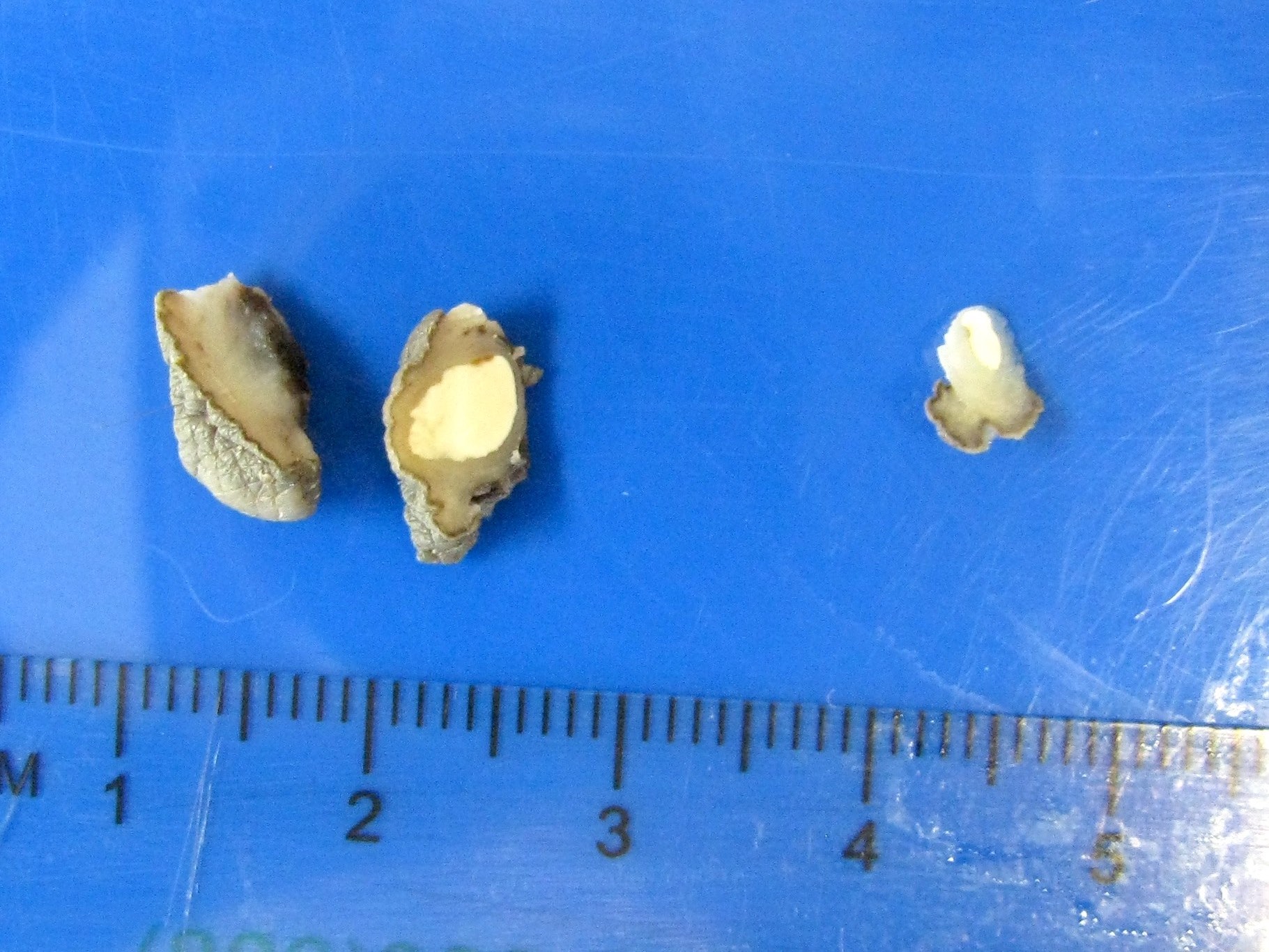
- Multinodular and skin covered masses with rock hard and pearly white surface (Diagn Cytopathol 2017;45:922, Dermatol Online J 2010;16:5)
- Surface erosion / ulceration may be present
- Cut surface is chalky to cheesy white solid
- Size of the nodules ranges from 1 to 50 mm; 50% of patients have lesions that are 6 to 10 mm (Dermatol Online J 2010;16:5)
Gross images
Contributed by Debra Zynger, M.D.
White nodule
Microscopic (histologic) description
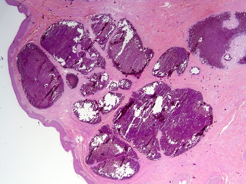
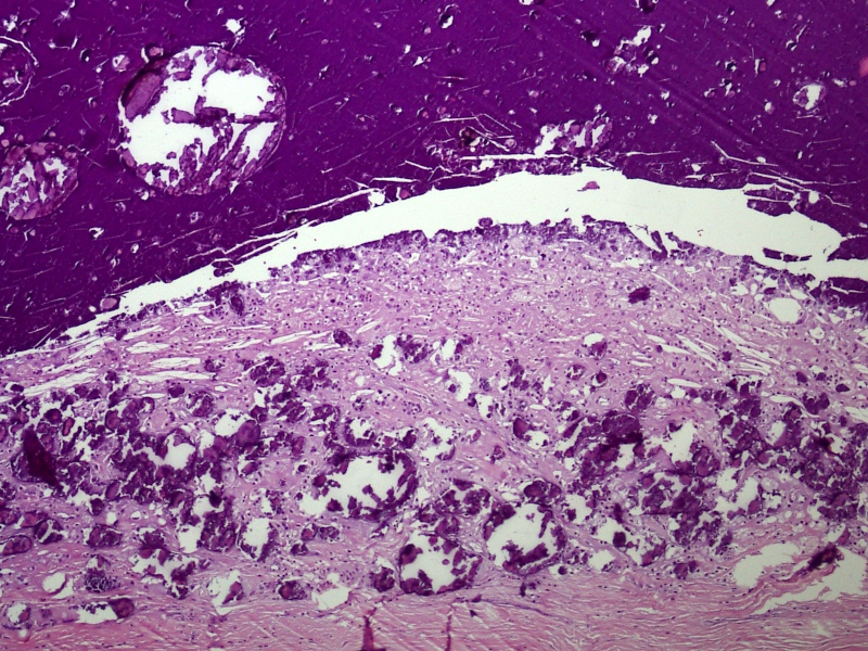
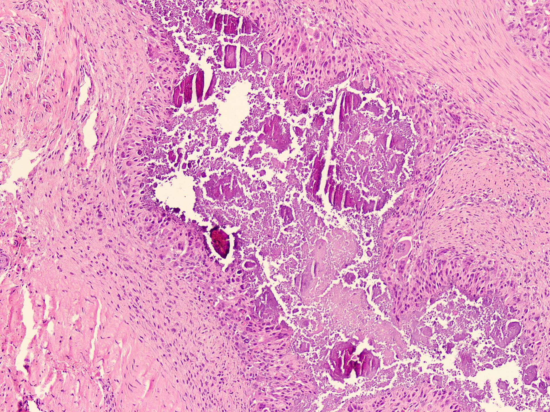
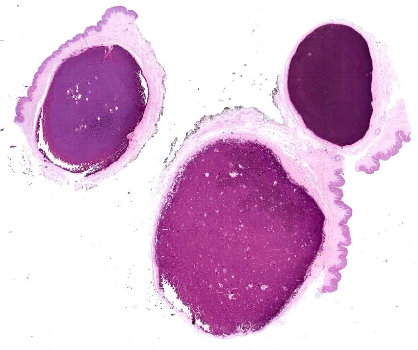
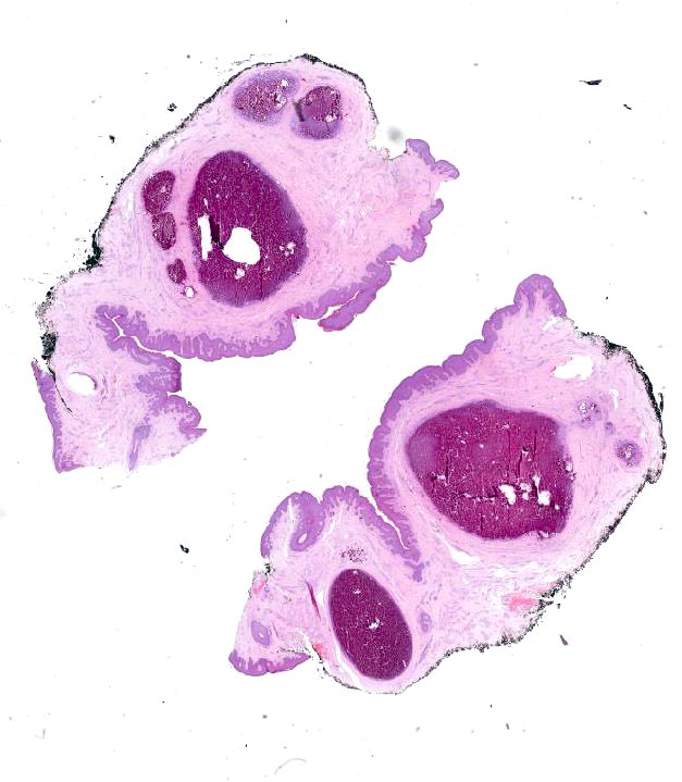
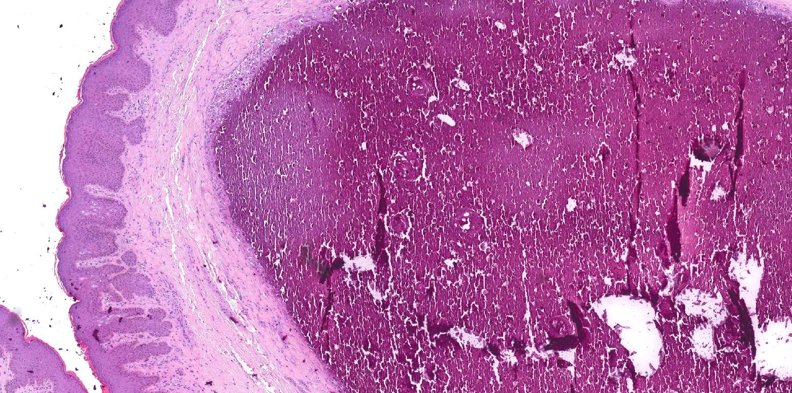
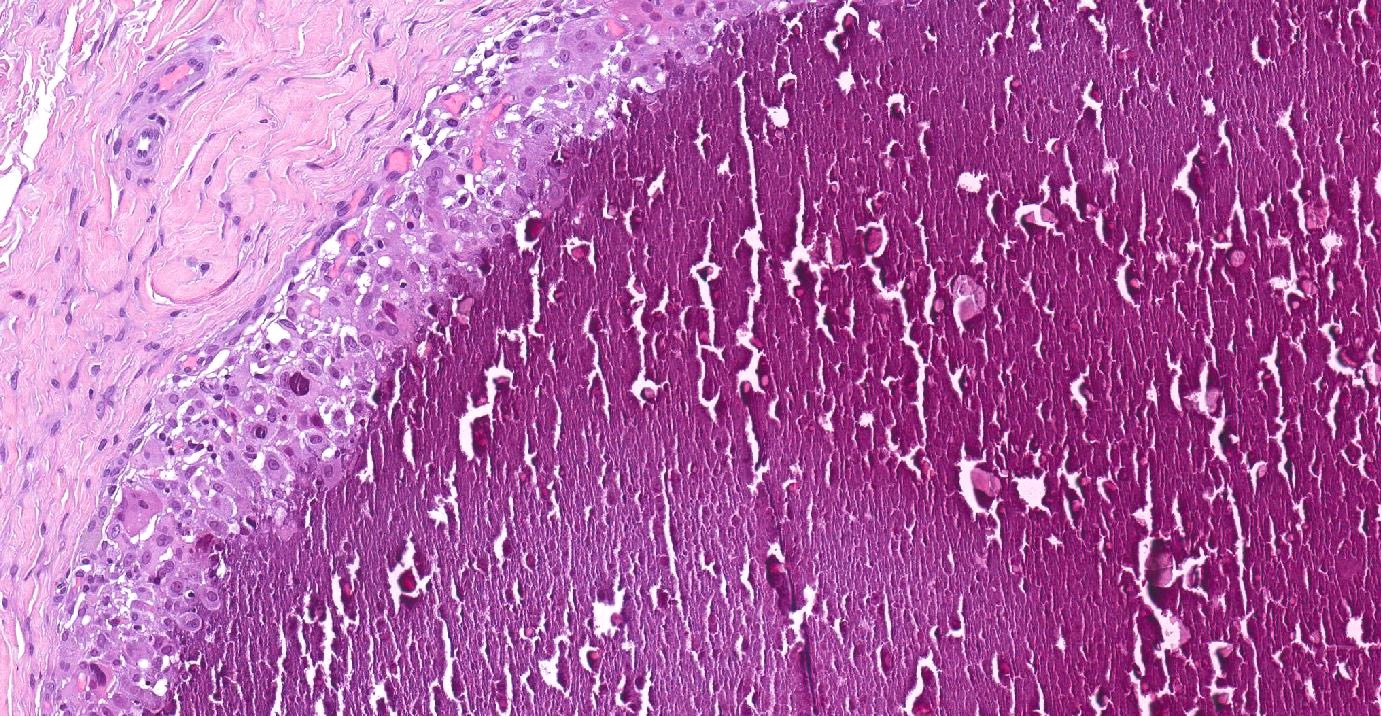
- Multiple varying sized, granular, fractured calcium deposits within the dermis and or subcutis (Arch Ital Urol Androl 2020;92:64)
- May be surrounded with fibroblast proliferation chronic inflammation and foreign body giant cell reaction (G Ital Dermatol Venereol 2015;150:495)
- Some cases may have associated remnants of epidermal inclusion cyst (G Ital Dermatol Venereol 2015;150:495, Am J Clin Exp Urol 2022;10:194)
- Overlying epidermis may show atrophy or ulceration
- Decalcification of the sections may be required to properly visualize the deposits on the slides
Microscopic (histologic) images
Contributed by Nasir Ud Din, M.B.B.S., Debra Zynger, M.D. and @katcollmd on Twitter
Skin tissue showing calcium deposits
Multiple calcium deposits
Calcium deposits with granulomatous reaction

Dermal calcium deposits with epidermis
Scrotal calcinosis
Scrotal calcinosis
Cytology description
- Cytological evaluation of scrotal calcinosis is usually not done due to its typical clinical appearance
- Aspirate is difficult to obtain as it is thick and shows sticky, chalky white material
- Smears reveal calcified, basophilic, amorphous, granular material along with refractile crystals
- These features are nonspecific and raise a differential of calcified epidermal inclusion cysts, dystrophic calcification following any injury or inflammation and scrotal calcinosis (Diagn Cytopathol 2017;45:922)
Cytology images
Contributed by Sleiman Khal, M.D. (Case #468)
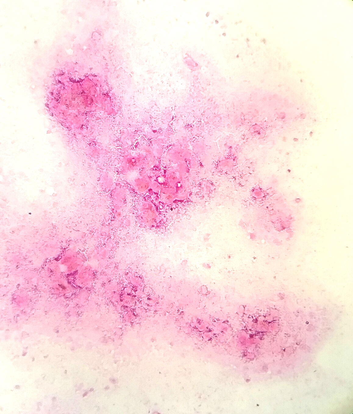
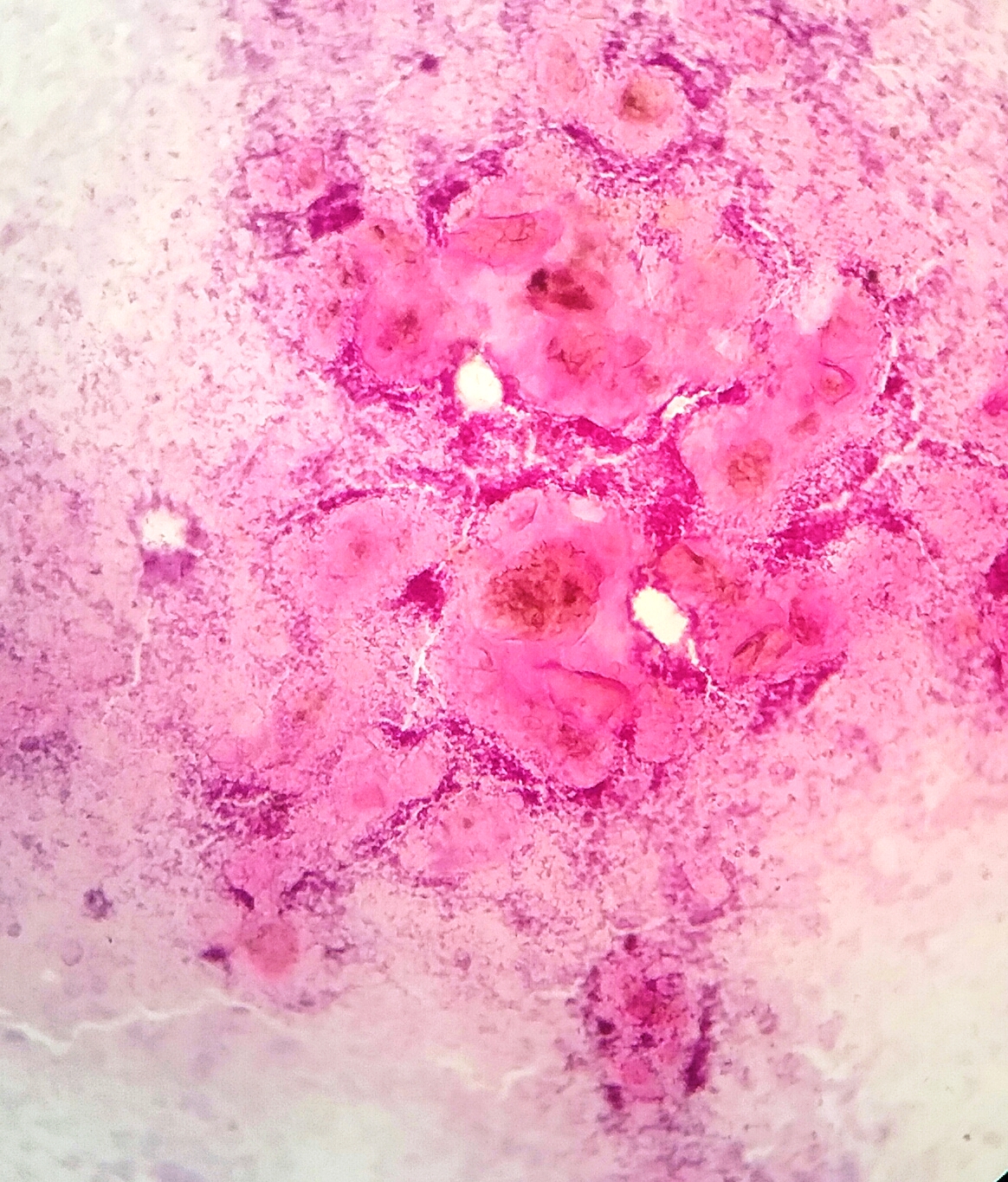
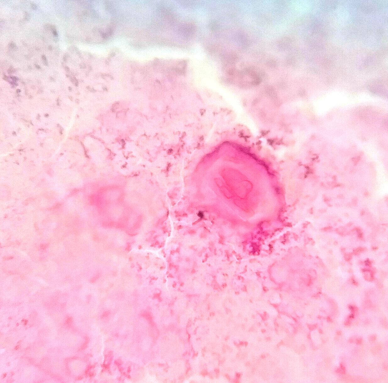
FNA smear of scrotal calcinosis
Images hosted on other servers:

FNA smear of scrotal calcinosis
Positive stains
- Immunohistochemistry is not done in most cases since it is a straightforward histologic diagnosis
- CK AE1 / AE3 may be utilized to highlight the presence of any component of epidermal inclusion cyst, seen in some cases (Dermatol Online J 2010;16:5)
Videos
Scrotal calcinosis: pathology mini tutorial
Sample pathology report
- Scrotal mass, excision biopsy:
- Scrotal calcinosis (see comment)
- Comment: Scrotal calcinosis is a nonneoplastic condition. Localized excision is considered adequate treatment.
Differential diagnosis
- Multiple epidermal inclusion cysts or pilar cysts in scrotum:
- Some cases of scrotal calcinosis may have remnants of an epithelial cyst
- True cysts are lined by stratified squamous epithelium
- Lumen showing keratinous material
- Idiopathic cutaneous calcinosis:
- Usually in the extremities
- Isolated scrotal involvement is rare
- Dystrophic calcification due to Onchocerca volvulus:
- Presence of parasite in tissue sections
Board review style question #1
Which of the following is true regarding scrotal calcinosis?
- Idiopathic in most cases
- More common in elderly
- Only affects Caucasians
- Related to hypercalcemia
- Usually a solitary lesion
Board review style answer #1
Board review style question #2
What is the most characteristic histological feature of scrotal calcinosis depicted in the photomicrograph shown above?
- Cyst formation
- Epidermal atrophy
- Granulomatous reaction
- Inflammation
- Multifocal deposits of calcium
Board review style answer #2
