

Penis & scrotum
Inflammatory
Melanosis and lentiginosis
Authors: Alcides Chaux, M.D., Antonio L. Cubilla, M.D.

Last author update: 1 February 2010
Last staff update: 21 September 2023
Copyright: 2002-2024, PathologyOutlines.com, Inc.
PubMed Search: (Melanosis OR lentiginosis) penis
Table of Contents
Definition / general | Clinical features | Clinical images | Microscopic (histologic) description | Microscopic (histologic) images | Differential diagnosisCite this page: Chaux A, Cubilla AL. Melanosis and lentiginosis. PathologyOutlines.com website. https://www.pathologyoutlines.com/topic/penscrotumlentiginousmel.html. Accessed April 19th, 2024.
Definition / general
- Penile melanosis and penile lentiginosis are benign pigmented lesions frequently found in glans and foreskin
- Penile melanosis shares clinicopathological features with Laugier-Hunziker syndrome of oral mucosa (eMedicine: Laugier-Hunziker Syndrome [Accessed 30 March 2018]) and vulvovaginal melanosis (J Am Acad Dermatol 1989;20:567)
Clinical features
- Benign, although associated with melanoma
Penile melanosis:
- Large, often single, flat and pigmented macule with irregular borders
- Pigmentation may be associated with Laugier-Hunziker syndrome (Int J Dermatol 2004;43:571)
Penile lentiginosis:
- Penile lentigines are 0.2 - 2 cm, oval to irregular lesions with uniform or variegated pigmentation
- Areas of depigmentation are characteristic
- Lesions are scattered on shaft or glans
- Clinically may resemble an atypical melanocytic lesion
- May be associated with Cowden disease (J Cutan Med Surg 2001;5:228), Bannayan-Riley-Ruvalcaba syndrome (J Am Acad Dermatol 2005;53:639)
Clinical images
AFIP images
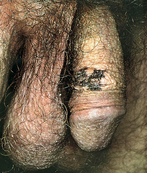
Melanosis
Microscopic (histologic) description
Penile melanosis:
Penile lentiginosis:
- Melanocytic hyperplasia, hyperpigmentation of basal epithelium and otherwise normal epithelium
Penile lentiginosis:
- Elongation of rete ridges with basal layer hyperpigmentation, slight melanocytic hyperplasia, epithelial hyperplasia and stromal melanophages, no atypia (J Am Acad Dermatol 1990;22:453)
- In hyperpigmented areas, there are increased number of melanocytes along the basal layer
- Lymphocytes, which are found in close apposition, destroy melanocytes and surrounding keratinocytes lack pigmentation (Pigment Cell Res 1992;5:404)
Microscopic (histologic) images
AFIP images
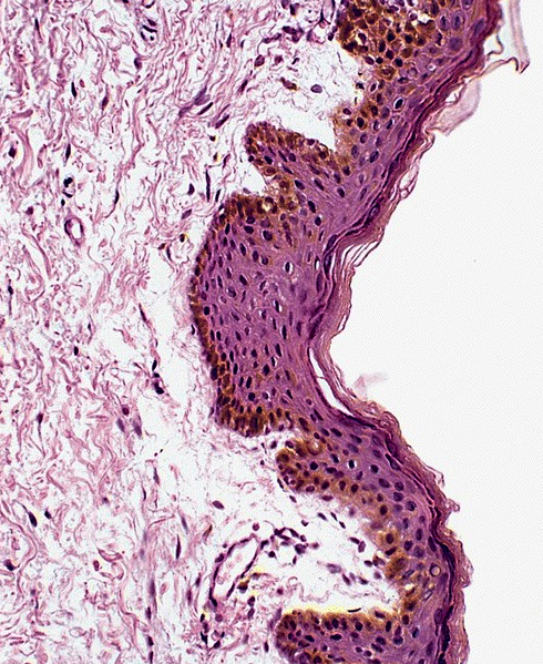
Melanosis
Differential diagnosis
- Congenital melanocytic nevus
- Melanoma: difficult to distinguish clinically, may need to biopsy (Urology 1976;7:323)
