

Penis & scrotum
Inflammatory
Squamous hyperplasia
Authors: Alcides Chaux, M.D., Antonio L. Cubilla, M.D.

Last author update: 1 February 2010
Last staff update: 7 December 2023
Copyright: 2002-2024, PathologyOutlines.com, Inc.
PubMed Search: Squamous hyperplasia penis
Table of Contents
Definition / general | Sites | Clinical features | Gross description | Microscopic (histologic) description | Microscopic (histologic) images | Differential diagnosisCite this page: Chaux A, Cubilla AL. Squamous hyperplasia. PathologyOutlines.com website. https://www.pathologyoutlines.com/topic/penscrotumpenssqhyper.html. Accessed April 24th, 2024.
Definition / general
- Benign thickening of squamous epithelium (more than 15 cell layers) without atypia
Sites
- May affect any penile anatomical compartment
Clinical features
- Most common epithelial change associated with keratinizing penile carcinoma
- Usually found adjacent to neoplastic changes (in situ or invasive carcinoma)
- Uncertain if reactive or precancerous (Anal Quant Cytol Histol 2007;29:185)
- Benign but associated with squamous cell carcinoma, particularly verrucous and low grade papillary subtypes (Int J Surg Pathol 2004;12:351)
Gross description
- Flat, smooth and slightly raised pearly white areas
Microscopic (histologic) description
- Hyperkeratosis, acanthosis and hypergranulosis but normal maturation of squamous epithelium
- Minimal to no parakeratosis
- No cytological atypia, no koilocytosis
- May be adjacent to carcinoma or merge with adjacent low grade carcinoma
Morphological patterns:
- Flat: most common type, linear interface between epithelium and lamina propria
- Papillary: serrated appearance at low power view, jagged interface with stroma
- Pseudoepitheliomatous: downward florid but superficial proliferation of regular squamous cell nests with peripheral palisading, often appearing detached but with no keratinization, no stromal reaction, no desmoplasia and no extension beyond lamina propria
- Verrucous: marked acanthosis with hyperkeratosis, slight papillomatosis and linear interface with stroma
Microscopic (histologic) images
Contributed by Alcides Chaux, M.D. and Antonio Cubilla, M.D.
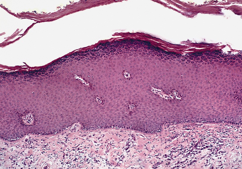
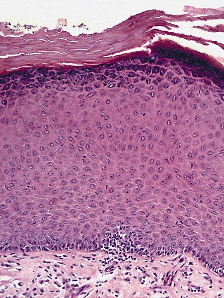
Flat: hyperkeratosis and acanthosis but also normal maturation without atypia
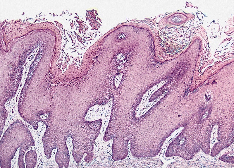
Papillary: hyperkeratosis, papillomatosis and acanthosis
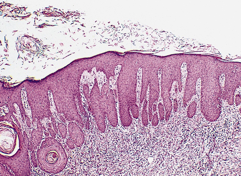
Pseudoepitheliomatous
AFIP images

Acanthosis, absence
of nuclear atypias
and hyperkeratosis
Differential diagnosis
- Penile intraepithelial neoplasia, differentiated type:
- Cytological atypia, more frequent parakeratosis
- Pseudohyperplastic carcinoma:
- Irregular nests, no peripheral palisading, evident stromal reaction and extension beyond lamina propria
- Squamous cell carcinoma with pseudohyperplastic features
- Verruciform carcinomas:
- Cytological atypia, evidence of stromal invasion
- Verruciform xanthoma:
- Lipid laden histiocytes (foamy cells) in lamina propria
