

Penis & scrotum
Inflammatory
Peyronie disease
Authors: Alcides Chaux, M.D., Antonio L. Cubilla, M.D., Komal Arora, M.D., Brian D. Stewart, M.D., Alessandra F. Nascimento, M.D.
Editor-in-Chief: Debra L. Zynger, M.D.

Last author update: 1 February 2010
Last staff update: 27 December 2023
Copyright: 2002-2024, PathologyOutlines.com, Inc.
PubMed Search: Peyronie disease[TI]
Table of Contents
Definition / general | Essential features | Terminology | ICD coding | Epidemiology | Sites | Etiology | Clinical features | Diagnosis | Radiology description | Radiology images | Case reports | Treatment | Clinical images | Gross description | Gross images | Microscopic (histologic) description | Microscopic (histologic) images | Positive stains | Negative stains | Electron microscopy description | Molecular / cytogenetics description | Videos | Sample pathology report | Differential diagnosisCite this page: Chaux A, Cubilla AL, Arora K, Stewart BD, Nascimento AF. Peyronie disease. PathologyOutlines.com website. https://www.pathologyoutlines.com/topic/penscrotumpeyronie.html. Accessed April 24th, 2024.
Definition / general
- Fibrous thickening of dermis and Buck’s fascia between corpora cavernosa and tunica albuginea, causing curvature towards side of lesion and restricting movement of these structures during erection
Essential features
- Proliferation of fibroblasts and myofibroblasts on or around the tunica albuginea of the penis, most often the dorsal aspect, that may lead to plaques with curvature of the penis towards the plaque
Terminology
- Induratio penis plastica, penile fibromatosis
Epidemiology
- Usually 40+ years, rare in younger population (J Androl 2003;24:27)
- Prevalence 3 - 9% (Int J Impot Res 2002;14:379)
Sites
- Tunica albuginea, most commonly the dorsal aspect
Etiology
- May be related to microtrauma (coital, urethral instrumentation), which releases fibroblast related cytokines
- May arise secondary to urethritis as a sclerosing inflammatory process
- Etiology may be related to Parc protein (BJU Int 2010;106:1706) or Wnt2 (J Sex Med 2012;9:1430)
Clinical features
- May be associated with carcinoid syndrome, Dupuytren contracture
- May be associated with beta blockers, thiazides (J Sex Med 2010;7:1529), hypertension, diabetes and immune reactions
- Fibrosis produces an abnormal curvature of the penis, which may cause pain during erection and intercourse
- In some cases, nodules adherent to tunica albuginea are noted over the mid dorsal line
- May be clinically mimicked by epithelioid sarcoma (Int J Impot Res 2003;15:378)
Diagnosis
- Usually clinical, however, occasionally may be a histologic confirmation
Radiology description
- Ultrasound
- Echogenic plaques, frequently calcifications
- MRI
- Thickened and hypointense plaques on T1 and T2 in and around tunica albuginea
- T2 is superior (Radiographics 2009;29:477)
Radiology images
Images hosted on other servers:

Penile deformation

Dorsal plaque
Case reports
- 59 year old man with penile ossification (Sao Paulo Med J 2007;125:124)
Treatment
- May need surgical treatment (Adv Urol 2008;2008:263450, Eur Urol 2009;55:1469, Curr Urol Rep 2011;12:444)
- Some nonsurgical options may stabilize disease (Asian J Androl 2008;10:79)
- Can respond to laser therapy and small amounts of irradiation and injected steroids (J Androl 2009;30:397, J Androl 2010;31:445) and interferon
- Prosthesis may be useful (Adv Urol 2008;2008:646052)
- May spontaneously regress (J Androl 2009;30:397)
Clinical images
AFIP images
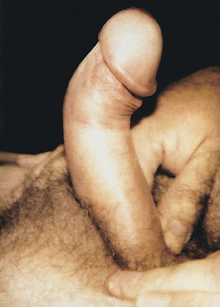
Penile curvature
Images hosted on other servers:


Penile curvature
Gross description
- Irregular areas of fibrosis in tunica albuginea with or without nodules in penile shaft
- Occasionally nodular areas of fibrosis may predominate
Gross images
AFIP images

Tunica albuginea
Microscopic (histologic) description
- Dense fibrous nodules, similar to those in Dupuytren contracture, fibromatosis and other desmoplastic conditions involving myofibroblasts
- More dense and less cellular than other types of superficial fibromatosis
- Disorganization of collagen of tunica albuginea with formation of nodules, often hyalinizing fibrosis
- Perivascular lymphoid infiltrate in early stages of disease in 1/3
- Fibrotic tunica albuginea with extension of fibrosis to corpus cavernosum
- Abnormal vessels with venous leakages
- Calcification or ossification may occur, linear band of calcification in 1/4 (J Urol 1997;157:282)
Microscopic (histologic) images
AFIP images
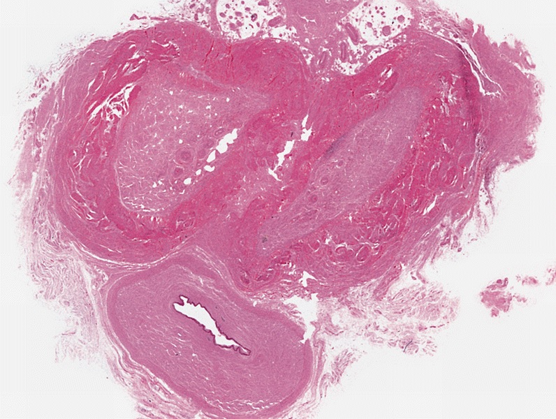
Thickening of tunica albuginea
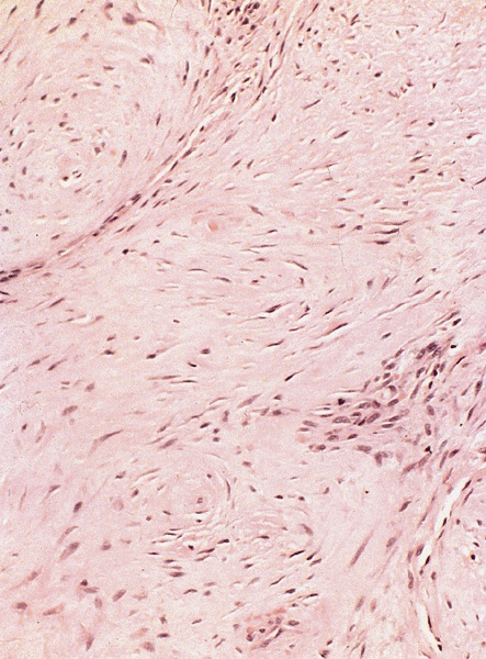
Fibrosis
Positive stains
- Vimentin, variable actin - muscle specific and smooth muscle actin, less frequently desmin
Electron microscopy description
- Penile plaques are composed of collagen fibrils, amorphous particulate material and fibroblasts (Int J Urol 1997;4:274)
Molecular / cytogenetics description
- No somatic mutations of beta catenin genes unlike desmoid fibromatosis (Mod Pathol 2001;14:695)
Videos
Penile fibromatosis
Sample pathology report
- Penis, plaque, excision:
- Features consistent with Peyronie disease
Differential diagnosis
- Spindle cell sarcomas (i.e. synovial sarcoma, malignant peripheral nerve sheath tumor, etc.)
- Epithelioid hemangioma (J Med Case Rep 2011;5:260)
- Epithelioid sarcoma: may clinically appear similar (Int J Impot Res 2003;15:378)
