

Penis & scrotum
Inflammatory
Plasma cell balanitis
Authors: Alcides Chaux, M.D., Antonio L. Cubilla, M.D.

Last author update: 1 February 2010
Last staff update: 20 February 2024 (update in progress)
Copyright: 2002-2024, PathologyOutlines.com, Inc.
PubMed Search: Plasma cell balanitis penis
Table of Contents
Definition / general | Terminology | Epidemiology | Etiology | Clinical features | Case reports | Treatment | Clinical images | Microscopic (histologic) descriptionCite this page: Chaux A, Cubilla AL. Plasma cell balanitis. PathologyOutlines.com website. https://www.pathologyoutlines.com/topic/penscrotumplasmacellbalanitis.html. Accessed April 19th, 2024.
Definition / general
- Inflammatory condition with prominent plasma cells (eMedicine: Balanitis Circumscripta Plasmacellularis [Accessed 2 April 2018])
Terminology
- Also known as balanitis circumscripta plasmacellularis, Zoon balanitis
- Balanitis: inflammation of glans
Epidemiology
- Middle aged and elderly uncircumcised men
Etiology
- Etiology unknown but probably reactive
Clinical features
- Disease tends to be chronic and may persist for months to years
- Solitary or rarely multiple brown-red plaques located in glans or inner foreskin
- Surface is shiny, slightly moist and stippled with minute red specks
- May simulate erythroplasia of Queyrat / Bowen disease
Case reports
- Middle aged man with erythroplasia of Queyrat (Int J STD AIDS 2008;19:861)
Treatment
- Circumcision (J Cutan Med Surg 2006;10:11), laser excision, topical antibiotics or steroids; possibly pimecrolimus 1% cream (Int J Dermatol 2008;47:198)
Clinical images
AFIP images
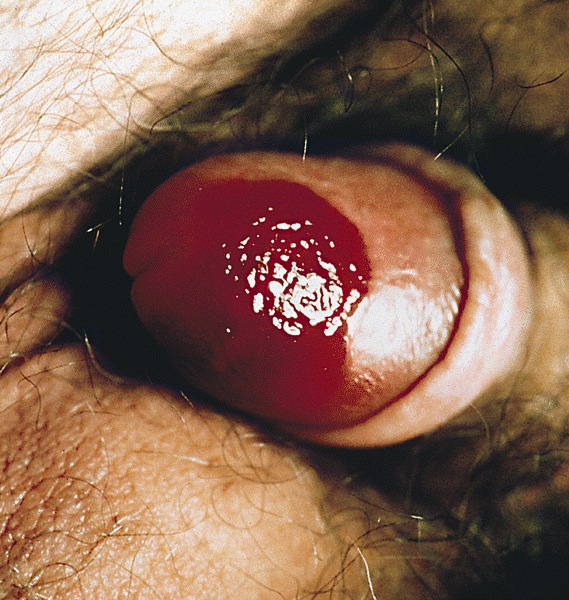
Glans mucosa
Microscopic (histologic) description
- Epidermal atrophy, band-like infiltrate of plasma cells in dermis, hemosiderin pigment laden macrophages (siderophages), edema and numerous capillaries
- Epithelium may show mild reactive changes which can simulate penile intraepithelial neoplasia
- Rarely plasma cells are scant / absent
