

Placenta
General
Placental development & hormones
Author: Mandolin S. Ziadie, M.D.
Last author update: 1 September 2011
Last staff update: 21 December 2021
Copyright: 2002-2024, PathologyOutlines.com, Inc.
PubMed Search: Placental development[title] "loattrfree full text"[sb]


Table of Contents
Implantation | First trimester | Second trimester | Third trimester | Mean placental weight by gestational age | Placental hormonesCite this page: Ziadie MS. Placental development & hormones. PathologyOutlines.com website. https://www.pathologyoutlines.com/topic/placentaplacentaldevel.html. Accessed April 20th, 2024.
Implantation
- Blastocyst implants on postovulation day 6 - 7; by day 10, ovum is implanted in stroma
- Trophoblasts proliferate and erode the maternal capillaries and venules to form the intervillous space
- Extraembryonic mesoderm grows into the primary villi and capillary formation occurs
- By 5 - 6 weeks, the villous vessels are formed
- At 8 weeks, they contain nucleated red blood cells (nRBC) which diminish to 10% by 10 weeks and are absent at 12 weeks
First trimester
Implantation and arterial plugs:
Villous morphology:
Microscopic (histologic) images:
Images hosted on other servers:

- Extravillous intermediate trophoblasts invade the endometrium while endovascular trophoblasts grow into arteries and form cellular plugs
Villous morphology:
- Early mesenchymal villi are large (170 microns) with scant connective tissue, few Hofbauer cells, no thick walled vessels
- They have a complete outer layer of syncytiotrophoblast and an inner cytotrophoblast layer
- During mid trimester, they mature to immature intermediate villi which have loose stroma with many Hofbauer cells and a complete trophoblastic coat
- Then transform to stem villi, which have denser stroma and thick walled vessels
- This process continues through the second trimester
Microscopic (histologic) images:
Images hosted on other servers:
Chorionic villi covered
by cytotrophoblast and
syncytiotrophoblast
Second trimester
Implantation and vascular remodeling:
Villous morphology:
Microscopic (histologic) images:
Images hosted on other servers:

- Invading trophoblasts extend into the myometrium while endovascular trophoblasts invade the arterial walls and destroy their endothelium and media, replacing them with fibrinoid material, creating a low pressure circulation
Villous morphology:
- Mesenchymal villi give rise to mature intermediate villi, from which terminal villi sprout
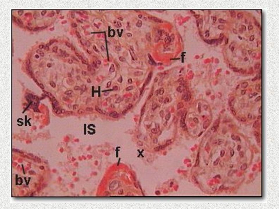
- Mature intermediate villi are large with loose stroma, capillaries, arterioles and venules
- Terminal villi appear near the end of the trimester and are much smaller (70 microns) with denser stroma surrounded primarily by syncytiotrophoblasts and a thin cytotrophoblast layer that may have syncytial knots
- Vessels are numerous (3 - 5 capillaries per villous) and in contact with the trophoblastic coat
Microscopic (histologic) images:
Images hosted on other servers:
"Syncytial knots" and pink fibrin
Third trimester
Villous morphology:
Microscopic (histologic) images:
Images hosted on other servers:


- Mature intermediate and terminal villi are now more prevalent and smaller than second trimester with thin stroma
- More have syncytial knots (approximately 30%) and vasculosyncytial membranes (fused fetal capillaries with syncytiotrophoblasts)
- Trophoblastic inclusions are common
Microscopic (histologic) images:
Images hosted on other servers:
Vascularized chorionic villi and hCG immunostain
Mean placental weight by gestational age
- Prior to 28 weeks: 253 grams
- 28 - 32 weeks: 314 grams
- 33 - 36 weeks: 391 grams
- 37 - 40 weeks: 456 grams
- > 40 weeks: 496 weeks
Placental hormones
Steroid hormones: estrogens and progesterone
Peptide hormones
Activin and inhibin:
Cytokine growth factors (TGF-alpha, TGF-beta, EGF):
Human chorionic adrenocorticotropin (hACTH):
Human chorionic gonadotropin (hCG) :
Human chorionic thyrotropin (hCT):
Human placental growth hormone:
Human placental lactogen (hPL) :
Insulin-like growth factors:
Placental alkaline phosphatase (PLAP):
Relaxin:
SP1:
- Trophoblasts synthesize estrogens and syncytiotrophoblasts synthesize progesterone, which maintains a noncontractile uterus and fosters development of an endometrium conducive to pregnancy
- By the end of the first trimester, placental production of these hormones replaces the corpus luteum
Peptide hormones
Activin and inhibin:
- Produced by trophoblast
- Regulate hCG production
Cytokine growth factors (TGF-alpha, TGF-beta, EGF):
- Produced by trophoblast
- Stimulates proliferation of trophoblast and production of fibronectin
Human chorionic adrenocorticotropin (hACTH):
- Small amounts produced
- Believed to function similar to ACTH
Human chorionic gonadotropin (hCG) :
- Glycoprotein similar in structure to pituitary LH
- Synthesized primarily by the villous syncytiotrophoblast
- Synthesis begins before implantation and is detectable 7 - 10 days after implantation, forming the basis for early pregnancy tests
- Peak levels reached at 8 - 10 weeks
- Maintains maternal corpus luteum that secretes progesterone and estrogens
Human chorionic thyrotropin (hCT):
- Small amounts produced, probably by the syncytiotrophoblast
- Believed to function similar to TSH
Human placental growth hormone:
- Differs from pituitary growth hormone by 13 amino acids
- Regulates maternal blood glucose levels so that the fetus has adequate nutrient supply
Human placental lactogen (hPL) :
- Polypeptide similar to growth hormone
- Synthesized by the villous syncytiotrophoblast
- First detectable by 4 weeks
- Peak levels at end of third trimester
- Acts as an insulin antagonist to influence growth, maternal mammary duct proliferation and lipid and carbohydrate metabolism
Insulin-like growth factors:
- Stimulate proliferation and differentiation of cytotrophoblast
Placental alkaline phosphatase (PLAP):
- Alkaline phosphatase normally produced by syncytiotrophoblast and primordial germ cells
- Also produced in seminoma, intratubular germ cell neoplasia, rarely in other non germ cell tumors
- May be involved in migration of primordial germ cells in developing fetus
Relaxin:
- Produced by villous cytotrophoblast
- Softens the cervix and pelvic ligaments in preparation for childbirth
SP1:
- Pregnancy specific beta-1 glycoprotein
- Present in syncytiotrophoblast and extravillous trophoblast
- Not in cytotrophoblast
