
Prostate gland & seminal vesicles
Seminal vesicles
Cystadenoma
Author: Daniel Athanazio, M.D., Ph.D.
Editorial Board Member: Bonnie Choy, M.D.
Deputy Editor-in-Chief: Maria Tretiakova, M.D., Ph.D.

Last author update: 27 March 2023
Last staff update: 27 March 2023
Copyright: 2022-2024, PathologyOutlines.com, Inc.
PubMed Search: Cystadenoma prostate

Table of Contents
Definition / general | Essential features | Terminology | ICD coding | Epidemiology | Sites | Pathophysiology | Etiology | Clinical features | Diagnosis | Radiology description | Radiology images | Prognostic factors | Case reports | Treatment | Clinical images | Gross description | Gross images | Microscopic (histologic) description | Microscopic (histologic) images | Positive stains | Negative stains | Sample pathology report | Differential diagnosis | Additional references | Board review style question #1 | Board review style answer #1 | Board review style question #2 | Board review style answer #2Cite this page: Athanazio D. Cystadenoma. PathologyOutlines.com website. https://www.pathologyoutlines.com/topic/prostatecystadenoma.html. Accessed April 20th, 2024.
Definition / general
- Rare, benign epithelial tumor composed of variably sized cysts lined by bland cuboidal cells
Essential features
- Distinguished from mixed epithelial and stromal tumor of the seminal vesicle by the absence of stromal hypercellularity
- Should be distinguished from prostatic stromal tumor of uncertain malignant potential entrapping glands
- Factors that favor seminal vesicle cystadenoma:
- Cystic tumor centered in the seminal vesicle
- No expression of prostatic differentiation markers
- Factors that favor seminal vesicle cystadenoma:
Terminology
- Some authors consider it as the expression of lowest grade (benign) of mixed epithelial and stromal tumor of the seminal vesicle (Adv Anat Pathol 2015;22:113)
ICD coding
- ICD-O: 8440/0 - cystadenoma, NOS
- ICD-11: 2F34 & XH5RJ2 - benign neoplasm of male genital organs & cystadenoma, NOS
Epidemiology
- Rare (< 30 described cases)
- Wide age distribution (23 - 66 years old)
Sites
- Centered within seminal vesicles
Pathophysiology
- Unknown
Etiology
- Unknown
Clinical features
- Obstructive urinary symptoms
- Some patients present with an asymptomatic mass or the masses are incidentally detected in imaging studies
Diagnosis
- Cystic or solid and cystic masses detected in imaging studies
Radiology description
- Cystic or solid and cystic masses centered within the seminal vesicles
Radiology images
Images hosted on other servers:

Mass of cystic component between bladder and rectum

Cystic mass with solid component connected to the seminal vesicle

Solid and cystic pelvic mass
Prognostic factors
- Benign neoplasm; local resection is considered curative
Case reports
- 31 year old man with an 8.8 cm cystic pelvic mass (Asian J Androl 2013;15:697)
- 48 year old man with a 3.5 cm mass in the right seminal vesicle (J Endourol Case Rep 2015;1:62)
- 49 year old man with a 12.0 cm solid and cystic pelvic mass (Pan Afr Med J 2017;28:149)
- 59 year old man with a 9.8 cm oval cyst between bladder and rectum (Chin Med J (Engl) 2018;131:2897)
- 71 year old man with a 5.5 cm oval cyst between bladder and rectum (Asian J Androl 2017;19:384)
Treatment
- Surgical resection
Clinical images
Images hosted on other servers:

Laparoscopic view
Gross description
- Cystic mass centered in the seminal vesicle with variable solid components
Gross images
Contributed by Daniel Athanazio, M.D., Ph.D.
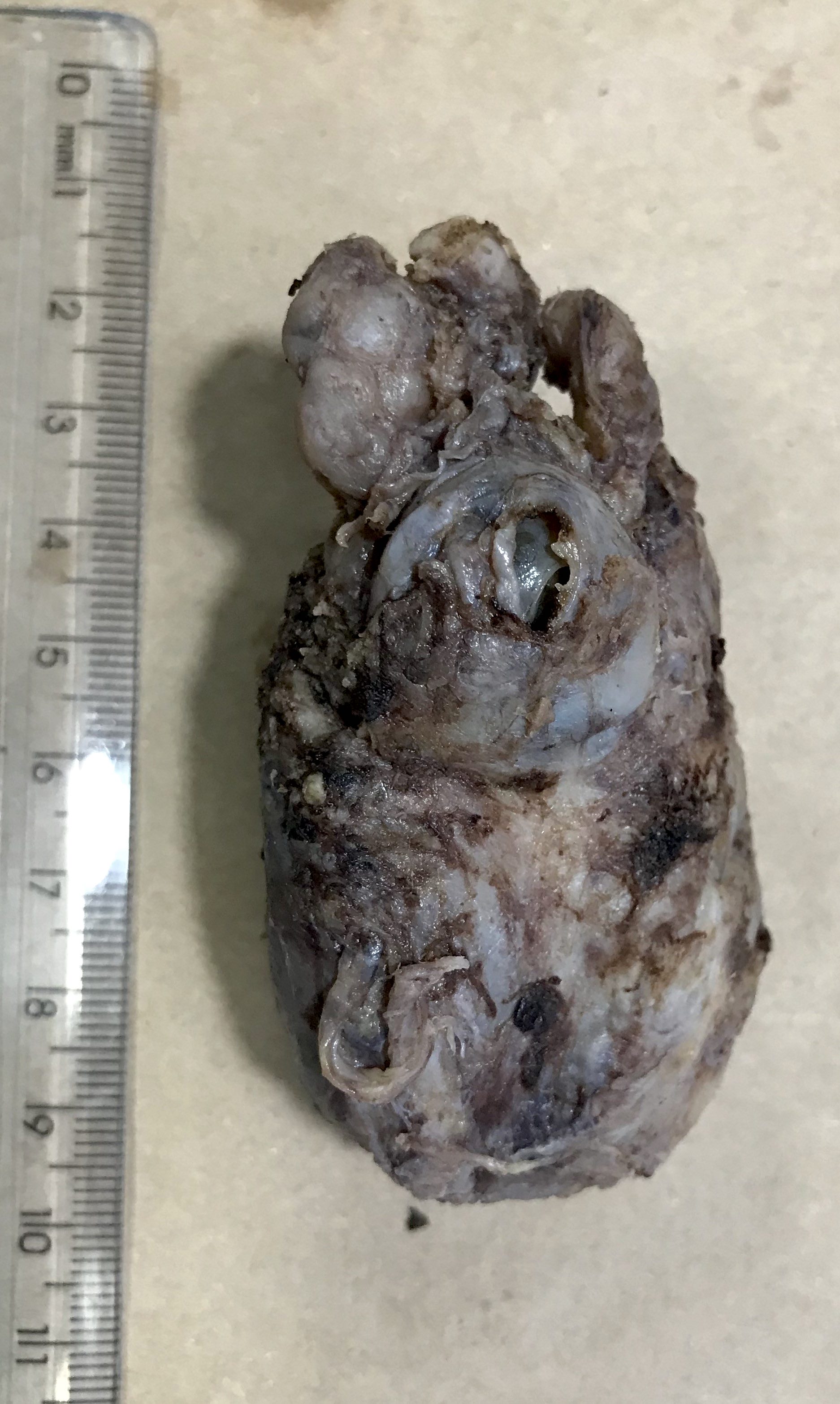
Cystic mass centered in the seminal vesicle
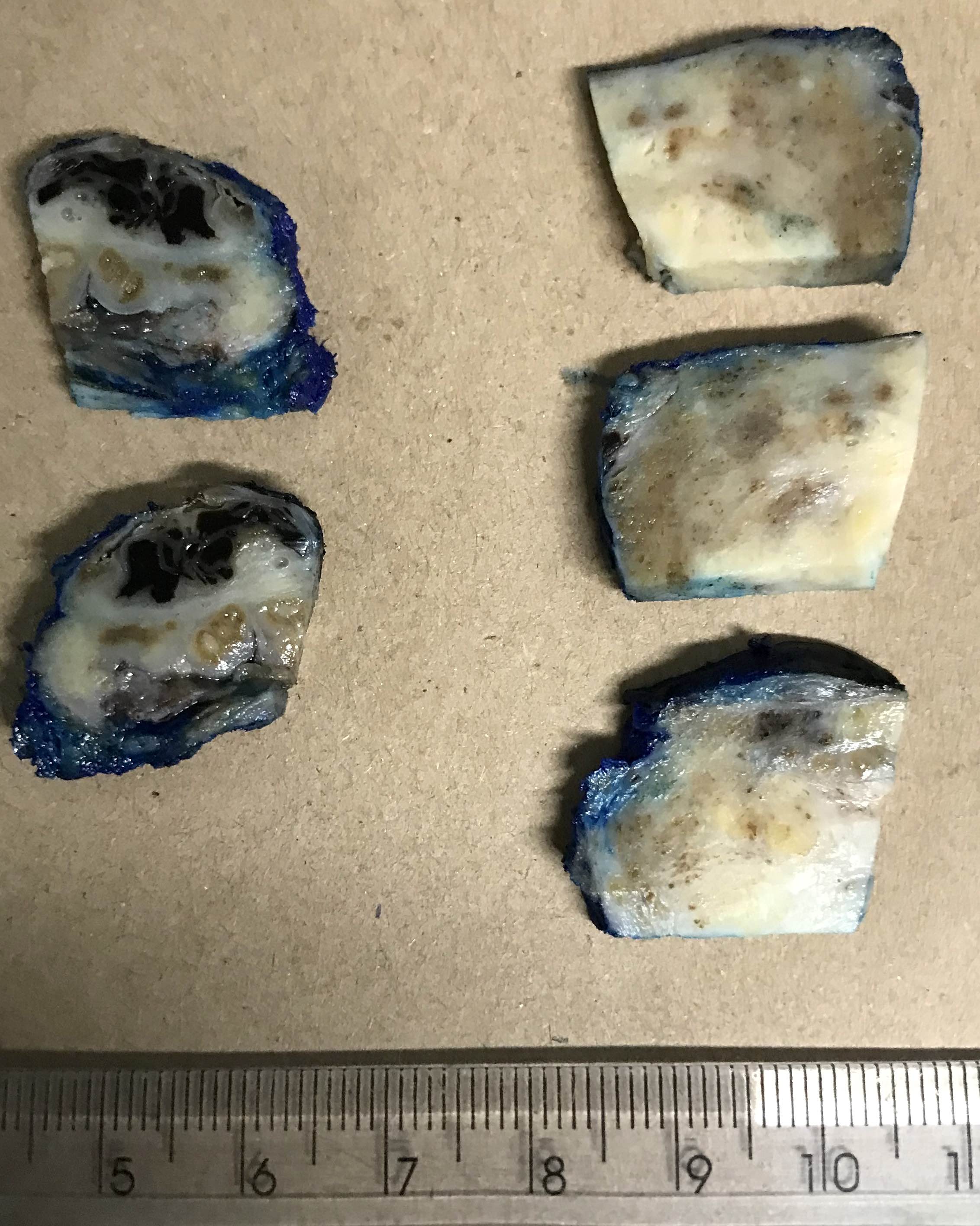
Cut surface
Images hosted on other servers:

Multilobulated cystic mass

Cut surface
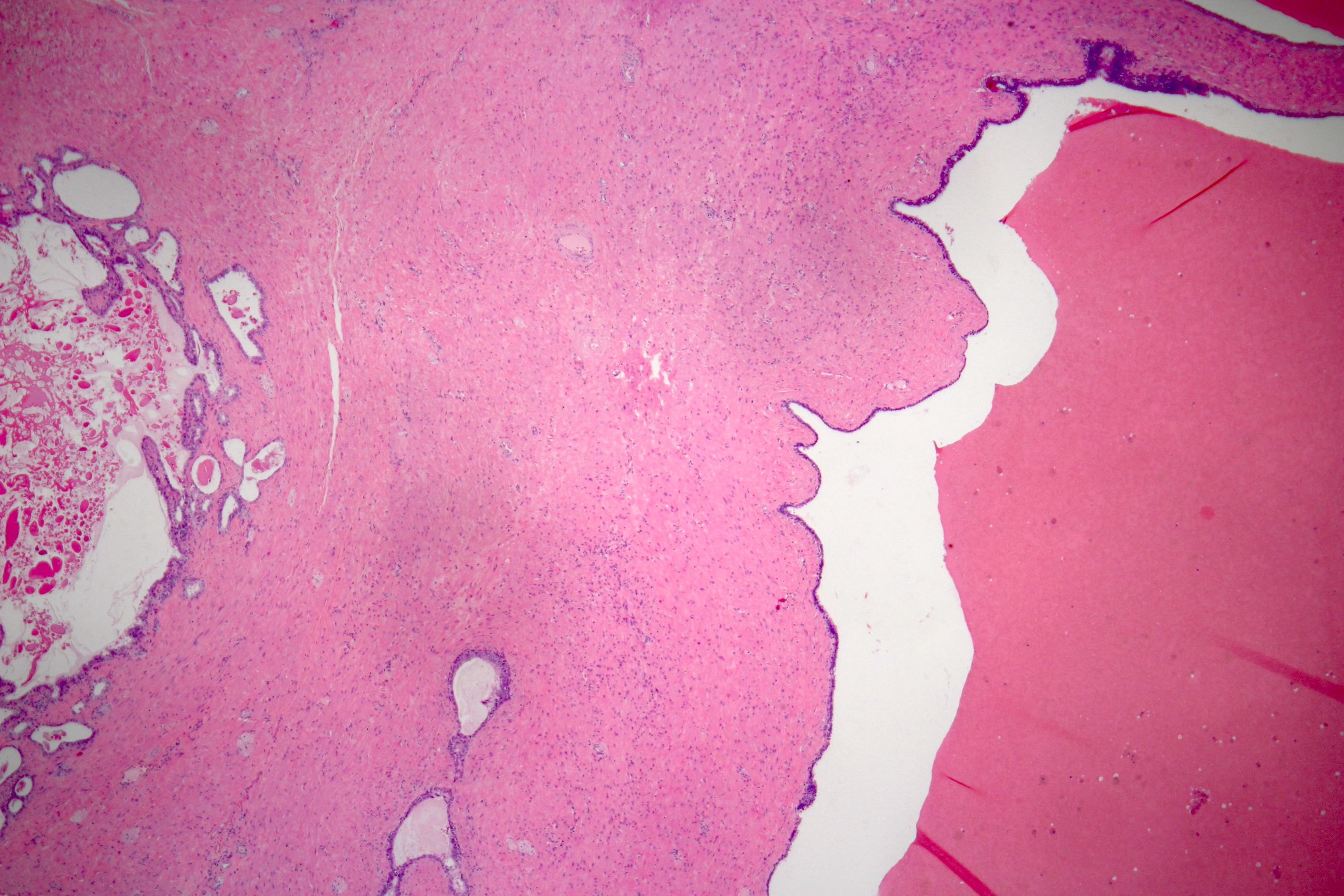
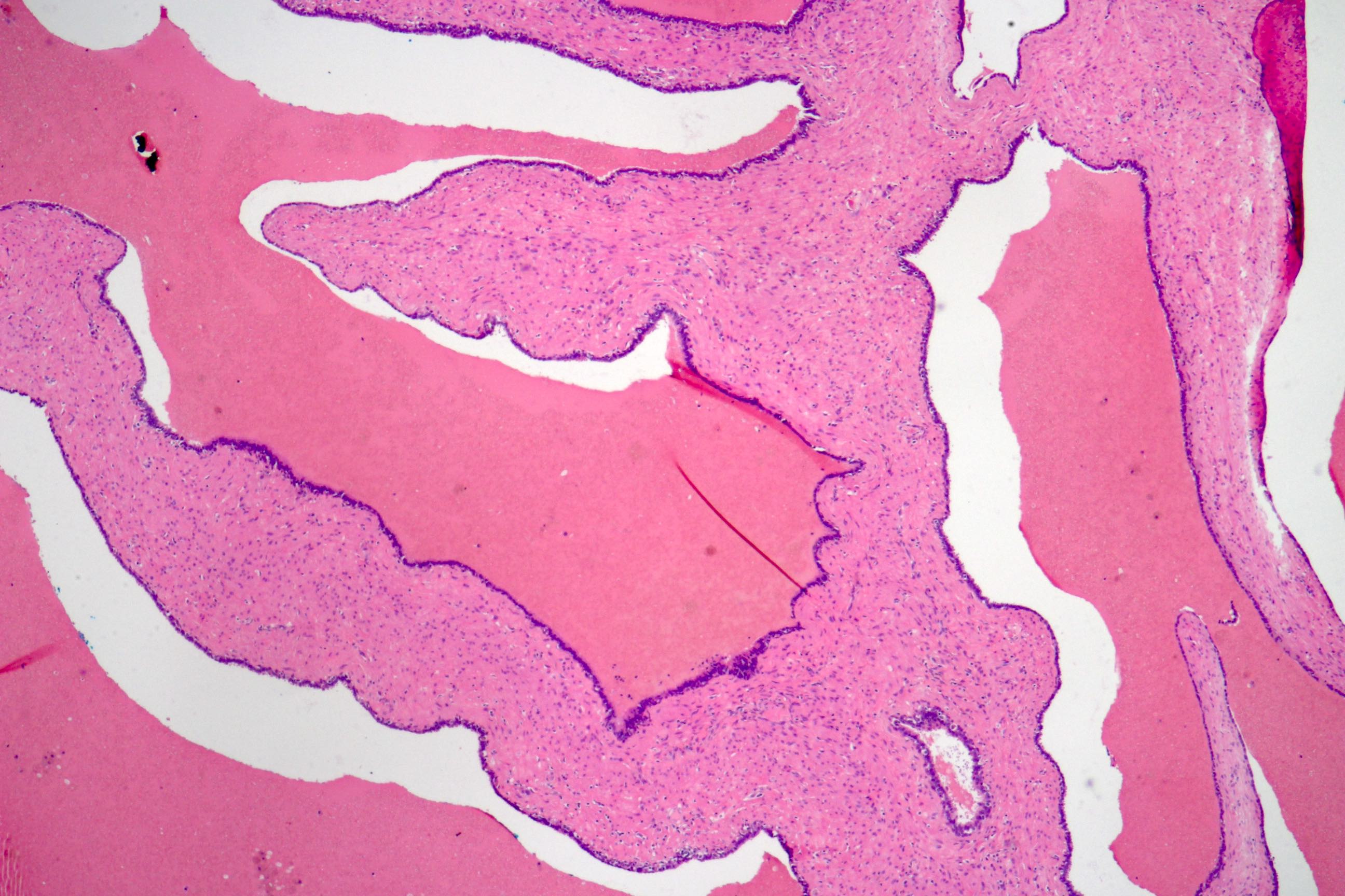
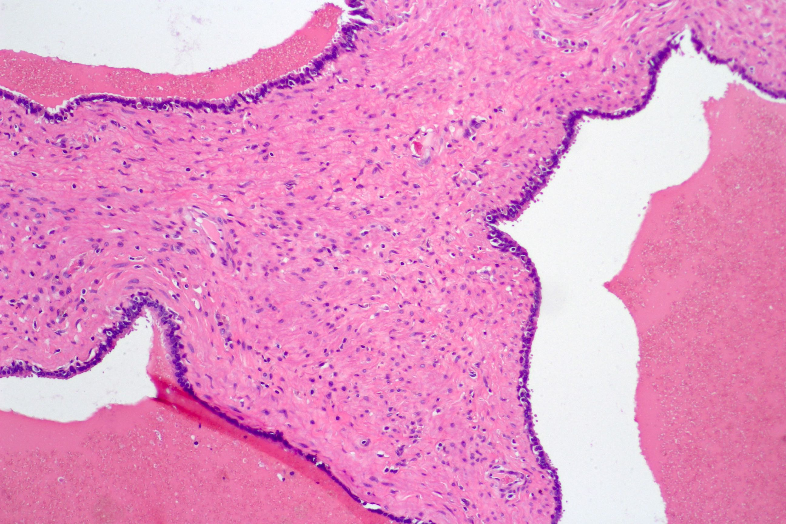
Microscopic (histologic) description
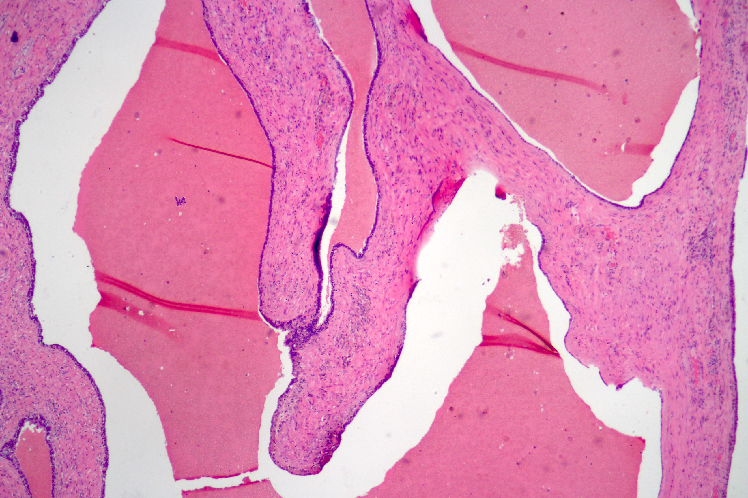
- Glandular spaces of varying sizes are lined by cuboidal cells without atypia (Adv Anat Pathol 2015;22:113)
- Lobular pattern, forming branching lumina and cysts that contain granular intraluminal secretions
- No hypercellularity or atypia in stromal cells
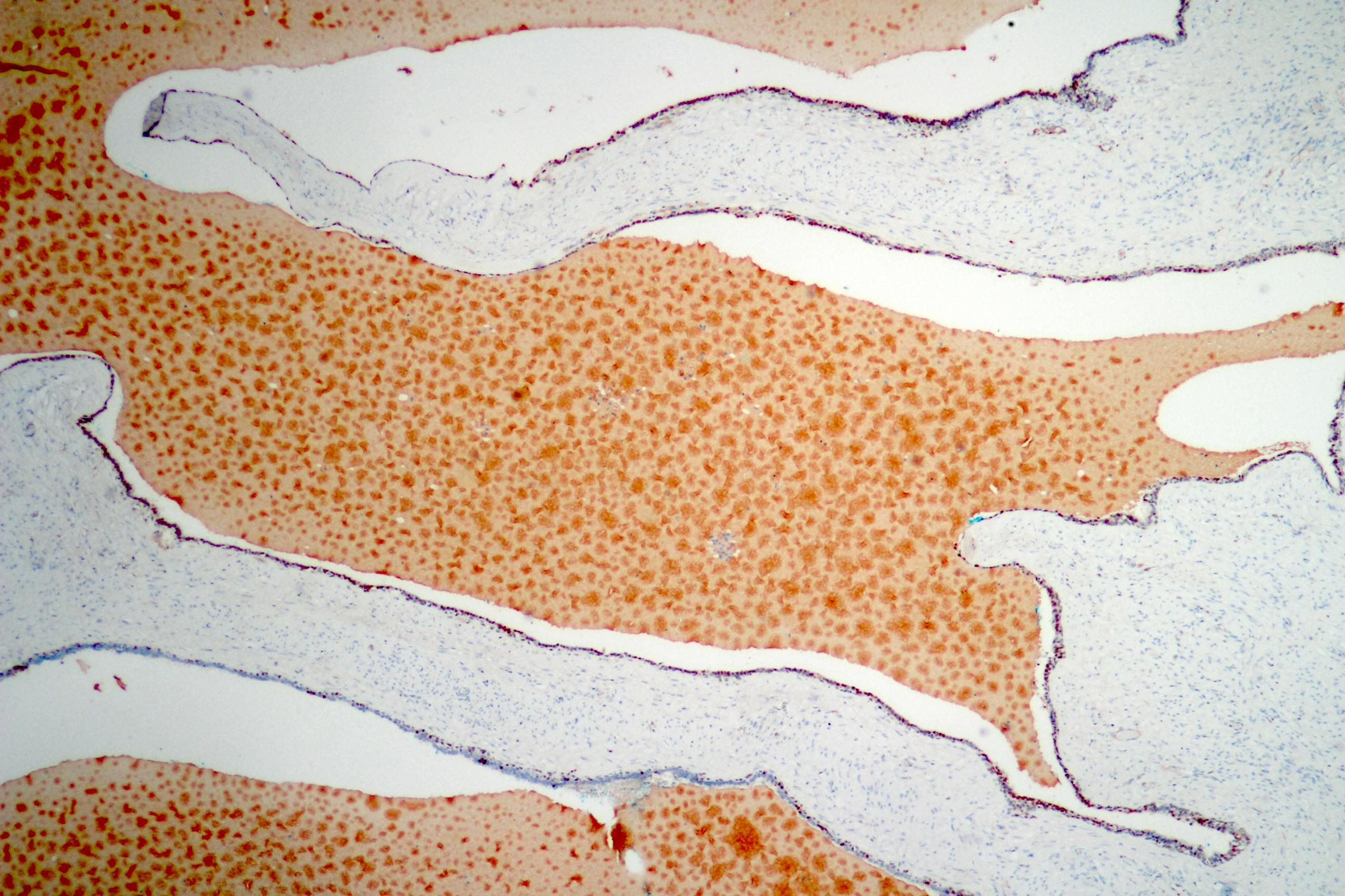
Microscopic (histologic) images
Contributed by Daniel Athanazio, M.D., Ph.D.
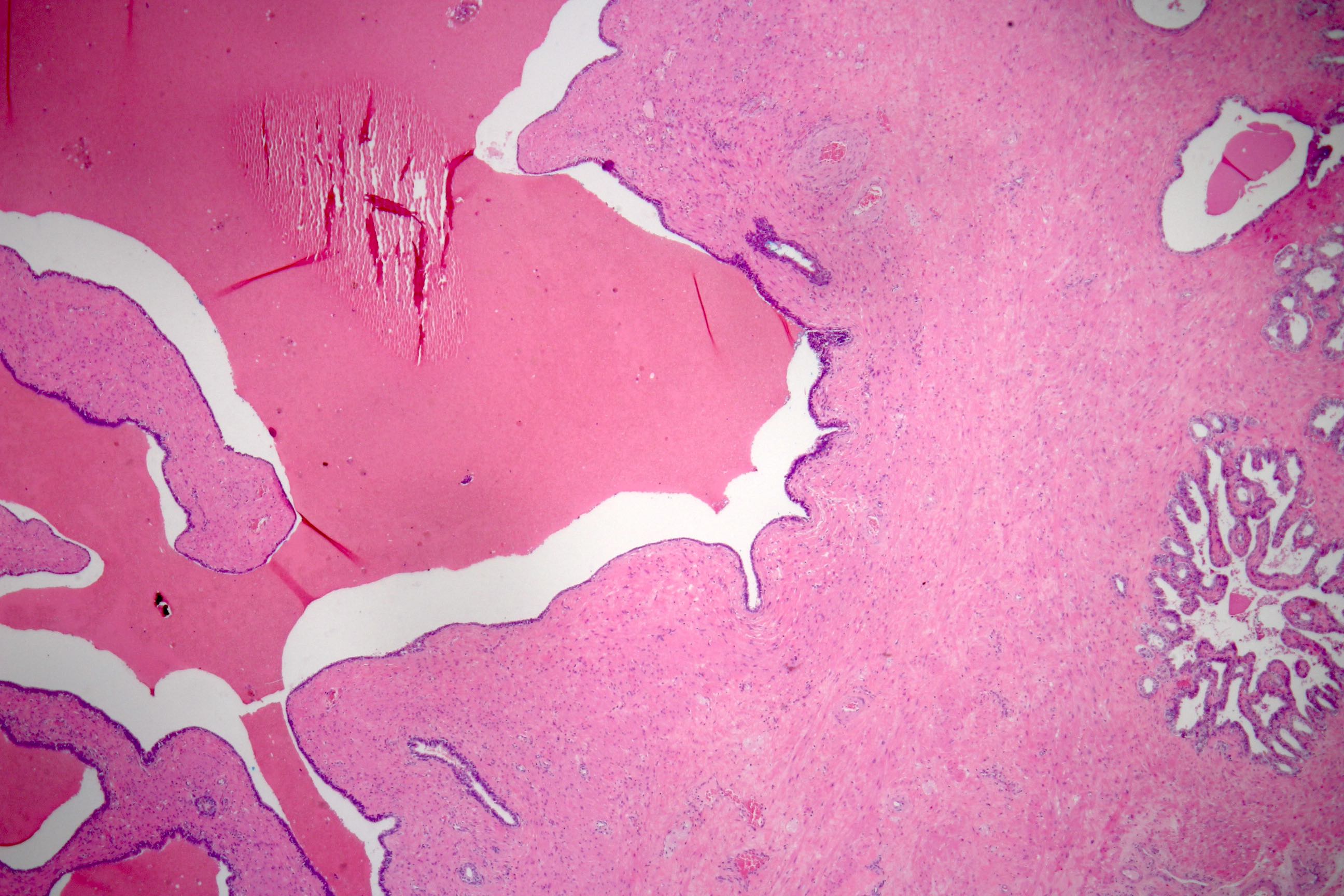
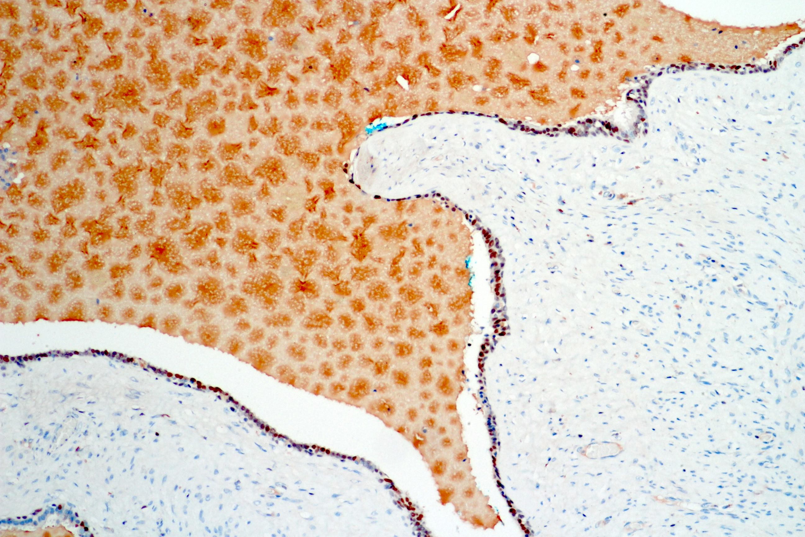
Cystic tumor
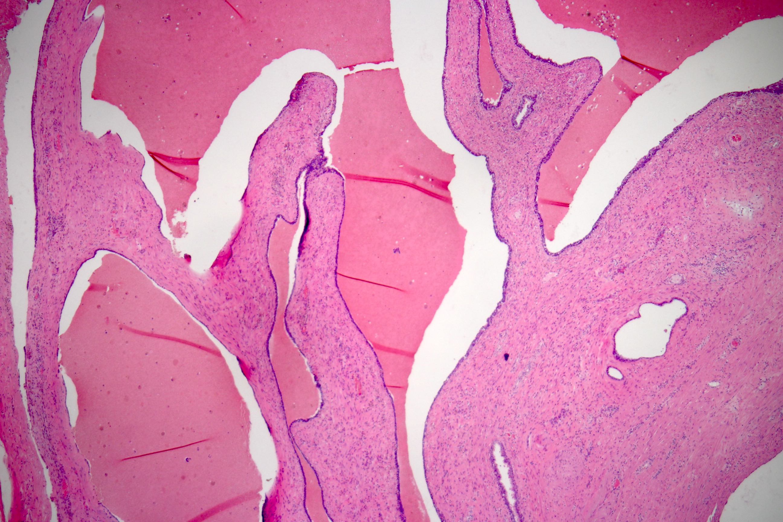
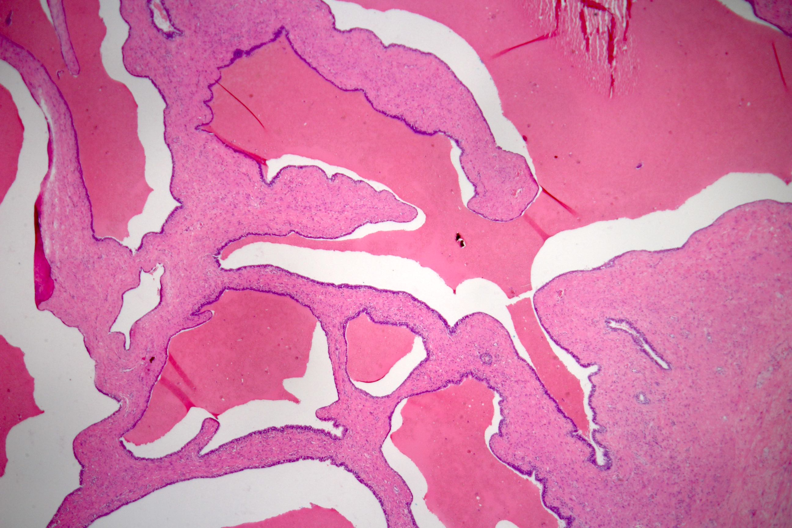
Finger-like projections

Finger-like projections
Leaf-like projections
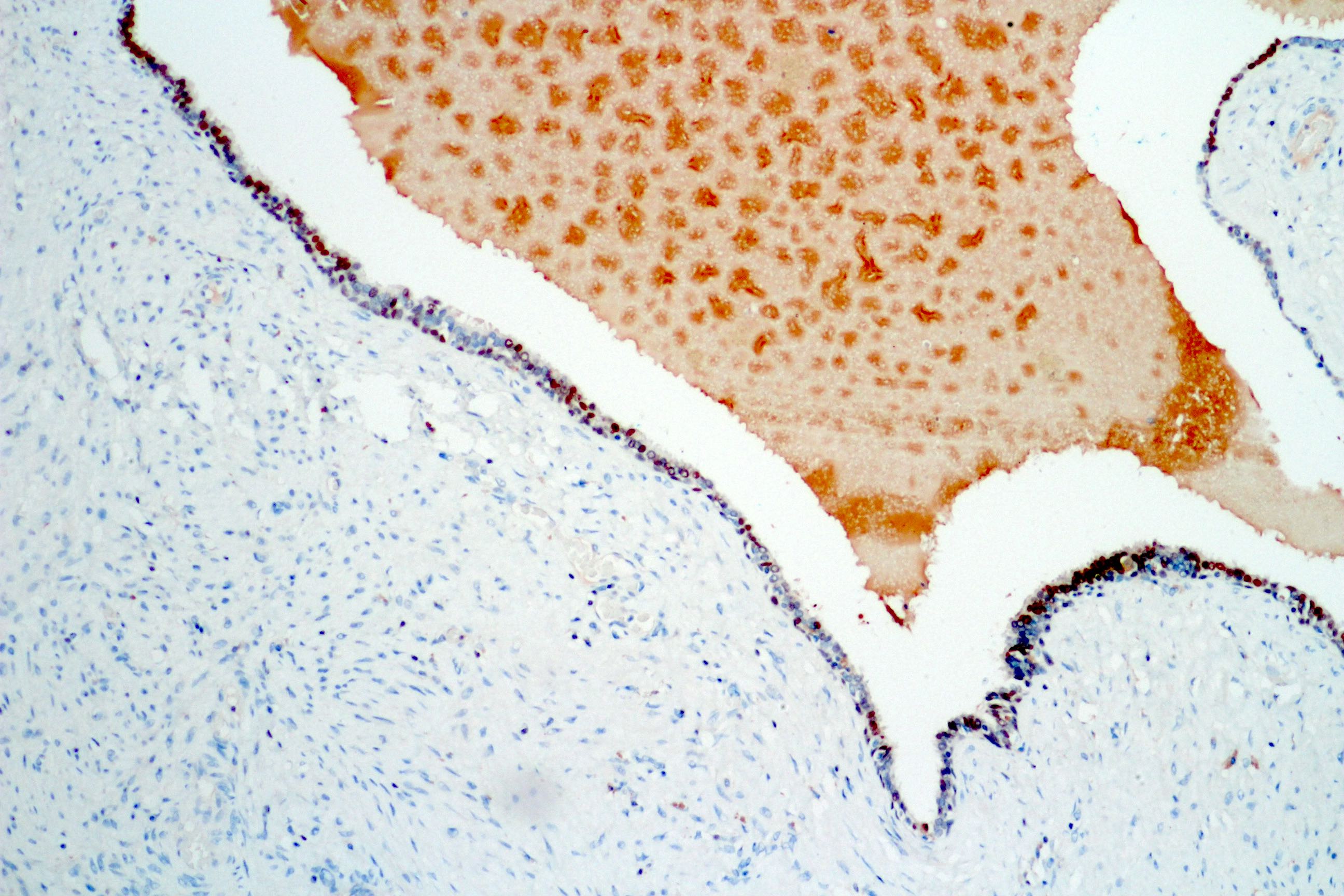
Nonproliferative stroma
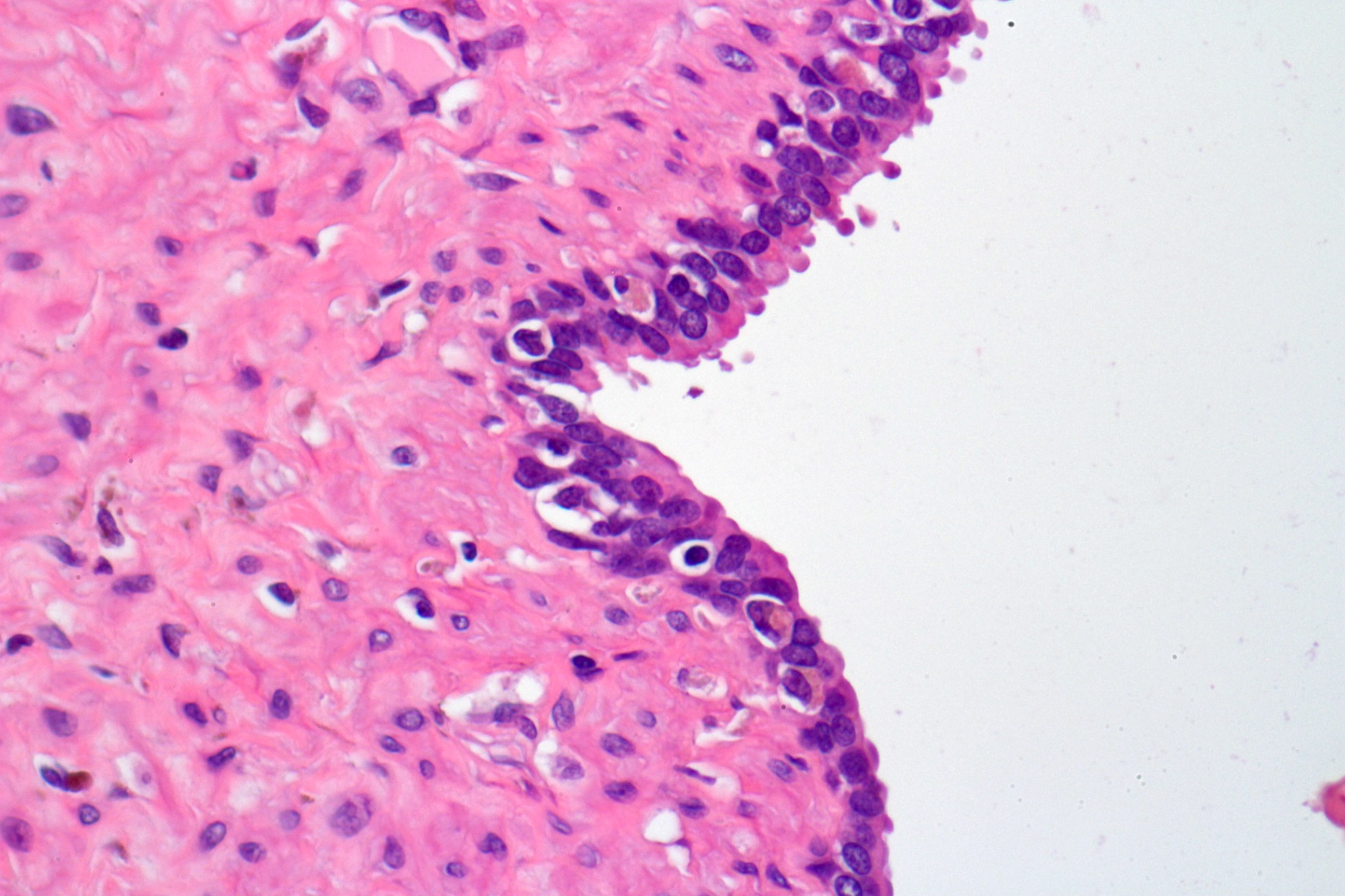
Bland epithelium
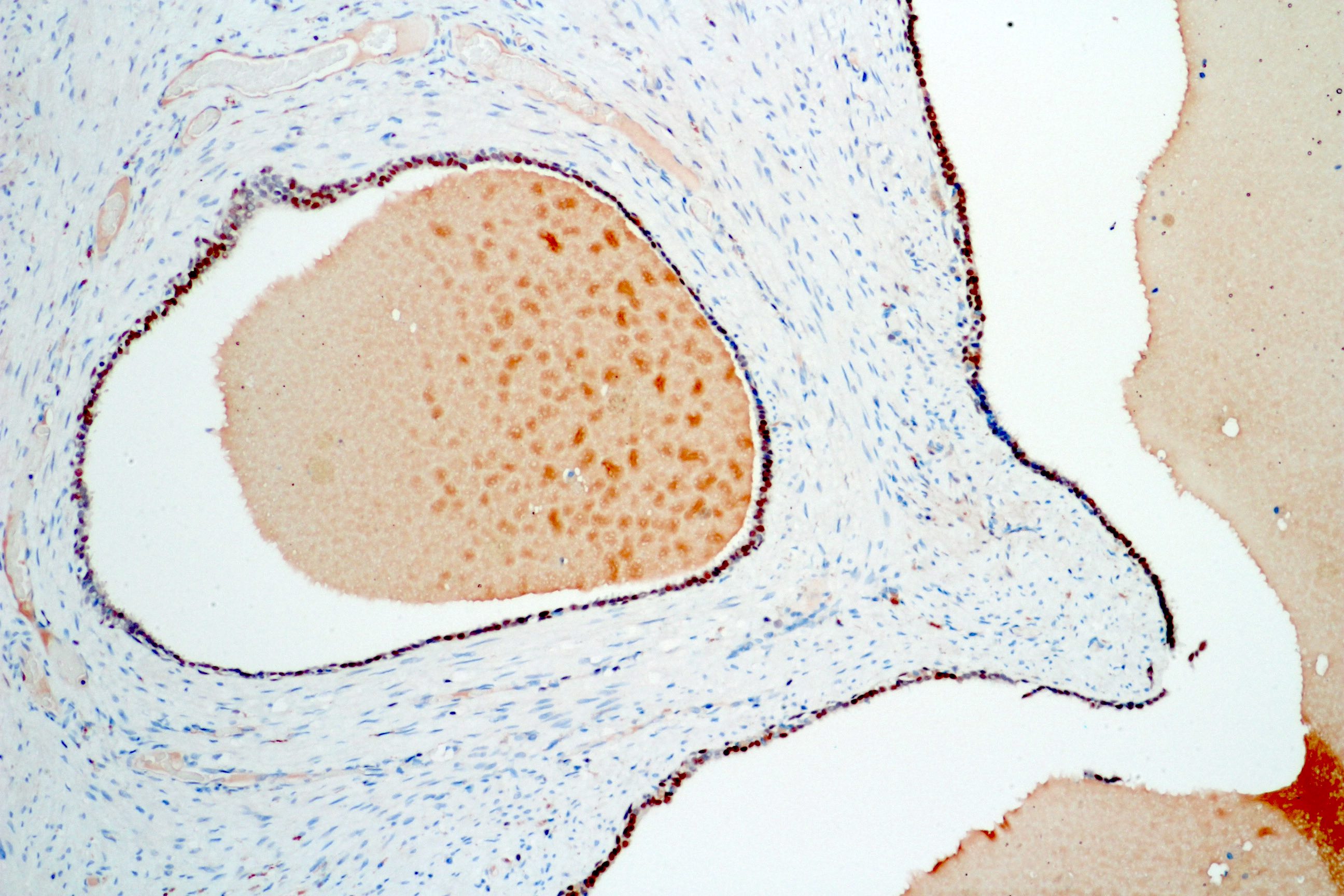
GATA3
GATA3

GATA3
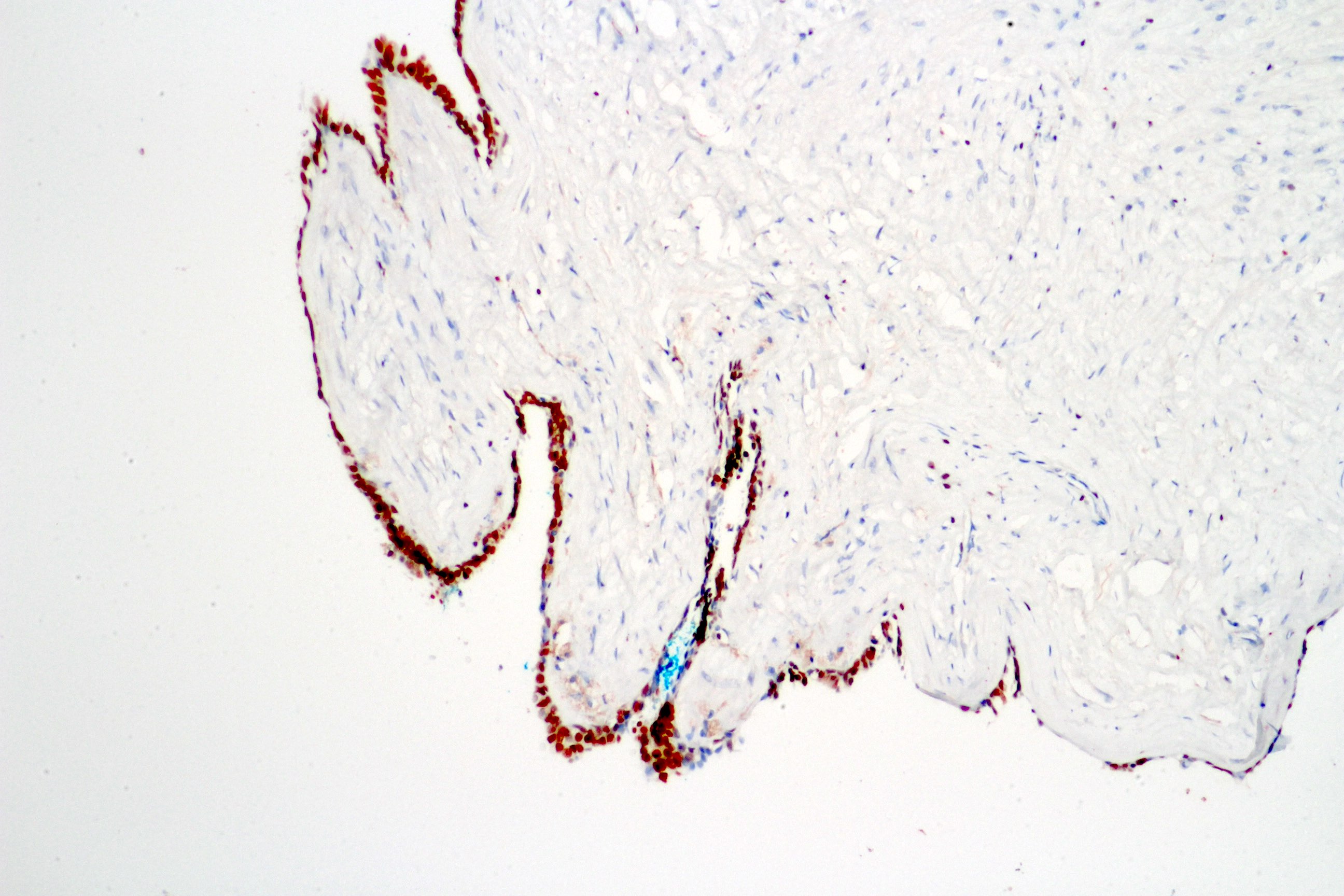
GATA3
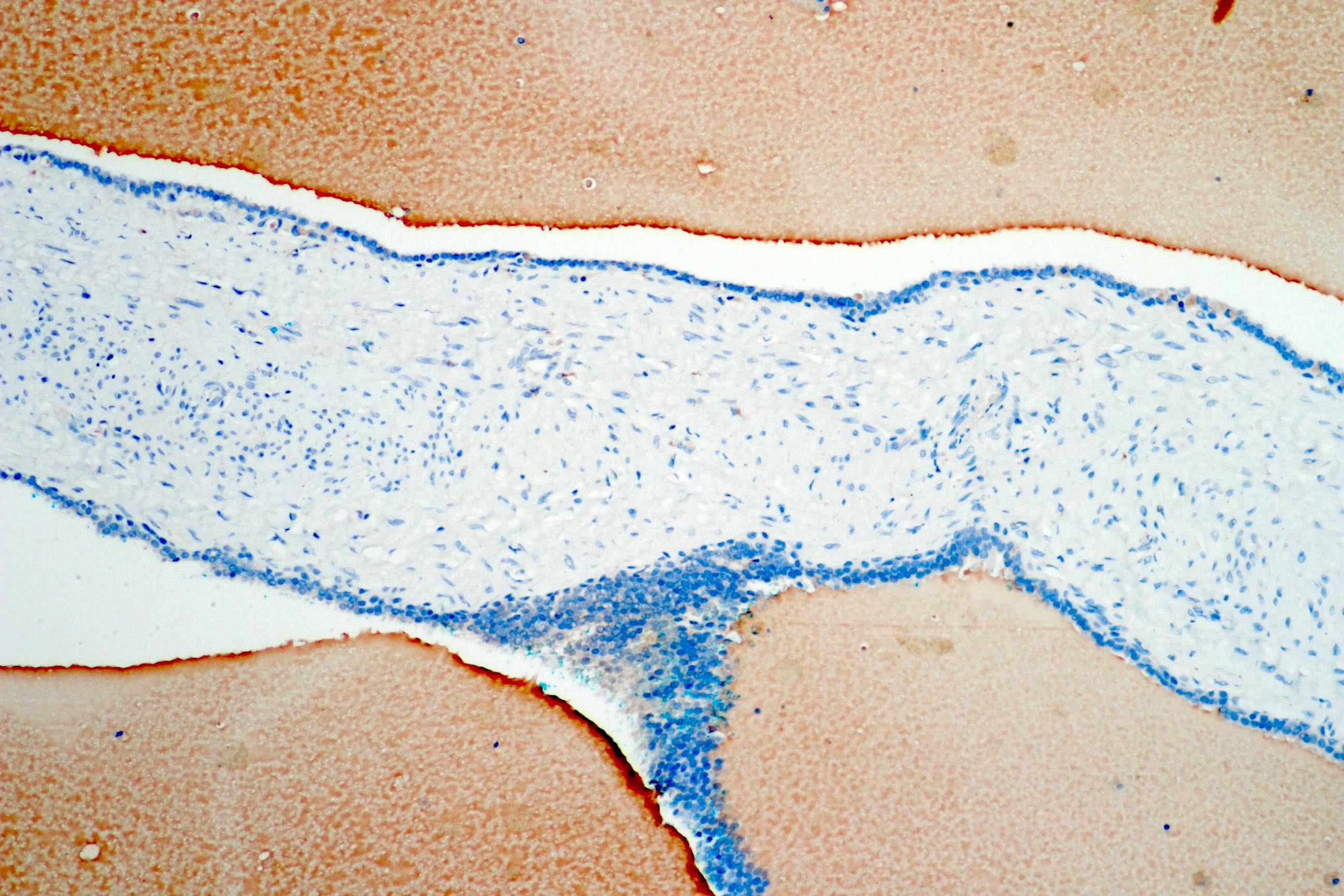
PSA negative
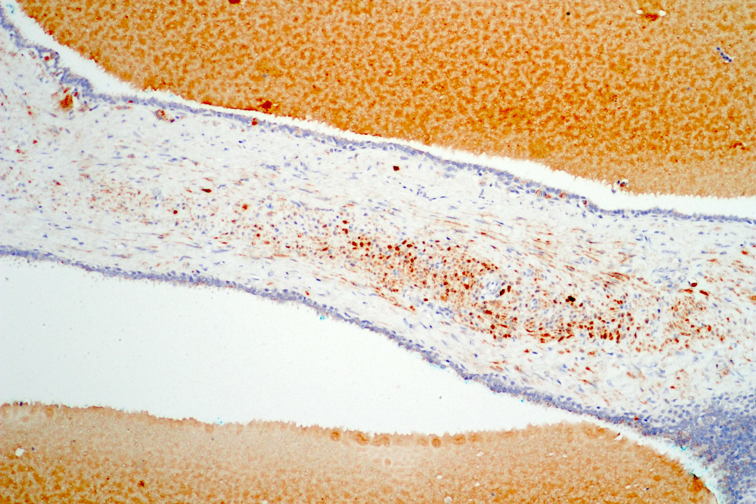
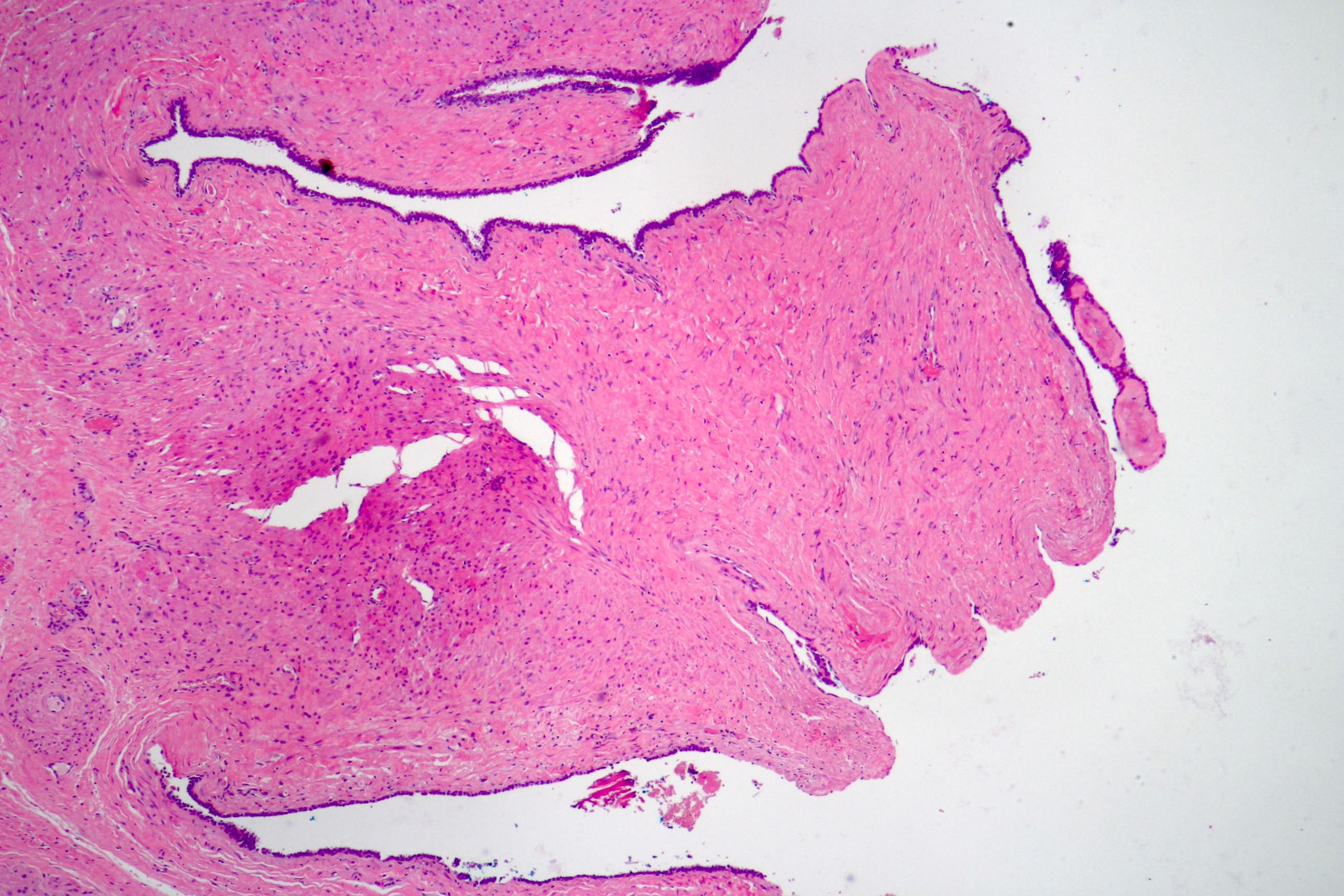
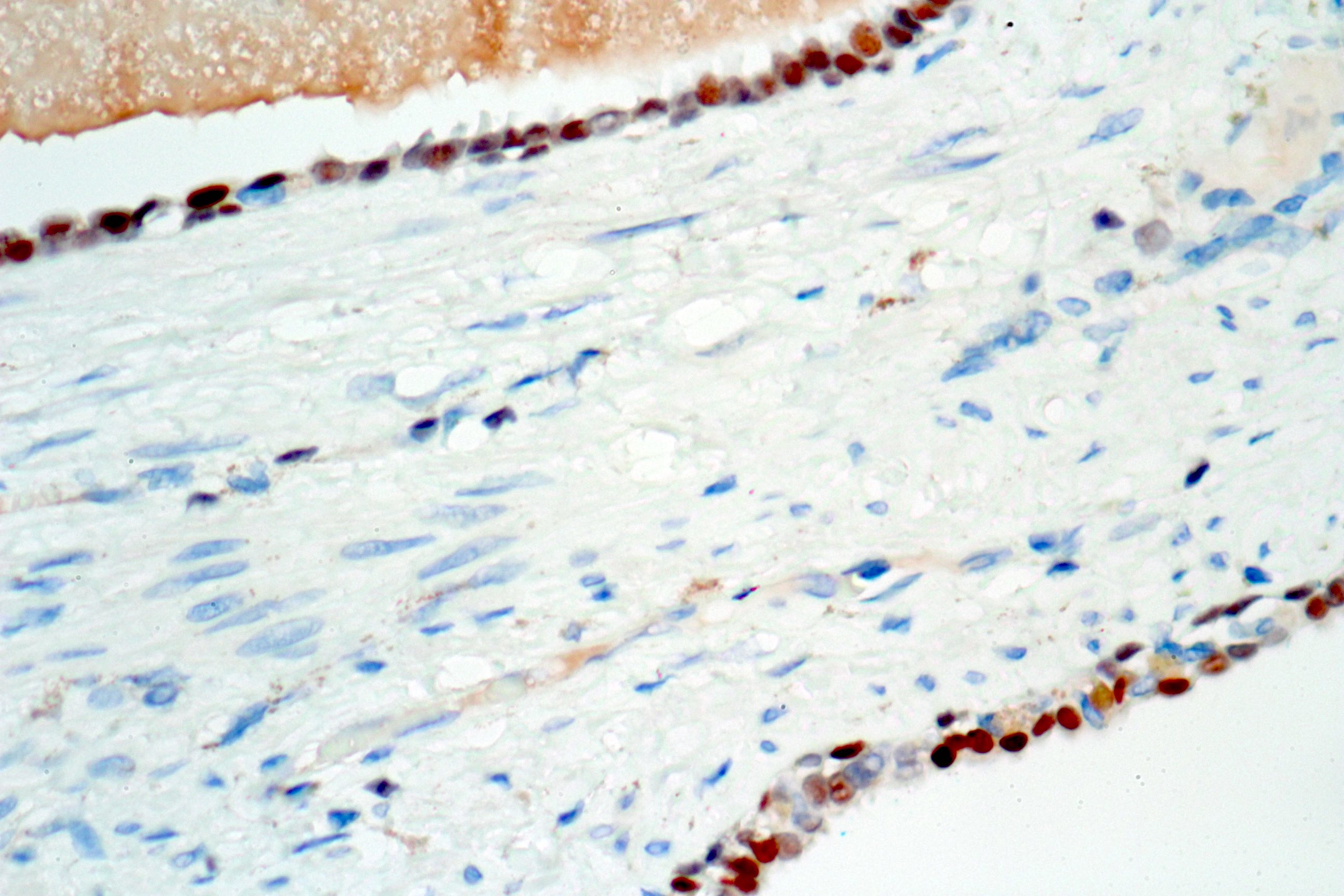
Calretinin negative
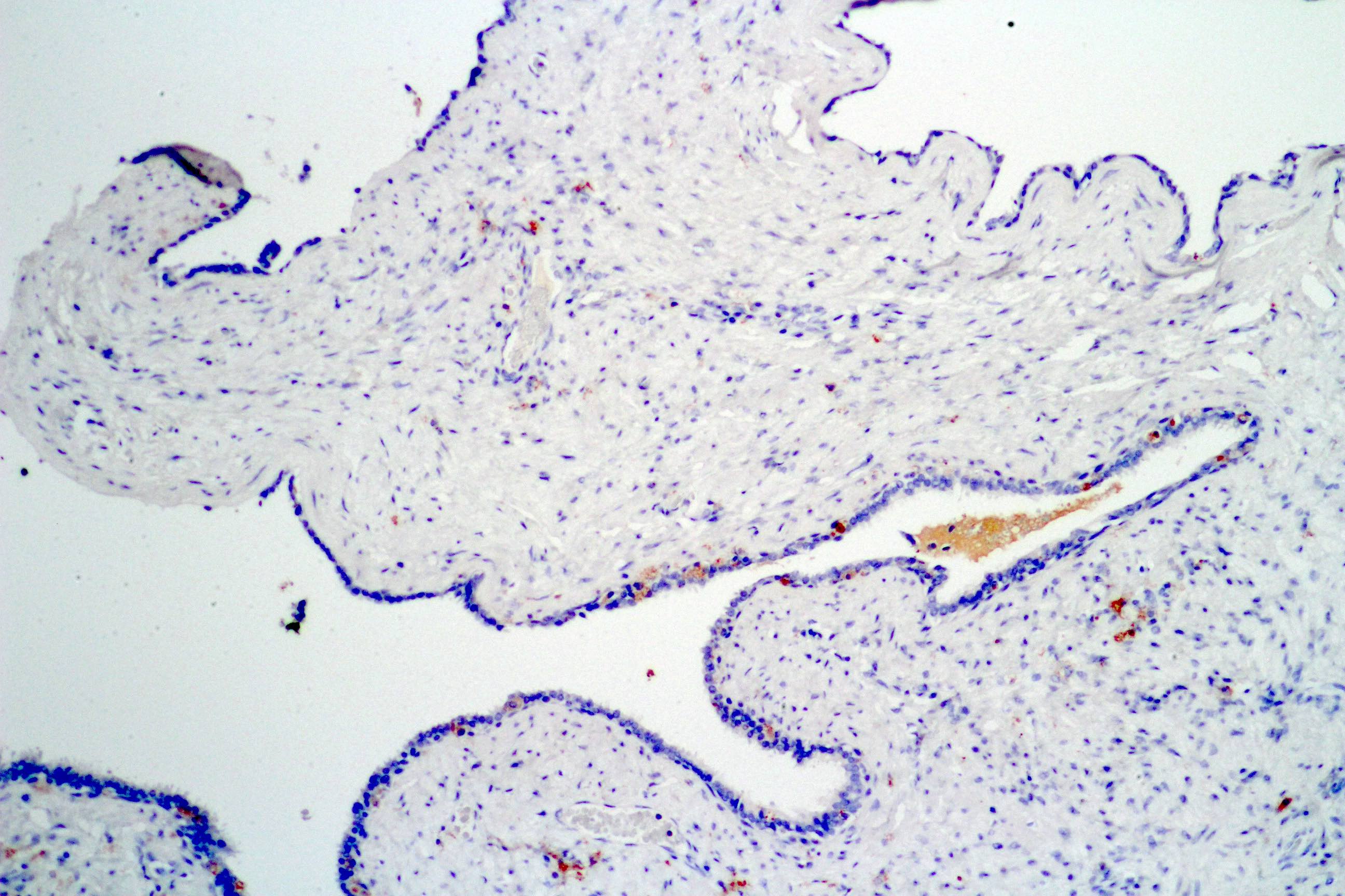
Prostein negative
Positive stains
- Epithelial lining stains for pancytokeratin and CK7
- Basal cells express high weight molecular keratins
- Stromal cells may stain for smooth muscle actin
- Normal epithelial lining of a seminal vesicle usually strongly expresses GATA3; however this marker is also positive in the prostate and should be interpreted with caution (Actas Urol Esp 2017;41:577)
- Normal and neoplastic seminal vesicle epithelium is strongly positive for PAX8 (Am J Surg Pathol 2011;35:1837, Hum Pathol 2017;69:123)
Negative stains
- Epithelial cells usually do not express PSA, prostein / P501S or calretinin
- Stromal cells are negative for S100
Sample pathology report
- Prostate and seminal vesicles, radical prostatectomy:
- Cystadenoma of the seminal vesicle (see comment)
- Comment: This is a benign neoplasia. Surgical resection is considered curative.
Differential diagnosis
- Mixed epithelial and stromal tumor of the seminal vesicle:
- Stromal hypercellularity
- Prostatic stromal tumor of uncertain malignant potential entrapping glands:
- Centered in the prostate, entrapped prostatic glands express PSA and other markers of prostatic differentiation
- Adenomatoid tumor:
- Expresses mesothelial markers such as calretinin
- Mesothelial cysts:
- Express mesothelial markers such as calretinin
- Cystic ductal adenocarcinoma of the prostate:
- Pseudostratified and atypical epithelial neoplastic cells
- Primary seminal vesicle carcinoma:
- Atypical cells
- Tumor centered in a seminal vesicle
- Adenocarcinomas usually show papillary glandular and trabecular growth patterns
- Exclusion of other primary sites and immunophenotype excluding primary prostate adenocarcinoma
Additional references
Board review style question #1
What major criterion favors the diagnosis of mixed epithelial and stromal tumor of the seminal vesicle over cystadenoma of the seminal vesicle?
- Expression of PSA in epithelial cells
- Expression of smooth muscle actin in stromal cells
- Stromal proliferation and hypercellularity
- Tumor grossly centered in the seminal vesicle
Board review style answer #1
C. Stromal proliferation and hypercellularity. Seminal vesicle cystadenoma shows no stromal hypercellularity (image shown above).
Comment Here
Reference: Cystadenoma
Comment Here
Reference: Cystadenoma
Board review style question #2
Which feature of immunohistochemistry favors the diagnosis of benign mesothelial cysts over cystadenoma of the seminal vesicle?
- Expression of calretinin in epithelial cells
- Expression of GATA3 in epithelial cells
- Expression of pankeratins in epithelial cells
- Expression of smooth muscle actin in stromal cells
Board review style answer #2
A. Expression of calretinin in epithelial cells. Seminal vesicle cystadenoma is negative for calretinin in the photo above.
Comment Here
Reference: Cystadenoma
Comment Here
Reference: Cystadenoma
