

Skin nontumor
Infectious disorders
Actinomycosis
Author: Abha Soni, D.O., M.P.H.
Editorial Board Member: Kiran Motaparthi, M.D.

Last author update: 24 June 2022
Last staff update: 24 January 2023
Copyright: 2003-2024, PathologyOutlines.com, Inc.
PubMed Search: Actinomycosis skin
Table of Contents
Definition / general | Essential features | Terminology | ICD coding | Epidemiology | Sites | Pathophysiology | Etiology | Clinical features | Diagnosis | Case reports | Treatment | Clinical images | Gross description | Gross images | Microscopic (histologic) description | Microscopic (histologic) images | Cytology description | Cytology images | Positive stains | Negative stains | Sample pathology report | Differential diagnosis | Additional references | Board review style question #1 | Board review style answer #1 | Board review style question #2 | Board review style answer #2Cite this page: Soni A. Actinomycosis. PathologyOutlines.com website. https://www.pathologyoutlines.com/topic/skinnontumoractinomycosis.html. Accessed April 19th, 2024.
Definition / general
- Rare suppurative and granulomatous infectious disease (Int J Surg Case Rep 2015;16:99)
- Caused by a group of anaerobic, gram positive, filamentous bacteria, which are normal flora in the oral cavity, gastrointestinal tract and female genital tract (Int J Surg Case Rep 2015;16:99)
- Clinically presents with abscess formation, tissue fibrosis, sinus drainage and occasionally, a soft tissue mass mimicking a tumor (Int J Surg Case Rep 2015;16:99)
Essential features
- Causative agent Actinomyces israelli
- Treated with high dose penicillin
- Splendore-Hoeppli phenomenon is a histologic feature
Terminology
- Most common causative agent is Actinomyces israelii
- Sulfur granules: small yellow granules found within the abscesses formed by Actinomyces infection (Ophthal Plast Reconstr Surg 1992;8:237)
- Lumpy jaw syndrome: large abscesses located on the head and neck, usually following dental disease and mandibular osteomyelitis (BMJ Case Rep 2015;2015:bcr2014206557 )
- Splendore-Hoeppli phenomenon (asteroid bodies): pink rim at the periphery of the colony, due to immunoglobulin and cell debris, which occurs around colonies of fungi, bacteria and parasites (J Oral Maxillofac Pathol 2018;22:161)
ICD coding
- ICD-10: A42.9 - actinomycosis, unspecified
Epidemiology
- Higher prevalence within areas of low socioeconomic status (BMC Infect Dis 2018;18:686)
- Use of intrauterine device contraceptive devices has increased the incidence of actinomycosis (Contraception 2007;75:S48)
Sites
- Cervicofacial (post dental infection), skin (posttraumatic injury creating an anaerobic environment), pelvic (post intrauterine device placement), abdominal (status post ruptured appendix or bowel perforation) and pulmonary (smokers with poor dental hygiene, aspiration of infective material) (Infect Drug Resist 2014;7:183)
Pathophysiology
- Predisposing factors include poor oral hygiene, trauma, male gender, diabetes mellitus, immunosuppression, alcoholism and malnutrition (Infect Drug Resist 2014;7:183)
- Filamentous bacteria are a normal commensal inhabitant of the oral and buccal cavities, gastrointestinal tract and female genitalia
- Etiology of infection in skin is commonly linked with a traumatic injury, human bite or a perforating injury, which creates an anaerobic environment for Actinomyces israelii to grow in (J Clin Diagn Res 2014;8:YD03)
Etiology
- Caused by Actinomyces israelii in humans and Actinomyces bovis in animals
Clinical features
- Localized pain, swelling and draining fistulas
Diagnosis
- Culture on chocolate agar media at 37 °C
- Gram stain is more sensitive than culture, especially if patient is on antibiotics
- Filamentous bacteria of actinomycosis can be identified on H&E and are highlighted by Gram stain (Infect Drug Resist 2014;7:183)
- Polymerase chain reaction (PCR) and nucleic acid probes are being developed for faster and more accurate identification
Case reports
- 16 year old boy with scalp actinomycosis presenting as a soft tissue tumor (Int J Surg Case Rep 2015;16:99)
- 32 year old woman with actinomycosis of the back and left axilla (J Clin Diagn Res 2014;8:YD03)
- 36 year old man with renal actinomycosis presenting as a urocutaneous fistula (Urol Case Rep 2019;28:101054)
- 42 year old man with primary cutaneous actinomycosis (AIDS Res Ther 2019;16:16)
- 55 year old woman with primary cutaneous actinomycosis of upper extremity (Trop Doct 2012;42:58)
- 82 year old man with actinomycosis of the upper lip (Ann Dermatol 2011;23:S131)
Treatment
- High dose penicillin is necessary to penetrate areas of fibrosis and suppuration / granules (Clin Infect Dis 2004;38:444)
- Drainage of abscesses or radical excision of sinus tracts
Clinical images
Contributed by Dhiraj B. Nikumbh, M.B.B.S., M.D.
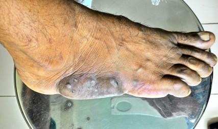
Multiple sinuses in foot
Images hosted on other servers:

Indurated lesion

Lesion over back

Left thigh lesion

Molar tooth appearance
Gross description
- Firm, swollen region on the skin with multiple draining abscesses and fistula tracts
- Pus draining yellow sulphur granules
Gross images
Images hosted on other servers:


Outer / inner surface and cross section of excised tissue
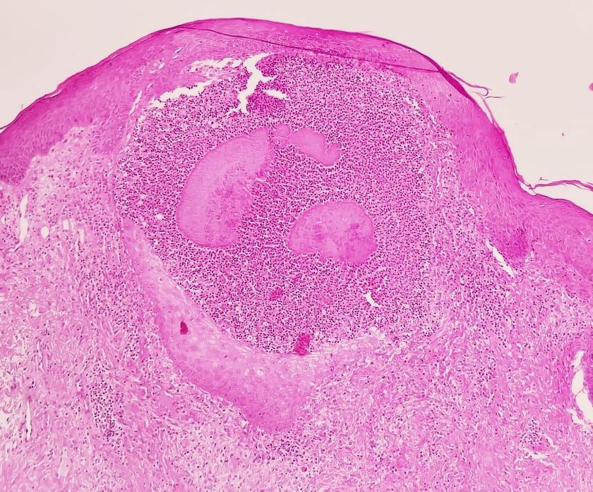
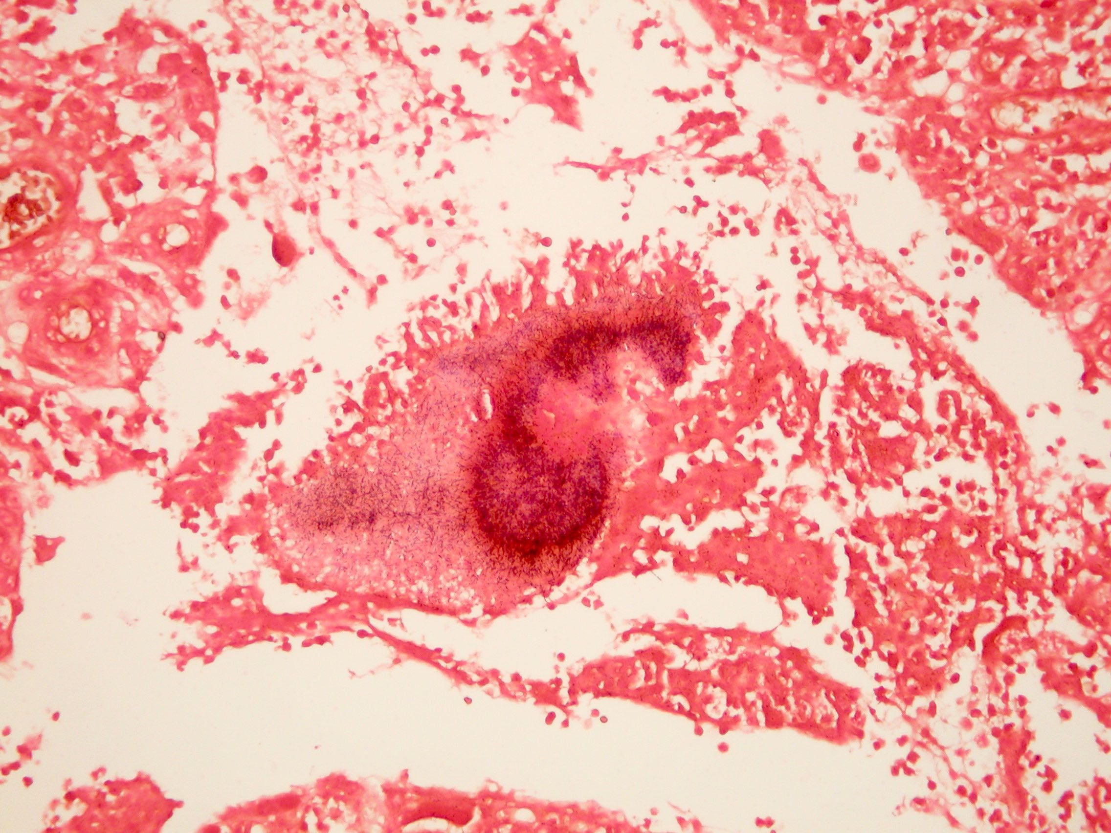
Microscopic (histologic) description
- Lymphocytes, neutrophils, giant cells and fibroblasts
- Bacterial colonies (sulphur granules) found at the center of the inflammatory reaction, composed of basophilic radiating filaments
Microscopic (histologic) images
Contributed by Kuroli Eniko, M.D., Yale Rosen, M.D. and Haitham Alfalah, M.D. (source: Wikipedia)
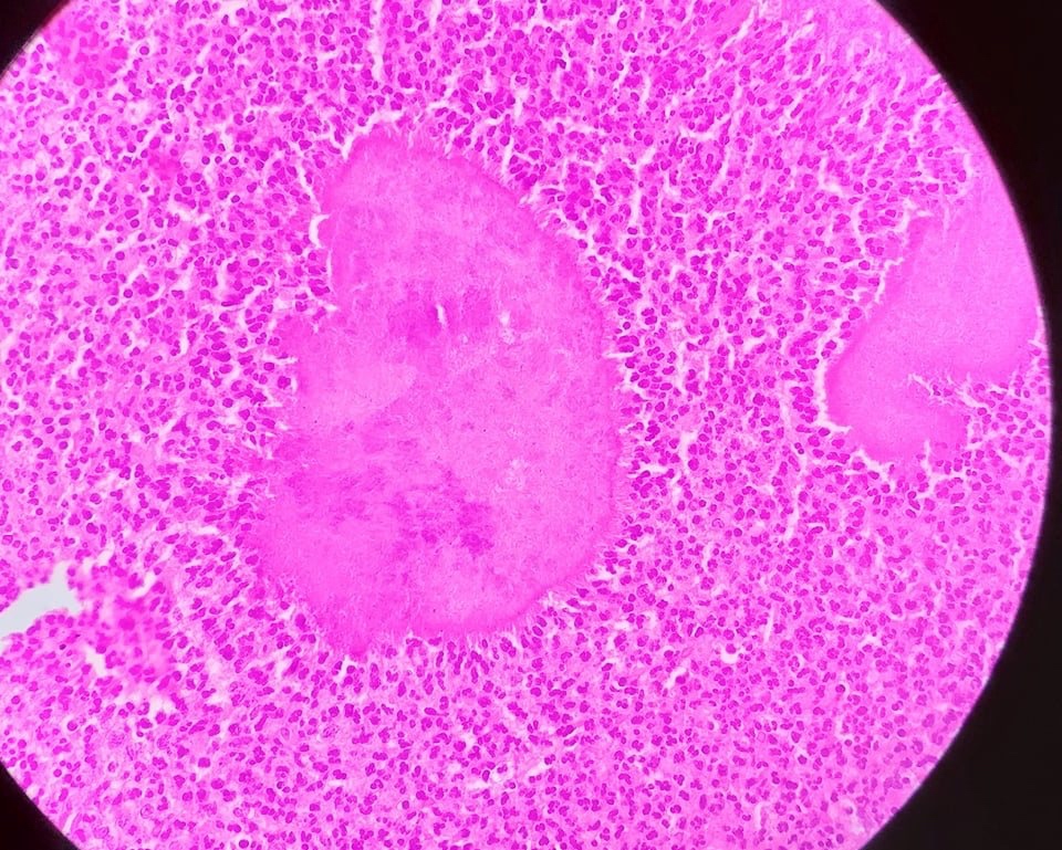
Suppurative and granulomatous inflammation
Dense sheets of neutrophils

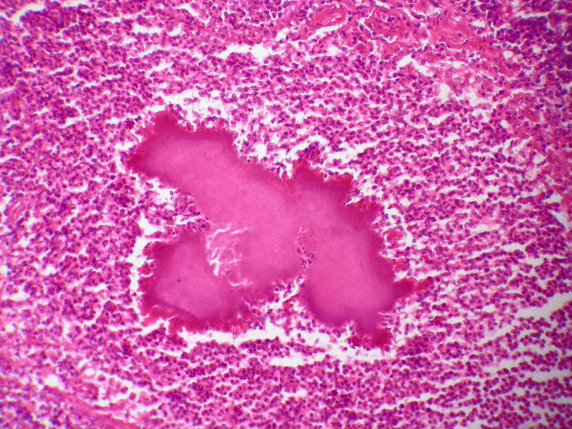
Colony of Actinomyces
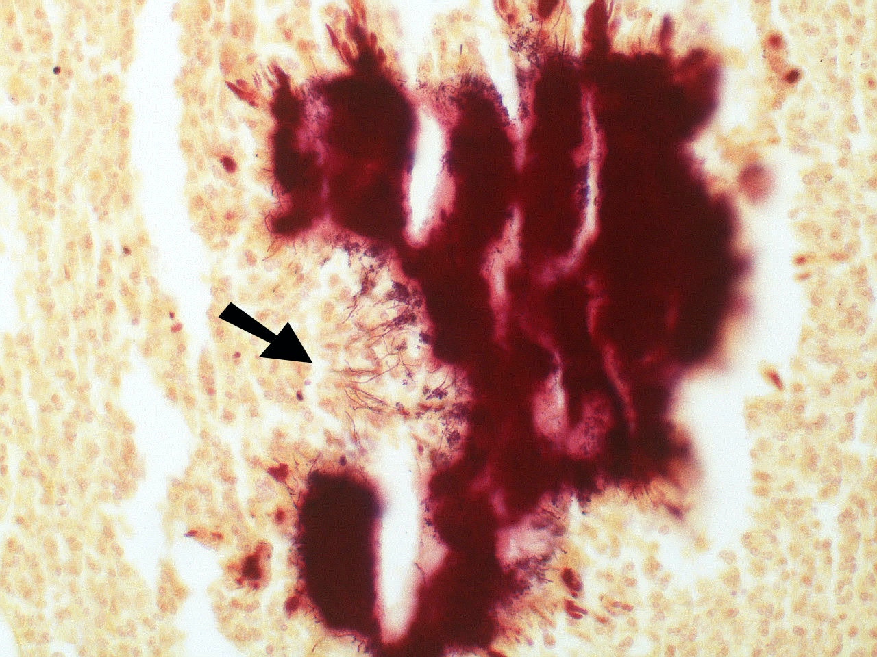
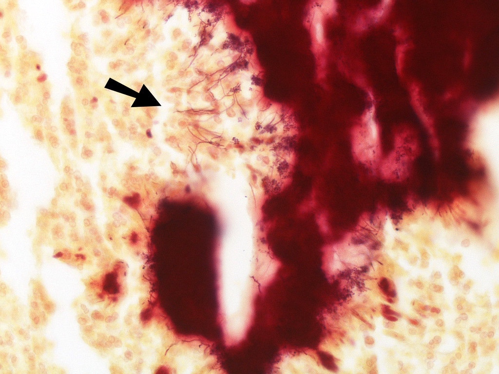
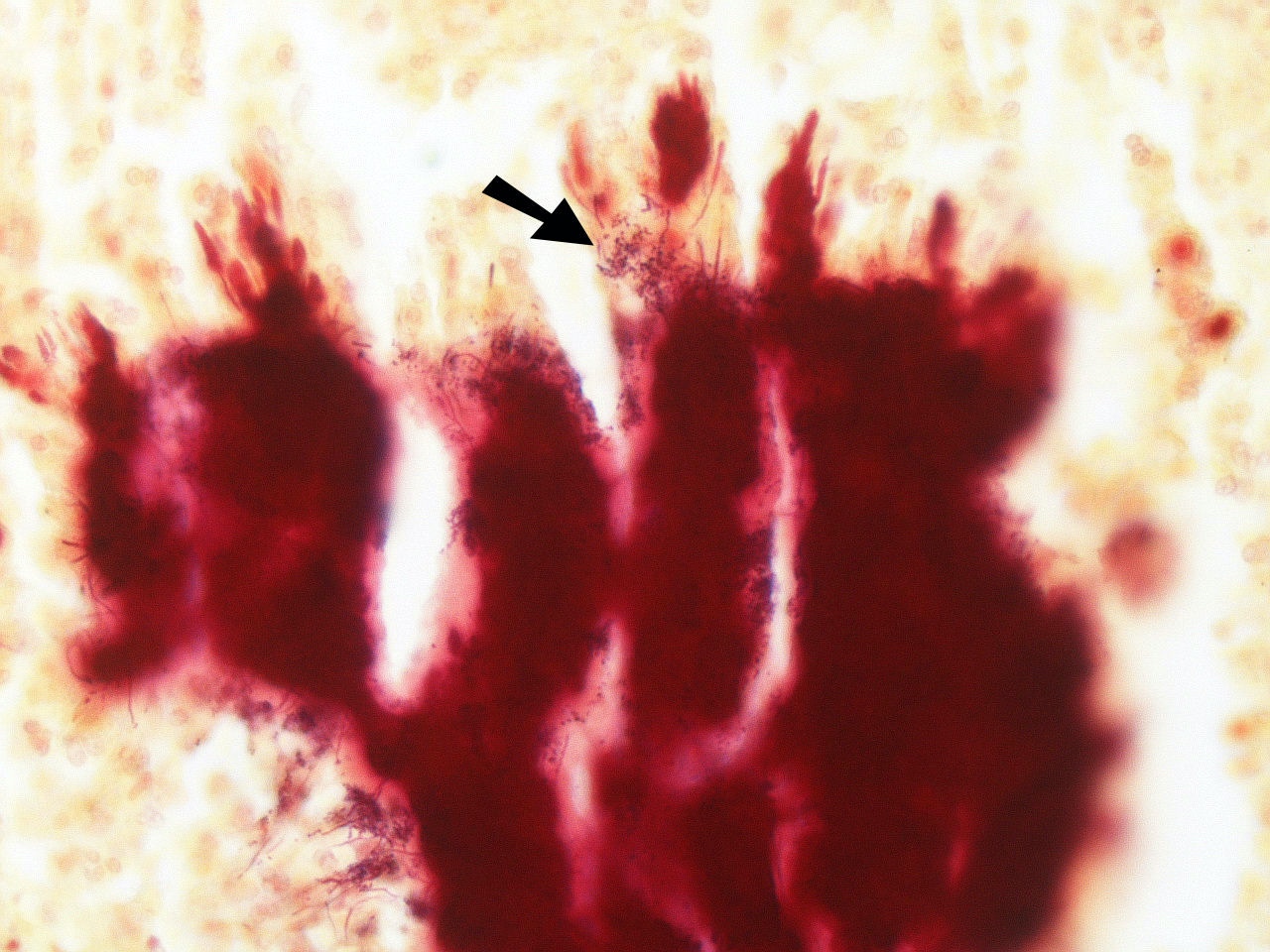
Thin gram positive filamentous organisms
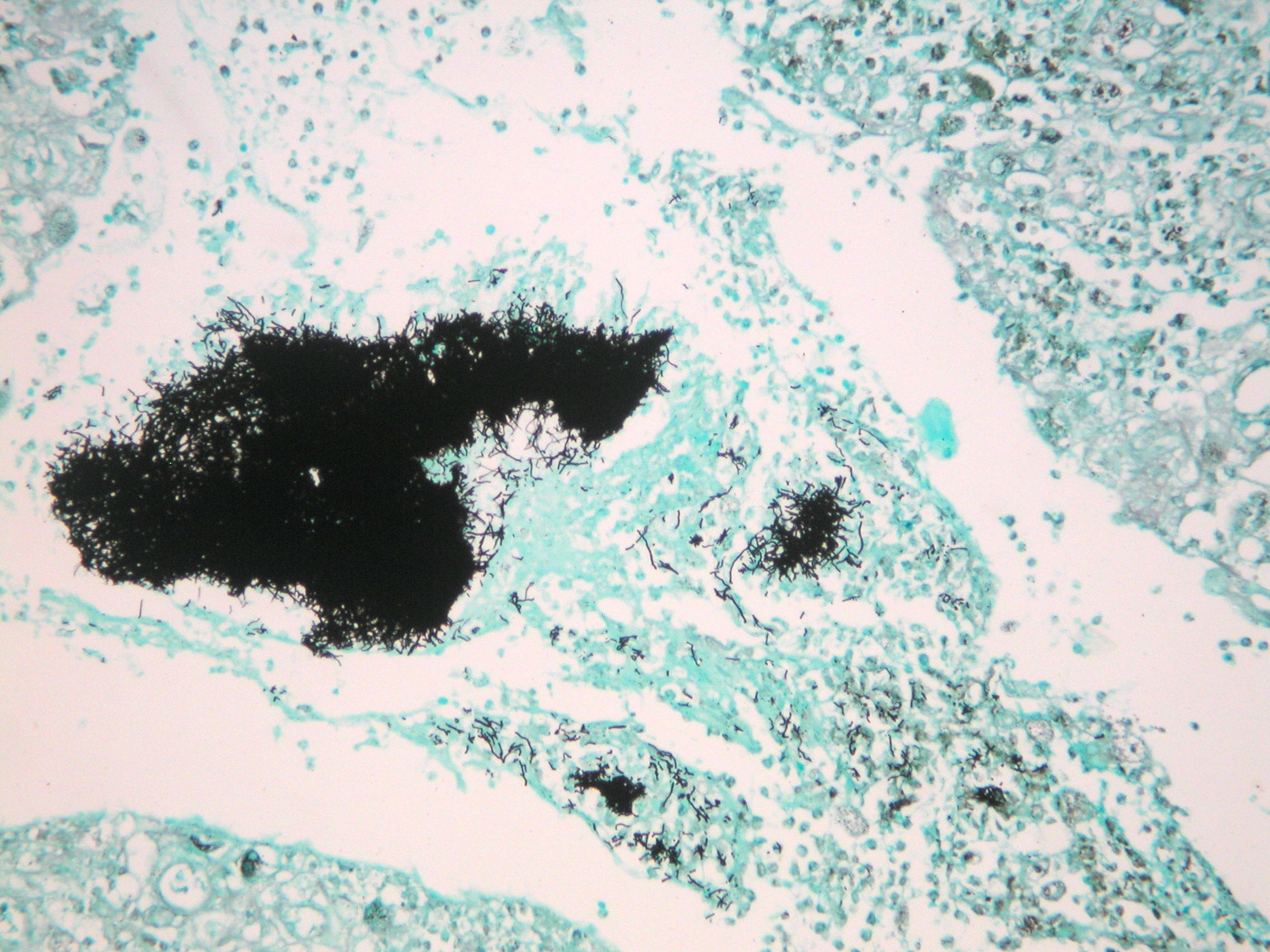
Grocott stain
Gram stain
Cytology description
- Mixed inflammatory infiltrate, foreign body multinucleated giant cells and clumps of filamentous organisms
Cytology images
Contributed by Dhiraj B. Nikumbh, M.B.B.S., M.D.
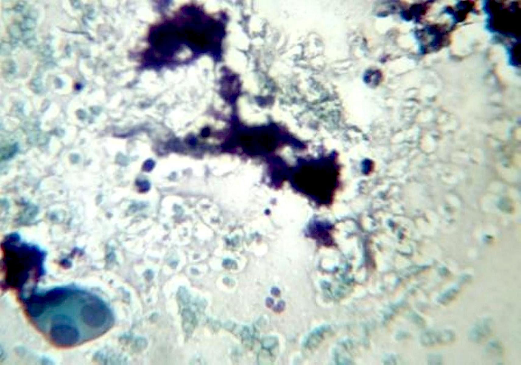
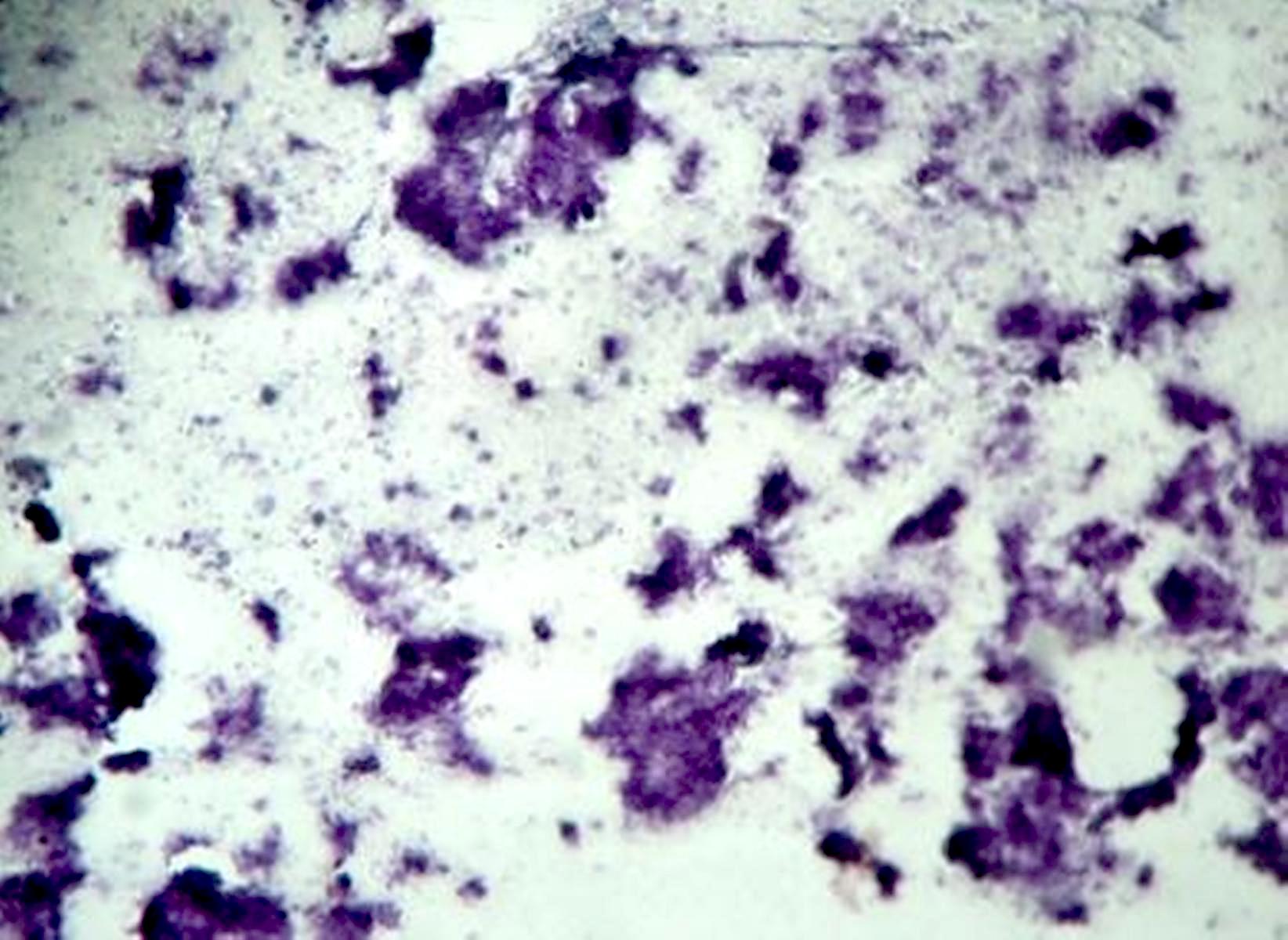
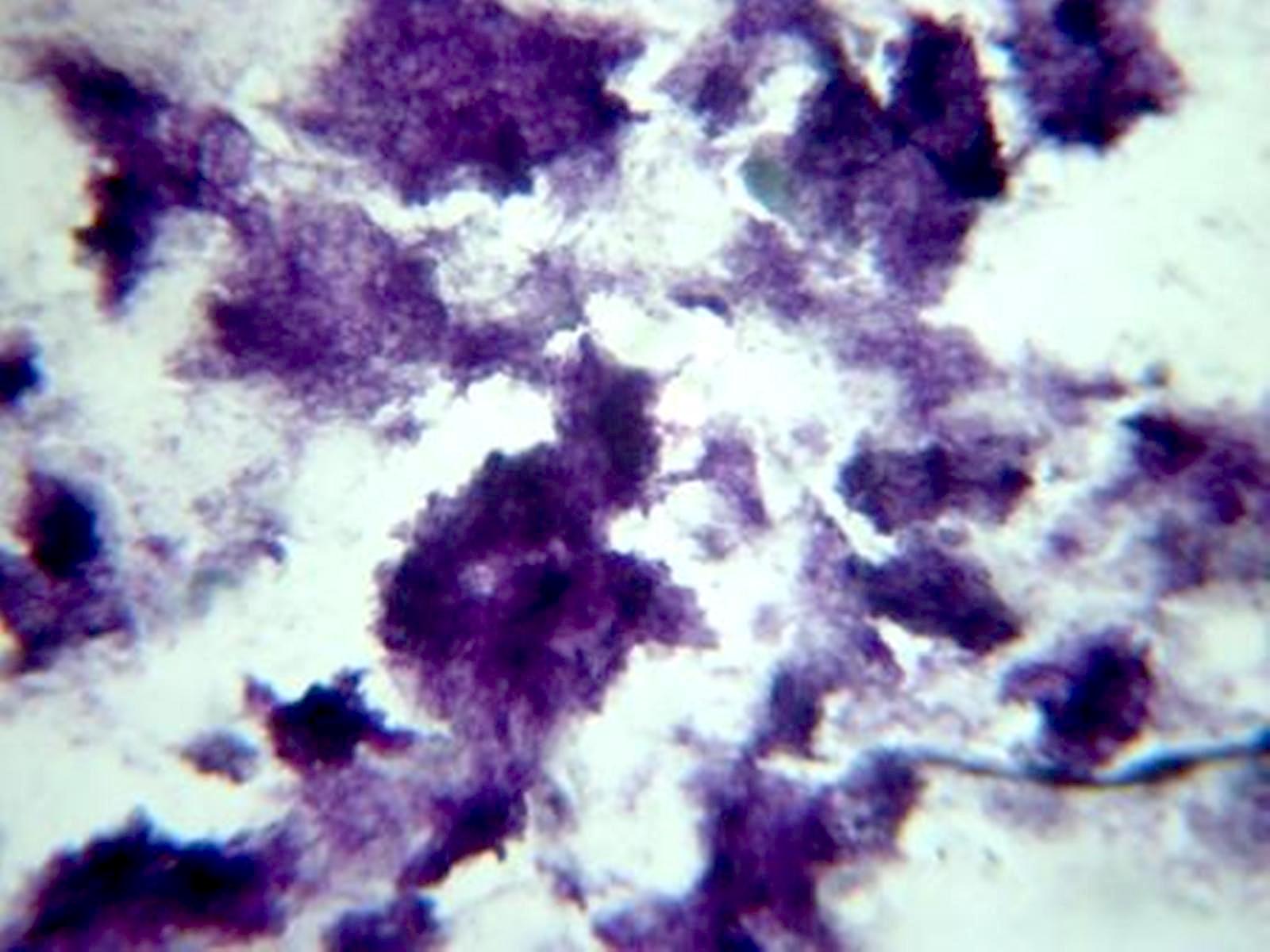
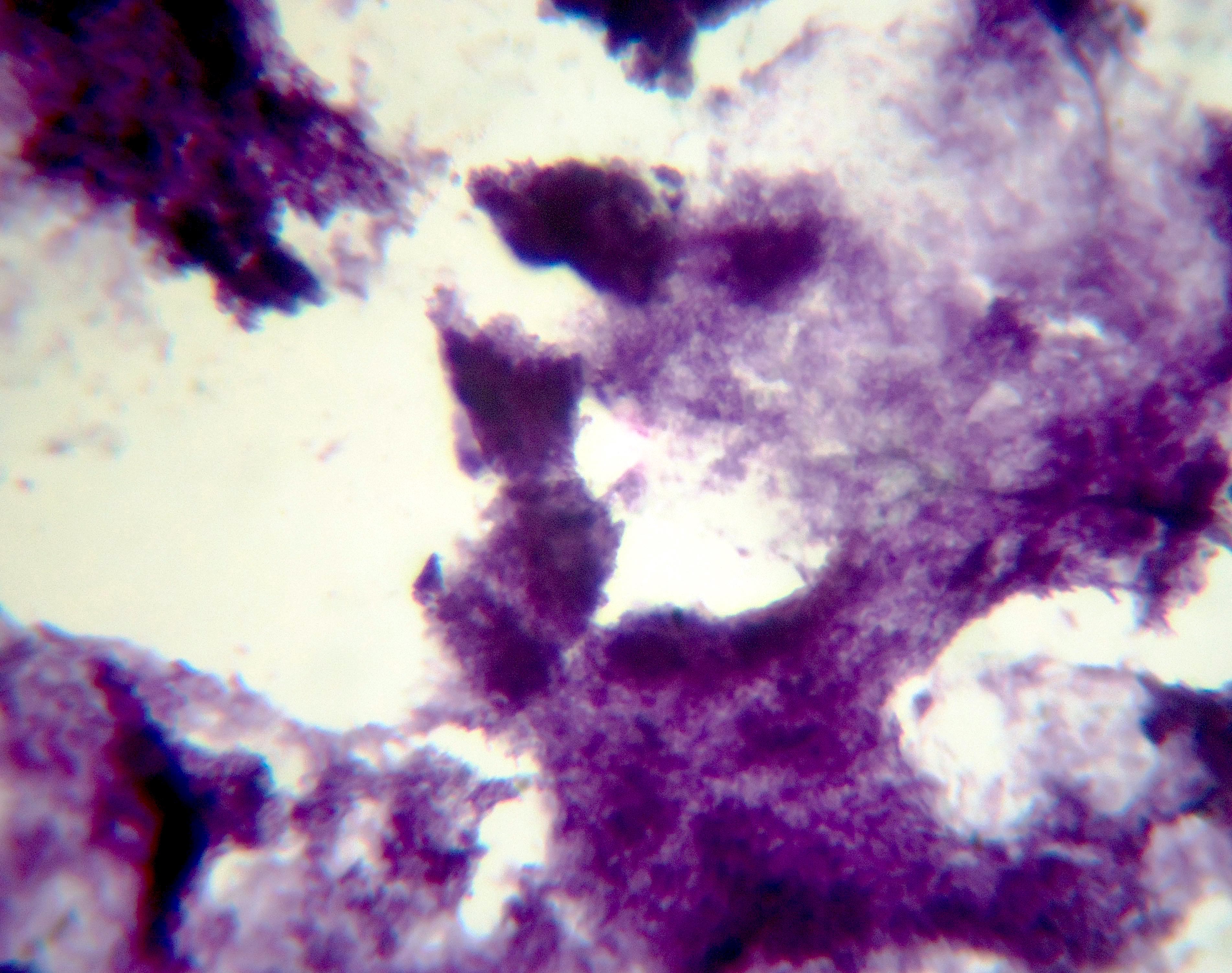
Mixed inflammatory infiltrate with clumps of fibrillar organisms
Images hosted on other servers:

Gram stain
Positive stains
- Gram positive
- GMS stain demonstrates filamentous bacteria, which do not stain with PAS but are visible on H&E
- Modified Ziehl-Neelsen stains (e.g., Fite-Faraco) (ScienceDirect: Nocardia [Accessed 24 June 2022])
Negative stains
- Acid fast stains
Sample pathology report
- Skin, biopsy:
- Primary cutaneous actinomycosis
Differential diagnosis
- Nocardiosis:
- Branching filamentous bacteria are partially acid fast positive
- Histologic appearance of Nocardia is similar to other actinomycetes family members; culture and biochemical testing is necessary for diagnosis / identification
- Fite-Faraco stain is helpful since positive in nocardiosis
- Tuberculosis:
- Characteristic histologic findings include caseating granulomas
- Detected by Ziehl-Neelsen stain and Mycobacterium culture
- Sporotrichosis:
- Paracoccidioidomycosis:
Additional references
Board review style question #1
What is the name of the unique histologic pattern that takes place in a cutaneous actinomycosis infections?
- Asteroid body reaction
- Flame figures
- Gout
- Splendore-Hoeppli phenomenon
Board review style answer #1
Board review style question #2
What type of granules are found within the abscesses formed by Actinomyces infection?
- Metachromatic granules
- Polyphosphate granules
- Polysaccharide granules
- Sulfur granules
Board review style answer #2
