

Skin nontumor
Alopecia
Alopecia mucinosa
Last author update: 1 July 2011
Last staff update: 30 June 2023 (update in progress)
Copyright: 2002-2024, PathologyOutlines.com, Inc.
PubMed Search: Alopecia mucinosa

Table of Contents
Epidemiology | Clinical features | Case reports | Microscopic (histologic) description | Microscopic (histologic) images | Positive stains | Differential diagnosis | Additional referencesCite this page: Hamodat M. Alopecia mucinosa. PathologyOutlines.com website. https://www.pathologyoutlines.com/topic/skinnontumoralopeciamucinosa.html. Accessed April 20th, 2024.
Epidemiology
- Coexisting lymphoma associated with very poor prognosis
Clinical features
- Also called follicular mucinosis
- Edematous and erythematous plaques of alopecia on head and neck
- Children: benign, self limited
- Adults: associated with cutaneous T cell lymphoma, Sezary syndrome, Hodgkin lymphoma, acute myeloblastic leukemia, chronic lymphocytic lymphoma and squamous cell carcinoma of the tongue
- Nodular or plaquelike lesion
- Patterns:
- Infiltrated plaque, solitary or multiple, associated with alopecia when in scalp or beard area
- Group of of follicular papules, either localized or extensively distributed on trunk and proximal limbs in addition to scalp and face
- Acneiform lesion with comedones, mucinorrhea (discharge of mucinous fluid from follicular ostia) and severe pruritis
- Clinical course: either spontaneous regression, chronic relapsing but benign course over many years or associated with lymphoma
Case reports
- 61 year old woman with SLE (Dermatol Online J 2010;16:7)
Microscopic (histologic) description
- Follicular infundibulum keratinocytes and outer root sheath are separated by pools of mucin
- Mixed infiltrate of lymphocytes, histiocytes and conspicuous eosinophils
- Marked follicular dilation with cyst formation and perifollicular scarring
- Both the dermis and and affected epithelium are typically infiltrated by lymphocytes, histiocytes and eosinophils
- In cases associated with lymphoma, atypical lymphocytes, convoluted lymphocytes, large transformed cells and mitotic figures may be seen
Microscopic (histologic) images
Contributed by Mowafak Hamodat, M.B.Ch.B., M.Sc.
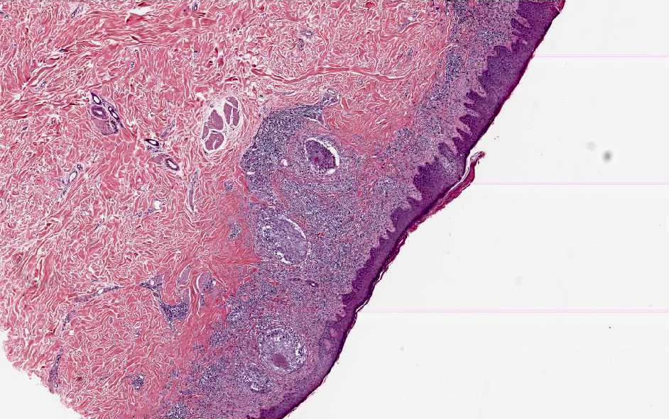
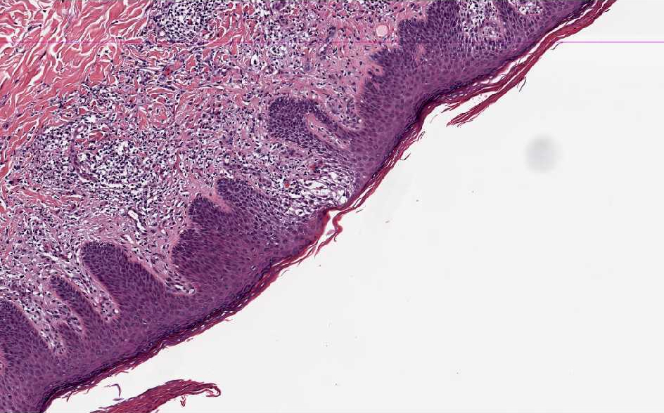
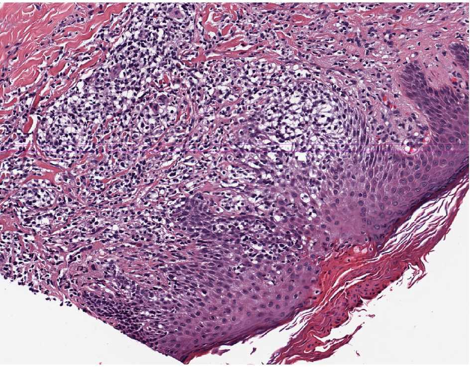
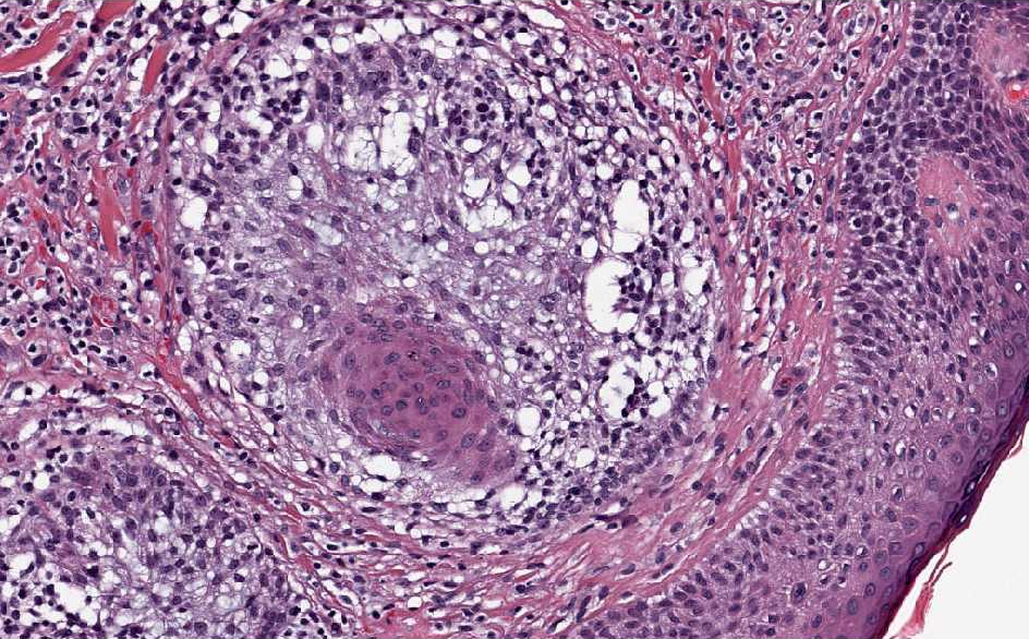
Various images
Images hosted on other servers:

Superficial and deep,
perivascular and perifollicular,
infiltrate of lymphocytes and
numerous eosinophils
Positive stains
- Alcian blue
Differential diagnosis
- Coexisting mycosis fungicides: has atypical or cerebriform lymphocytes, bandlike infiltrate in upper dermis, no / minimal eosinophils; obtain multiple biopsies as needed
- Note: TCR gene rearrangement present in 50% of patients whether associated with tumor or not
Additional references
