

Skin nontumor
Vesiculobullous and acantholytic reaction patterns
Mechanical blister
Author: Narina Grove, M.D., M.A.

Last author update: 1 September 2016
Last staff update: 15 February 2024
Copyright: 2003-2024, PathologyOutlines.com, Inc.
PubMed Search: Blood blister
Table of Contents
Definition / general | Essential features | Terminology | ICD coding | Epidemiology | Sites | Pathophysiology | Etiology | Diagrams / tables | Clinical features | Diagnosis | Treatment | Clinical images | Gross description | Microscopic (histologic) description | Microscopic (histologic) images | Electron microscopy description | Differential diagnosis | Additional referencesCite this page: Grove N. Mechanical blister. PathologyOutlines.com website. https://www.pathologyoutlines.com/topic/skinnontumorbloodblister.html. Accessed April 24th, 2024.
Definition / general
- Essentially identical to friction blister with the extension of cytolysis through the basal layer resulting in a hemorrhagic blister
- Intraepidermal cleavage due to cytolysis and necrosis of keratinocytes in the upper stratum malpighii
Essential features
- Mechanical blister associated with manual labor
- Cytolysis extends through the basal layer causing hemorrhage
- Mostly clinical diagnosis
- Symptomatic treatment
Terminology
- Friction blister: blister caused by shearing force, which subsequently fills with clear fluid
- Blood blister: blister caused by excessive force, which fills with blood
- Hemorrhagic bullae: larger blisters (> 0.5 cm), often multiple and associated with underlying disease process and more often deeper (suprabasilar or subepidermal)
ICD coding
- S90.82 - blister (nonthermal) of foot
Epidemiology
- Common in athletes, dancers or those wearing poorly fitting shoes
- Associated with manual labor
Sites
- Hands and feet; sites subjected to repetitive friction
Pathophysiology
- Shearing forces within the epidermis cause friction blisters in the areas where epidermis is thick and firmly attached to the underlying tissue
Etiology
- Prolonged walking or repetitive actions
Diagrams / tables
Images hosted on other servers:
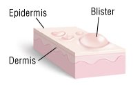
Friction blister
Clinical features
- Hemorrhagic blister usually occurring on exposed skin
- Most commonly occurring on hands and feet
Diagnosis
- Typically, a clinical diagnosis
- If solitary, diagnosis is usually a simple blood blister, rarely biopsied
- Multiple blood blisters of unknown origin may indicate an underlying process (see "differential diagnosis" below)
Treatment
- Symptomatic treatment
Clinical images
Images hosted on other servers:


Blood blisters
Gross description
- Cutaneous vesicle or bulla filled with sanguinous fluid
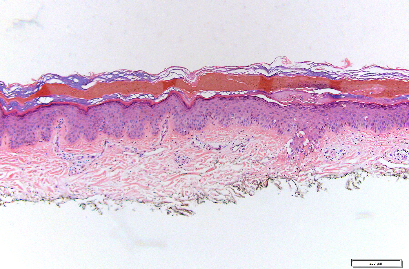
Microscopic (histologic) description
- Intraepidermal split
- The roof of the blister is composed of the stratum corneum, variable stratum granulosum and amorphous cellular debris
- Most of the degenerated keratinocytes are pale and are located at the floor of the cleft
- The deeper part of the epidermis consists of undamaged cells
- In severe cases, the cytolysis may extend through the basal layer leading to hemorrhagic blisters
Microscopic (histologic) images
Contributed by Sara C. Shalin, M.D., Ph.D.
Images hosted on other servers:

Friction blister
Electron microscopy description
- Clumped tonofilaments, intracellular edema, small vacuoles at the cell periphery, areas devoid of organelles
Differential diagnosis
-
Common
- Friction blister
- Trauma
- Heparin induced bullous hemorrhagic dermatosis (J Am Acad Dermatol 2006;54:S5)
-
Less common
- Amyloidosis
- Bullous hemorrhagic Darier disease
- Bullous melanoma
- Henoch-Schonlein purpura
- Systemic disease
Additional references
