

Skin nontumor
Infectious disorders
Malakoplakia
Last author update: 1 March 2016
Last staff update: 16 November 2020
Copyright: 2003-2024, PathologyOutlines.com, Inc.
PubMed Search: Malakoplakia [title] skin


Table of Contents
Definition / general | Terminology | Etiology | Clinical features | Diagnosis | Case reports | Treatment | Clinical images | Microscopic (histologic) description | Microscopic (histologic) images | Positive stains | Electron microscopy description | Additional referencesCite this page: Soni A, Slominski A. Malakoplakia. PathologyOutlines.com website. https://www.pathologyoutlines.com/topic/skinnontumormalakoplakia.html. Accessed April 25th, 2024.
Definition / general
- Greek derivation: Malako-soft and Plako-plaque
- Chronic granulomatous inflammation
- Most commonly occurs in the genitourinary tract
- Rarely occurs in skin (< 100 cases reported, Arch Pathol Lab Med 2008;132:113)
- Co-involvement of the gastrointestinal tract and other visceral organs also rarely occurs
Terminology
- Described initially by Von Hansseman in 1901 and by Michaelis and Gutmann in 1902 (Pathol Annu 1981;16:103)
Etiology
- Likely due to inadequate killing of bacteria by macrophages or monocytes that exhibit defective phagolysosomal activity (eMedicine - Malakoplakia)
- Partially digested bacteria accumulate in monocytes or macrophages, leading to deposition of calcium and iron on residual bacterial glycolipid
- Basophilic inclusion structure that contains calcium is called Michaelis-Gutmann body, and is considered pathognomonic
- Patients typically suffer from immunosuppression (HIV, cancer, lymphoma, post-transplant) or autoimmune diseases (SLE, rheumatoid arthritis)
- Most cases are associated with E.coli
Clinical features
- Most often affects the urinary tract, but may affect GI tract, lymph nodes, brain, bone, adrenals and skin
- May present with papules, plaques, polyps, ulcers and sinuses
- Skin lesions are non progressive but persistent firm nodules up to 2 cm in diameter
- Skin colored, yellow or pink
- May contain a central dimple or draining sinus
Diagnosis
- Histologic diagnosis required
Case reports
- 23 year old man (Dermatol Online J 2010;16:10)
- 51 year old man (An Bras Dermatol 2013;88:432)
- Malakoplakia of the face (Br J Oral Maxillofac Surg 2010;48:55)
- Cutaneous malakoplakia in an HIV+ patient (Int J STD AIDS 2007;18:435)
Treatment
- Antibiotics that concentrate in macrophages (quinolones, trimethoprim-sulfamethoxazole) are associated with a high cure rate
- Antibiotic therapy against E. coli with surgery is most effective
Clinical images
Images hosted on other servers:

Purple plaque

After treatment
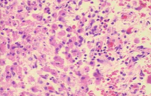
Microscopic (histologic) description
- Confluent sheets of histiocytes (von Hanseman cells) with eosinophilic granular cytoplasm and eccentric nuclei
- Cells contain characteristic basophilic bodies containing calcium (von Kossa+)
- Round, sometimes laminated structures are known as Michaelis - Gutmann bodies
- The targetoid pattern is accentuated by staining with PAS
- Also histiocytes, neutrophils, plasma cells, lymphocytes and granulation tissue
Microscopic (histologic) images
Images hosted on other servers:

Michaelis-Gutmann body

Calcified structures

Periodic acid Schiff stain

PAS, Von Kossa, Prussian blue
Positive stains
- PAS, Perls (iron) stain and von Kossa (calcium) stain the Michaelis-Gutmann bodies
- Microorganisms may stain with anti-bCG antibodies (J Am Acad Dermatol 2000;43;351)
Electron microscopy description
- Histiocytes containing phagolysosomes with intact or partially digested bacteria
- The granules within the macrophages, which stain with PAS, contain engorged lysosomes that contain bacteria debris
