

Skin nontumor
Vesiculobullous and acantholytic reaction patterns
Pemphigoid gestationis
Last author update: 1 July 2011
Last staff update: 17 November 2021
Copyright: 2002-2024, PathologyOutlines.com, Inc.
PubMed Search: Pemphigoid gestationis [title]

Table of Contents
Definition / general | Terminology | Epidemiology | Sites | Etiology | Clinical features | Prognostic factors | Case reports | Treatment | Clinical images | Microscopic (histologic) description | Microscopic (histologic) images | Positive stains | Differential diagnosis | Additional referencesCite this page: Hamodat M. Pemphigoid gestationis. PathologyOutlines.com website. https://www.pathologyoutlines.com/topic/skinnontumorpemphigoidgestationis.html. Accessed April 18th, 2024.
Definition / general
- Rare, self limiting, autoimmune, subepidermal bullous disease, occurring during or soon after pregnancy or in women taking oral contraceptives
Terminology
- Formerly called herpes gestationis due to herpetiform nature of blisters but disease is NOT related to herpes infection
- Called pruritus gravidarum when occurs without significant cutaneous stigmata
Epidemiology
- Occurs in 1 per 50,000 pregnancies
- Rarely complicates hydatidiform mole and gestational choriocarcinoma
- Rarely present in postpartum period
- May follow a change in sexual partner
Sites
- Pruritic lesions of abdomen, chest, back and extremities
Etiology
- Due to circulating autoantibodies against placental collagen XVII (BP180, BPAG2) a hemidesmosomal transmembrane protein and less frequently BP230 (J Cell Biochem 1999;72:356)
Clinical features
- Usually urticarial papules, also blisters and rash
- Usually resolves within weeks to months after delivery
- Tends to recur with subsequent pregnancy
- Associated with premature delivery, small for gestational age infants
Prognostic factors
- Poor prognostic factors: onset in first or second trimester and presence of blisters (Br J Dermatol 2009;160:1222)
Case reports
- 29 year old woman with involvement of mother and newborn (Arch Gynecol Obstet 2009;279:235)
Treatment
- Oral and topical corticosteroids (J Am Acad Dermatol 2006;55:823)
Clinical images
Contributed by Mark R. Wick, M.D.
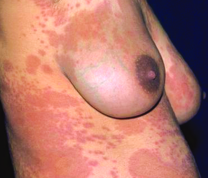
Breast skin
Images hosted on other servers:

36 year old woman at 24 weeks gestation

Annular erythematous-edematous lesions

Exudative and erythematous lesions

Various images
Microscopic (histologic) description
- Similar to bullous pemphigoid - subepidermal blister, with eosinophils in lumen
- Marked edema in papillary dermis
- Perivascular infiltrate consists of lymphocytes, histiocytes and large numbers of eosinophils
- Eosinophilic spongiosis may be seen
Microscopic (histologic) images
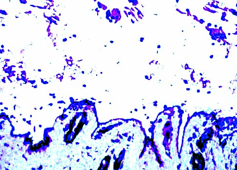
Contributed by Mark R. Wick, M.D.
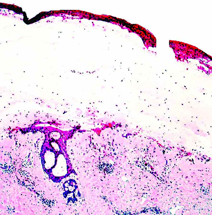
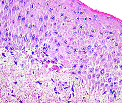

Breast skin
Collagen IV stain
IgG stain
Images hosted on other servers:

H&E and C3

H&E, subepidermal bulla with eosinophils

Perivascular infiltrate of lymphocytes and eosinophils

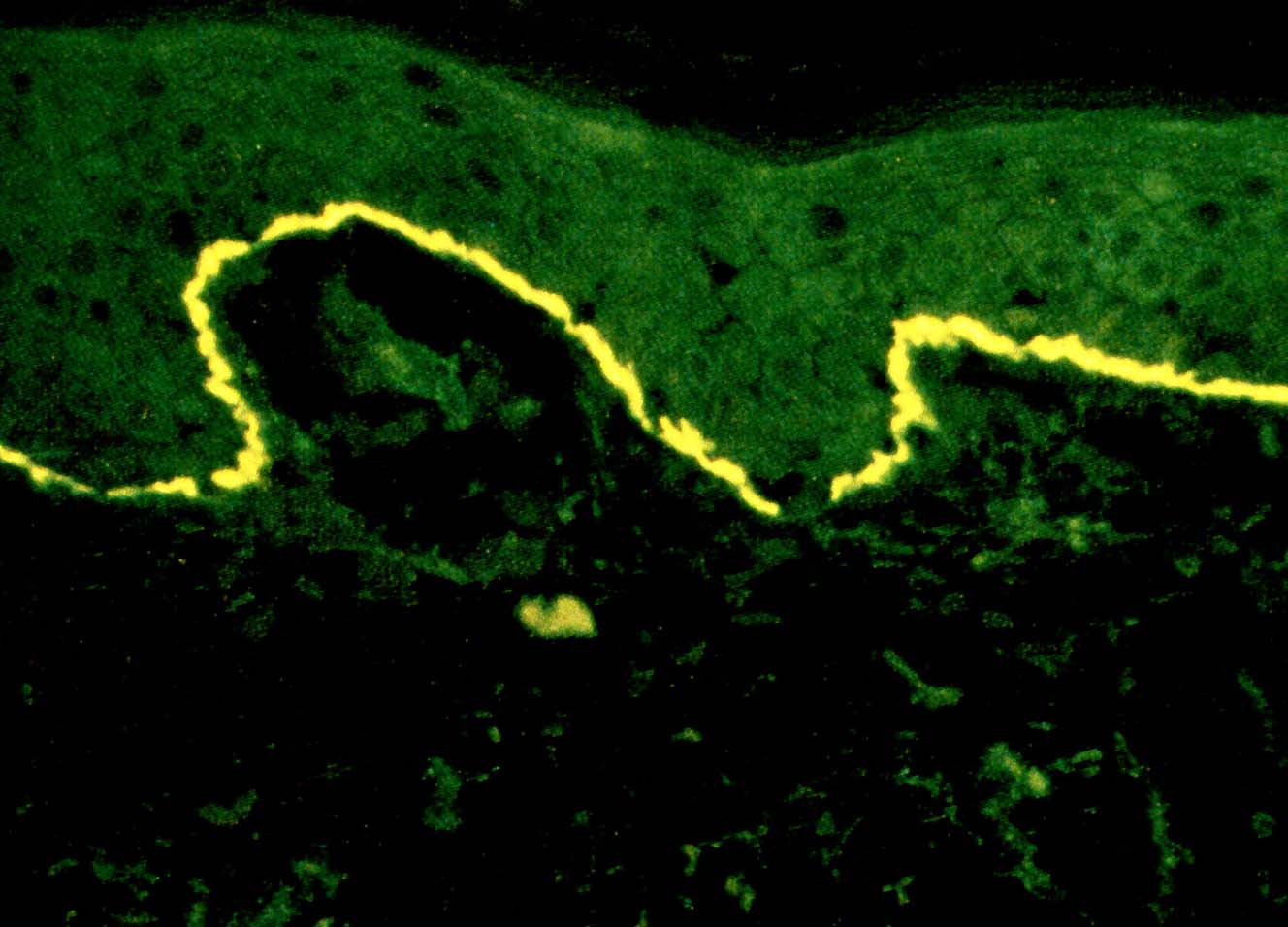
Linear C3 staining
Positive stains
- Linear C3 deposits along cutaneous basement membrane; variable IgG deposition (Eur J Obstet Gynecol Reprod Biol 2009;145:138)
- ELISA may be more sensitive and specific than indirect immunofluorescence (Int J Dermatol 2008;47:1245)
Differential diagnosis
- Pruritic urticarial papules of pregnancy: typically begins in stretch mark areas of abdomen and usually ends within 2 weeks after delivery; no antibody deposition
- Pregnancy prurigo: usually develops in the third trimester of pregnancy, presents with pruritic papules and nodules; histologic changes are those of low grade nonspecific spongiotic dermatitis
Additional references
