

Skin nontumor
Neutrophilic and eosinophilic dermatoses
Acute febrile neutrophilic dermatosis (Sweet syndrome)
Last author update: 1 August 2011
Last staff update: 8 February 2024
Copyright: 2002-2024, PathologyOutlines.com, Inc.
PubMed Search: Sweet syndrome [title]

Table of Contents
Definition / general | Treatment | Microscopic (histologic) description | Microscopic (histologic) images | Differential diagnosis | Additional referencesCite this page: Hamodat M Acute febrile neutrophilic dermatosis (Sweet syndrome). PathologyOutlines.com website. https://www.pathologyoutlines.com/topic/skinnontumorsweetssyndrome.html. Accessed April 24th, 2024.
Definition / general
- Also called acute febrile neutrophilic dermatosis
- Abrupt onset of tender or painful erythematous plaques and nodules on the face and extremities and less commonly on the trunk, in association with fever (usually), malaise and a neutrophil leukocytosis
- Associated with AML, less often with solid malignancies
- Often females, any age but rare in childhood
- Unknown etiology, but may represent immunological hypersensitivity reaction
Treatment
- Most cases respond to oral corticosteroids
- Thalidomide was successful in one patient who failed to respond to metronidazole, dapsone and methotrexate; IV immunoglobulin was used in a child with concurrent immunodeficiency
- Also response to 5-azacytidine in patient with myelodysplastic syndrome
- A case of histiocytoid Sweet's syndrome was responsive to dapsone; dapsone has also been used with systemic corticosteroids in a patient who was HIV positive
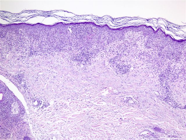
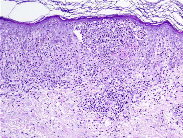
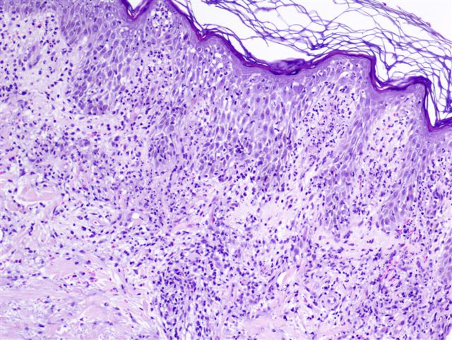
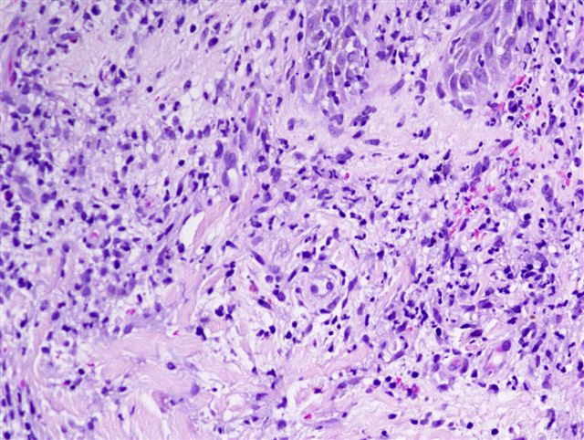
Microscopic (histologic) description
- Intense neutrophilic dermal infiltrate in reticular dermis, may be perivascular, diffuse and surround sweat glands; edema with marked leukocytoclasia; marked papillary edema
- Occasional presence of dermal papillary microabscesses can result in confusion with dermatitis herpetiformis
- Epidermis is normal, occasionally slight spongiosis, vesiculation with spongioform pustule
- Necrotic keratinocytes may be present
- Variable eosinophils, lymphocytes and histiocytes
- Blood vessels are dilated and show endothelial swelling
Microscopic (histologic) images
Contributed by Angel Fernandez-Flores, M.D., Ph.D.
Differential diagnosis
- Behçet disease may be associated with lesions similar to Sweet syndrome
- Gram stain and PAS exclude infection
- Granuloma faciale: fibrinoid necrosis is minimal but eosinophils are prominent
- Late lesions of erythema elavatum diutinum and granuloma faciale show fibrosis, not seen in Sweet Syndrome
- Pyoderma gangrenosum: has ulcer, no leukorrhexis
- Rheumatoid neutrophilic dermatitis, neutrophil rich variant of anaplastic large cell lymphoma
- Presence of fibrinoid vascular changes distinguishes necrotizing vasculitis such as leukocytoclastic vasculitis, erythema elevatum diutinum and granuloma faciale from Sweet syndrome
Additional references
