

Skin melanocytic tumor
Melanoma
Lentigo maligna melanoma
Resident / Fellow Advisory Board: Caroline I.M. Underwood, M.D.


Last author update: 7 February 2022
Last staff update: 24 January 2023
Copyright: 2002-2024, PathologyOutlines.com, Inc.
PubMed Search: Lentigo maligna melanoma "free full text"[sb]
Table of Contents
Definition / general | Essential features | Terminology | ICD coding | Epidemiology | Sites | Pathophysiology | Etiology | Clinical features | Diagnosis | Prognostic factors | Case reports | Treatment | Clinical images | Microscopic (histologic) description | Microscopic (histologic) images | Positive stains | Negative stains | Molecular / cytogenetics description | Sample pathology report | Differential diagnosis | Board review style question #1 | Board review style answer #1 | Board review style question #2 | Board review style answer #2Cite this page: Gillam J, Crimmins J, Mochel M. Lentigo maligna melanoma. PathologyOutlines.com website. https://www.pathologyoutlines.com/topic/skintumormelanocyticlentigomaligna.html. Accessed May 4th, 2024.
Definition / general
- Subtype of melanoma arising on chronically sun damaged skin and appearing as an irregular pigmented macule, corresponding to an intraepidermal proliferation of atypical melanocytes; over time, may develop foci that are indurated, papular or nodular, indicating tumorigenic growth (Am J Pathol 1969;55:39)
- Lentigo maligna (LM) typically refers to the in situ form of this disease, while lentigo maligna melanoma (LMM) designates invasive disease (JAMA Dermatol 2019;155:782)
Essential features
- Presents as a flat, growing, irregularly pigmented lesion on chronically sun damaged skin, which may develop a raised, papular or nodular focus, indicating tumorigenic growth
- Microscopically, a proliferation of intraepidermal melanocytes overlying solar elastosis and exhibiting crowded growth along the basal epidermis; irregular distribution of nests and effacement of epidermal rete with or without an underlying dermally invasive component
- Immunohistochemistry for melanocytic markers (MelanA / MART1, SOX10, MITF, HMB45) may assist in identification of diagnostic architectural features and may distinguish the lesion from mimics
- Prognosis is correlated with presence and depth of invasion, mitotic rate among invasive cells and presence / absence of ulceration
- Prognosis is excellent if noninvasive and completely excised
- Treatments: excision (gold standard), adjuvant topical therapies and radiation (adjuvant / unresectable setting)
Terminology
- Chronic sun damage (CSD) associated melanoma
- Hutchinson melanotic freckle
ICD coding
Epidemiology
- Older patients, usually over 50 (Annu Rev Pathol 2014;9:239)
- Develops at sites of chronic, continuous, cumulative sun exposure
Sites
- Face, neck, ears, scalp (if not shielded by hair), forearms, dorsal hands
Pathophysiology
- Acquisition of oncogenic genetic mutations by chronic ultraviolet light exposure (Annu Rev Pathol 2014;9:239)
- Flat, spreading, pigmented radial growth phase eventually gives rise to invasive, tumorigenic vertical growth phase with metastatic potential
Etiology
- Chronic ultraviolet light exposure
Clinical features
- Growing, irregularly pigmented lesion on chronically sun damaged skin
- Development of a raised, papular or nodular focus indicates tumorigenic / vertical growth phase
- Reference: Am J Pathol 1969;55:39
Diagnosis
- Skin exam revealing classic features, as described above
- Dermoscopy
- Asymmetric hyperpigmented follicular openings, pigmented rhomboidal structures, annular granular pattern (J Am Acad Dermatol 2000;42:25, Br J Dermatol 2012;167:280)
- Pseudonetwork and homogeneous areas (Clin Cosmet Investig Dermatol 2019;12:403)
- Biopsy with diagnostic histopathologic features, as described below
Prognostic factors
- Lentigo maligna (melanoma in situ of lentigo maligna type)
- Excellent prognosis after excision if no invasive component (Br J Dermatol 2014;171:1605)
- Lentigo maligna melanoma (malignant melanoma of lentigo maligna type)
- Controlling for other parameters (depth, ulceration, etc.); prognosis overlaps that of other melanoma subtypes
- Poor prognostic factors include greater Breslow depth (distance from granular zone to deepest invasive melanoma cell), presence of ulceration, high mitotic rate, presence of microsatellite, satellite or in transit metastases, positive sentinel node and distant metastases (e.g., lung, liver, brain) (CA Cancer J Clin 2017;67:472)
Case reports
- 70 year old woman with a gradually enlarging pigmented macule on her face (Dermatology 2016;232:24)
- 76 year old man with an evolving pigmented lesion on the occipital scalp (J Cutan Pathol 2020;47:1155)
- 85 year old man with a pigmented right malar lesion (An Bras Dermatol 2017;92:565)
Treatment
- Complete excision, accomplished via wide local excision, staged surgical excision or Mohs micrographic surgery (Dermatol Surg 2011;37:1210)
- Excisions may utilize staged Mohs micrographic surgery (slow Mohs) with rush processing, examination of surgical margins and closure upon report of negative margins
- Mohs surgeons may also utilize frozen sections with melanocytic immunohistochemistry for margin assessment (J Am Acad Dermatol 2021;84:196)
- For in situ disease, topical therapies (including imiquimod) may be useful in the adjuvant setting or as primary treatment if unresectable (J Am Acad Dermatol 2015;72:1047)
- Radiotherapy (Radiat Oncol 2020;15:174)
- Consideration of sentinel lymph node biopsy (CA Cancer J Clin 2017;67:472)
Clinical images
Contributed by Julia Nunley, M.D.
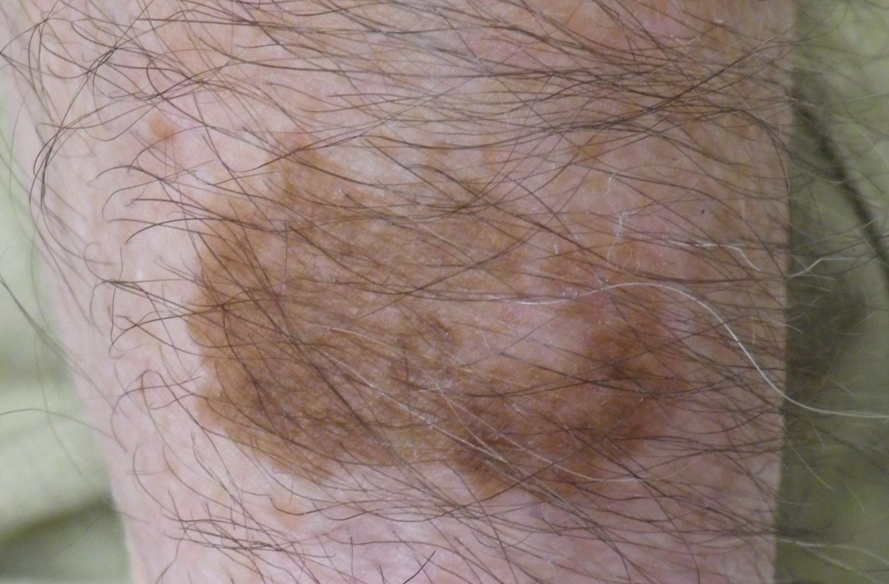
Lentigo maligna involving forearm
Images hosted on other servers:

Asymmetric
and irregularly
pigmented
macule
Microscopic (histologic) description
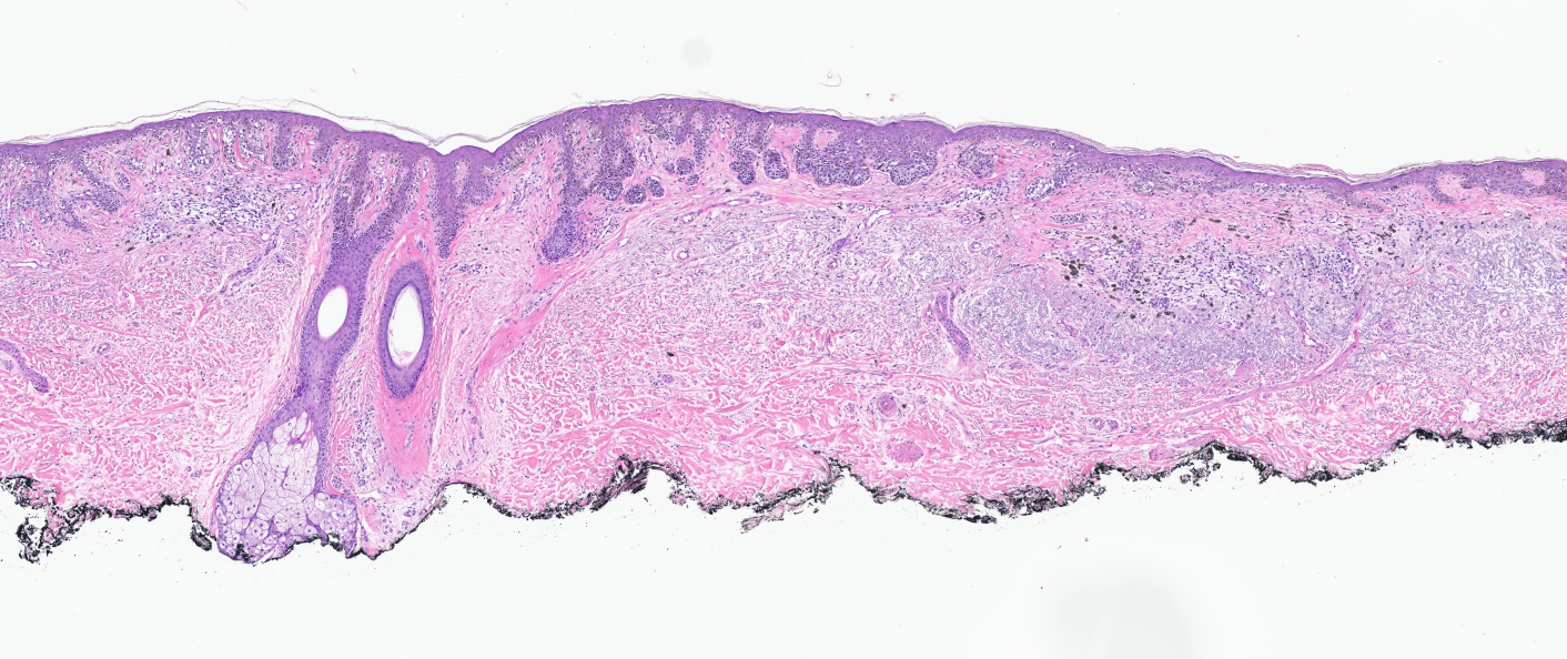
- Proliferation of intraepidermal (single and nested) melanocytes overlying solar elastosis
- Melanocytes demonstrate crowded growth along the basal epidermis
- Associated epidermal alterations, including loss (effacement) of epidermal rete and associated irregular epidermal hyperplasia
- Pagetoid scatter (melanocytes above the basal layer)
- Involvement of adnexal epithelium
- Melanocytic cytology is variable, ranging from small cells with dark nuclei and scant cytoplasm to epithelioid pigmented melanocytes, to spindled melanocytes
- Invasive component, if present, consists of single or nested melanocytes in the dermis with similar cytologic features to those in the in situ component (Cancer Res 1969;29:705, Am J Pathol 1969;55:39)
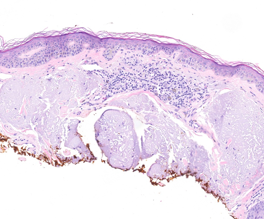
Microscopic (histologic) images
Contributed by Joseph Gillam, M.D., Jennifer Crimmins, M.D. and Mark Mochel, M.D.
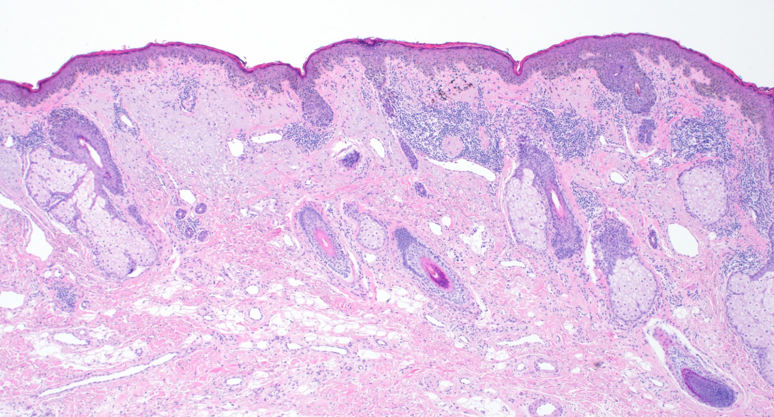
Broad intraepidermal proliferation of melanocytes
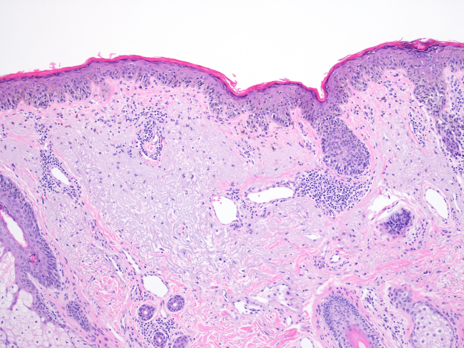
Crowded, atypical intraepidermal melanocytes
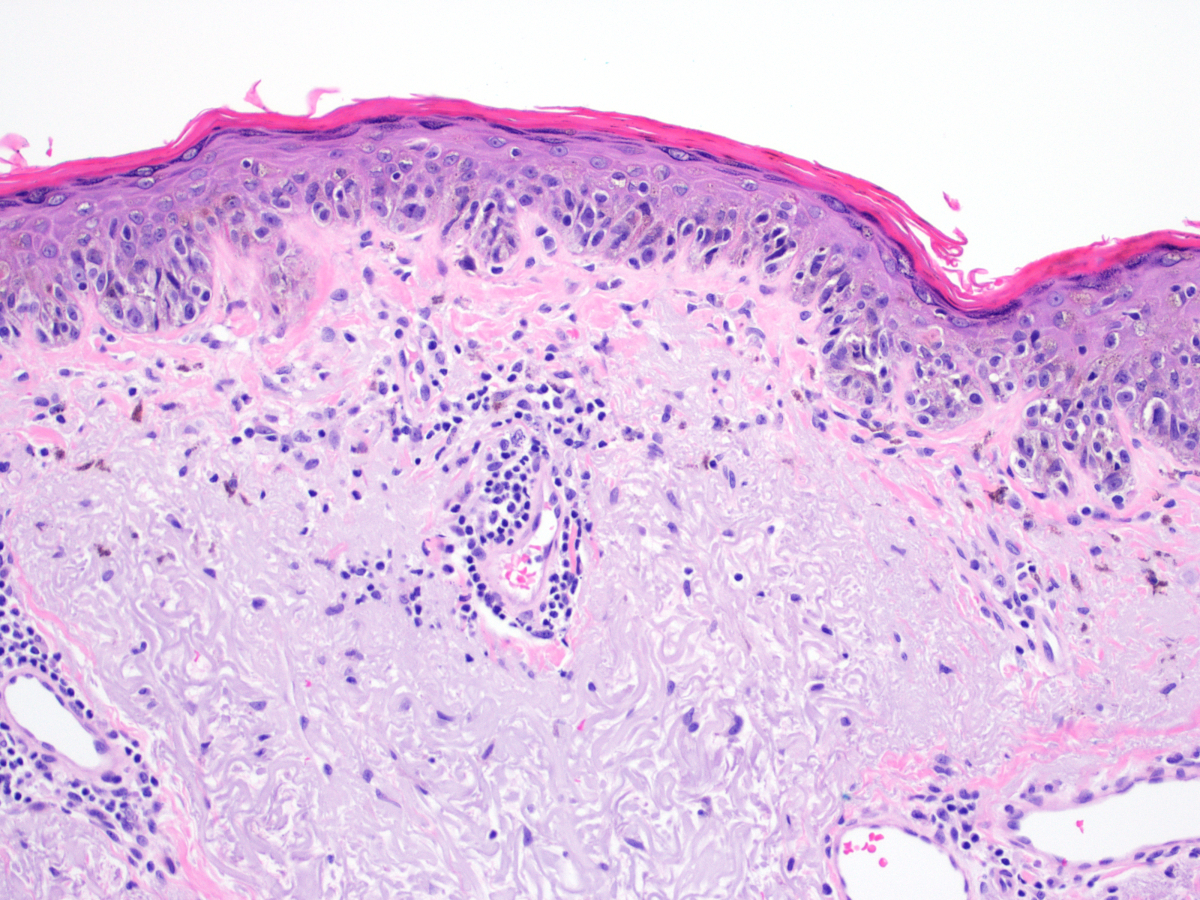
Atypical intraepidermal melanocytes
Broad compound proliferation of melanocytes
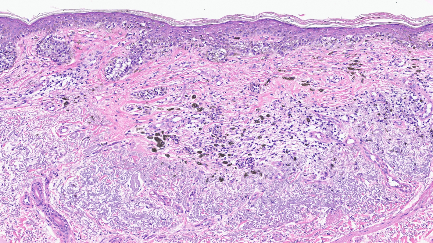
Dermal nests with fibrosis
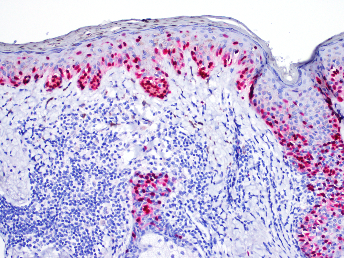
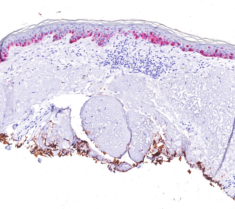
SOX10 highlighting pagetoid growth
Positive stains
- Immunohistochemistry for MelanA / MART1, SOX10 and MITF may assist in diagnosis by highlighting the arrangement of melanocytes in the epidermis and by assisting in identification of dermally invasive cells (Am J Dermatopathol 2014;36:387, Am J Dermatopathol 2014;36:124)
Negative stains
- Cytokeratins and p63
Molecular / cytogenetics description
- Activating BRAF mutations are less common than in superficial spreading (nonchronic sun damage) melanomas (N Engl J Med 2005;353:2135)
- Of cases with BRAF mutations, many are non-V600E (e.g., V600K) (Clin Cancer Res 2012;18:3242)
- High tumor mutation burden (Cells 2021;10:2320)
- Genetic alterations identified (Annu Rev Pathol 2014;9:239):
- Inactivating mutations of NF1
- Activating mutations of NRAS
- Copy number increase of CCND1
- Activating mutations of KIT
- TP53 and TERT mutations
Sample pathology report
- Skin, right temple, biopsy:
- Melanoma in situ, lentigo maligna type (see comment)
- Comment: Sections reveal a poorly circumscribed intraepidermal proliferation of atypical melanocytes with crowded growth along the basal epidermis, irregular distribution of nests and pagetoid scatter.
Differential diagnosis
- Lentigo:
- While intraepidermal melanocytes within a lentigo are increased in number, these melanocytes will lack nesting, crowding at the basal layer (contiguity) and pagetoid ascent of melanocytes
- Melanocytic hyperplasia of sun damaged skin:
- Lacks nesting, crowding at the basal layer (contiguity) and pagetoid ascent of melanocytes (Am J Dermatopathol 1996;18:560)
- Dysplastic nevus:
- On skin with chronic sun damage (indicated by significant solar elastosis), this diagnosis should be made with extreme caution
- Lesions with features of atypical / dysplastic / Clark nevus in this setting have a high likelihood of representing melanoma or melanoma in situ (J Cutan Pathol 2005;32:405)
- Squamous cell carcinoma in situ:
- Shares pagetoid ascent of atypical cells with melanoma in situ / lentigo maligna; in contrast to melanoma in situ, does not form rounded nests below the basal layer, is composed of polygonal cells with eosinophilic cytoplasm and tends to show suprabasilar confluence, leaving a residual distinctive layer of native basal epidermal keratinocytes (eyeliner sign) (J Cutan Pathol 2018;45:734, J Cutan Pathol 2016;43:24)
- Immunohistochemical stains can be utilized in challenging cases
- p63+
- MelanA / MART1-, SOX10-
Board review style question #1
A 76 year old retired chemist with light skin presents with a pigmented lesion on his right cheek. Physical examination reveals a 1.5 x 1.0 cm tan to brown macule with irregular borders. A biopsy is performed. What is the diagnosis?
- Dysplastic nevus
- Lentigo maligna
- Solar lentigo
- Squamous cell carcinoma in situ
Board review style answer #1
Board review style question #2
Which of the following behaviors represents the greatest risk factor for the development of this lesion, photographed above (H&E and MelanA)?
- Decades of midday yardwork
- Electronic cigarette use (vaping)
- Rare ethanol consumption
- Workplace exposures to organic chemicals
Board review style answer #2
