

Skin nonmelanocytic tumor
Cysts
Epidermal (epidermoid) type
Authors: V. Claire Vaughan, M.D., Joshua Wisell, M.D.
Editorial Board Member: Hillary Rose Elwood, M.D.
Editor-in-Chief: Debra L. Zynger, M.D.


Last author update: 9 May 2019
Last staff update: 1 November 2023
Copyright: 2002-2024, PathologyOutlines.com, Inc.
PubMed Search: Epidermal cyst / epidermoid cyst
Table of Contents
Definition / general | Essential features | Terminology | ICD coding | Epidemiology | Sites | Pathophysiology | Diagrams / tables | Clinical features | Diagnosis | Laboratory | Radiology description | Radiology images | Case reports | Treatment | Clinical images | Gross description | Gross images | Microscopic (histologic) description | Microscopic (histologic) images | Cytology description | Cytology images | Sample pathology report | Differential diagnosis | Additional references | Board review style question #1 | Board review style answer #1 | Board review style question #2 | Board review style answer #2Cite this page: Vaughan VC, Wisell J. Epidermal (epidermoid) type. PathologyOutlines.com website. https://www.pathologyoutlines.com/topic/skintumornonmelanocytickeratinouscystepidermal.html. Accessed April 19th, 2024.
Definition / general
- Benign skin tumor
- Cystic mass containing keratin
Essential features
- Cystic mass with soft white keratin contents
- Histologically cystic mass with squamous epithelium and keratin flakes
Terminology
- Epidermoid cyst, epidermal cyst, epidermal inclusion cyst, infundibular cyst
ICD coding
- ICD-10: L72.0 - epidermal cyst
Epidemiology
- Young and middle aged adults are most often affected (Int J Trichology 2017;9:108)
- M = F
Sites
- Face, neck, trunk, perineal area, cerebellopontine angle
- Less commonly spine, intrapancreatic accessory spleen
Pathophysiology
- Follicular orifice becomes plugged with bacteria and keratin, leading to cystic dilation and entrapment of keratin debris
- Presence of multiple epidermal inclusion cysts has been documented in Gardner syndrome, a variant of familial adenomatous polyposis with benign osteomas and intestinal fibromatoses
- Less frequently, patients may have lipomas, pilomatrixomas (including epidermoid cysts with pilomatrical lining) or leiomyomas
- Multiple and large epidermoid cysts may occur with the use immunosuppressants in the posttransplantation setting, for example, with cyclosporine or tacrolimus (Cutis 1992;50:36, Ann Dermatol 2011;23:S182)
- May complicate penetrating trauma to skin, such as a sewing needle, with resultant implantation of squamous epithelium into the dermis (Turk J Pediatr 2011;53:108)
Diagrams / tables
Images hosted on other servers:

Age distribution

Site distribution
Clinical features
- Present as smooth dome shaped swellings varying in size from a few millimeters to a few centimeters (Ann Dermatol 2017;29:33)
- Usually occur on the face, neck or trunk but can occur anywhere
- Overlying skin may be taut with the pressure of the cyst and have a central punctum
- Generally occurs in postpubertal individuals
- Freely mobile unless ruptured, in which case a foreign body giant cell reaction may make them more adherent to surrounding connective tissue
- May become painful and inflamed with external manipulation
- Cyst contents white, cheesy macerated keratin that may have an odor
Diagnosis
- Often based on clinical examination
- Pathological diagnosis relies on recognizing the cyst wall and contents
- In cases of extensive foreign body giant cell reaction or fibrosis, the classic features may not be visualized as readily and a diligent search for keratin flakes may lead to diagnosis
Laboratory
- No specific laboratory findings
Radiology description
- Ultrasound:
- Subcutaneous rounded structure that is mostly anechoic or hypoechoic with focal present inner echoes (pseudotestis appearance) representing cystic debris
- MRI:
- Fluid-like enhancement
- High intensity of cyst contents on T2 weighted images and peripheral cyst wall enhancement with T1 gadolinium enhancement
- Cyst rupture results in irregular enhancement (AJR Am J Roentgenol 2006;186:961)
Radiology images
Images hosted on other servers:

Lumbar spine epidermoid cyst

Abdominal epidermal inclusion cyst
Case reports
- 7 year old boy with epidermal inclusion cyst infected by molluscum contagiosum virus (J Dtsch Dermatol Ges 2018;16:1143)
- 40 year old woman with recurrent spinal epidermoid cysts with atypical hyperplasia (Medicine 2017;96:e8950)
- 51 year old man with squamous cell carcinoma arising in a 30 year old perineal epidermal inclusion cyst (World J Surg Oncol 2018;16:155)
- 63 year old man with epidermal inclusion cyst containing a splinter with nonpigmented fungi (J Cutan Pathol 2018;45:954)
- 73 year old man with squamous cell carcinoma arising in an epidermoid cyst with human papillomavirus changes (Clin Exp Dermatol 2018;43:201)
Treatment
- Complete excision of the cyst is curative
Clinical images
Contributed by Jeremy Hugh, M.D.
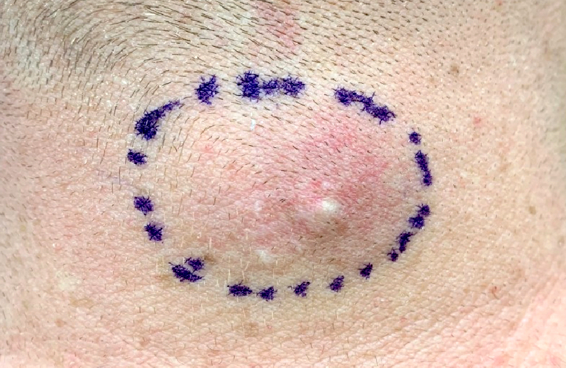
White subcutaneous nodule
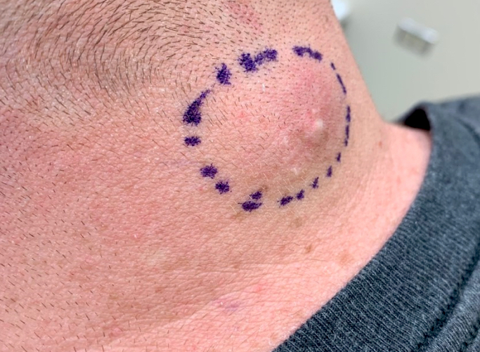
White nodule in hair bearing area
Images hosted on other servers:

Cheesy contents of
cystectomy in
mastoid region after
penetrating trauma
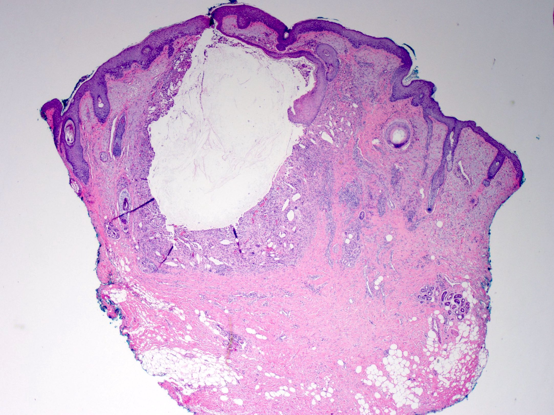
Gross description
- Pearly glistening cyst with creamy contents
Gross images
Contributed by the University of Colorado Department of Pathology
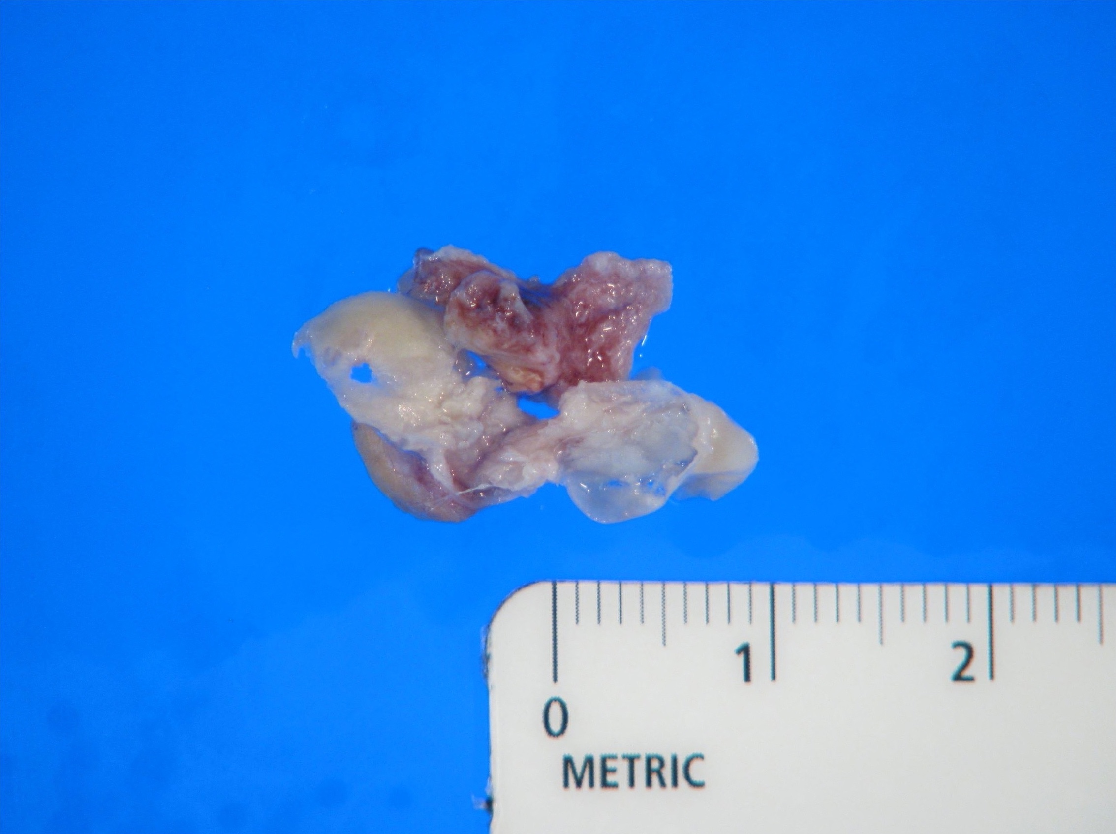
Disrupted cyst wall and adjacent soft tissue
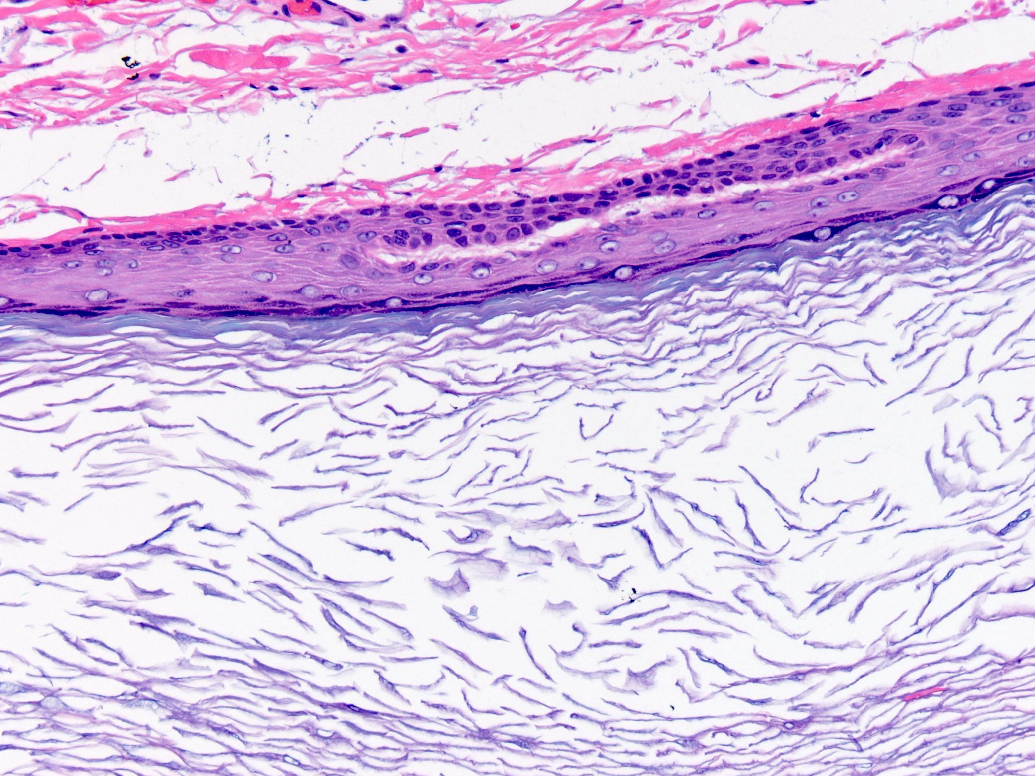
Microscopic (histologic) description
- Subepidermal cyst that may or may not open into the overlying epidermis
- Cyst lining composed of stratified squamous epithelium with a granular layer
- Cyst wall does not contain eccrine glands, sebaceous glands or hair follicles
- Cyst contents composed of abundant keratin flakes
- Foreign body giant cell reaction is frequently present in ruptured cysts
- Melanoma, basal cell carcinoma and squamous cell carcinoma have been reported in association with epidermal inclusion cysts (J Am Acad Dermatol 2013;68:e6, J Cutan Med Surg 2015;19:105)
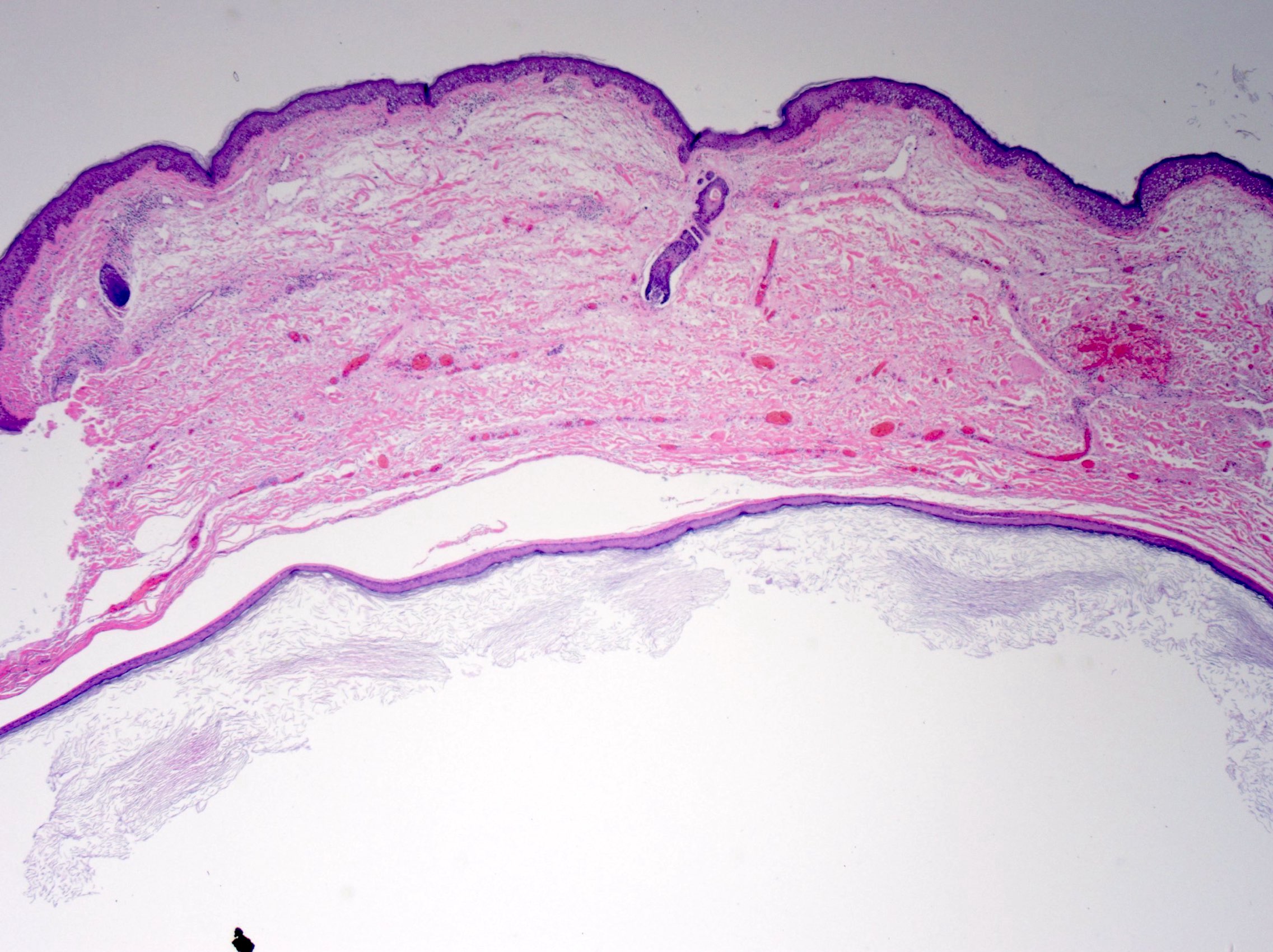
Microscopic (histologic) images
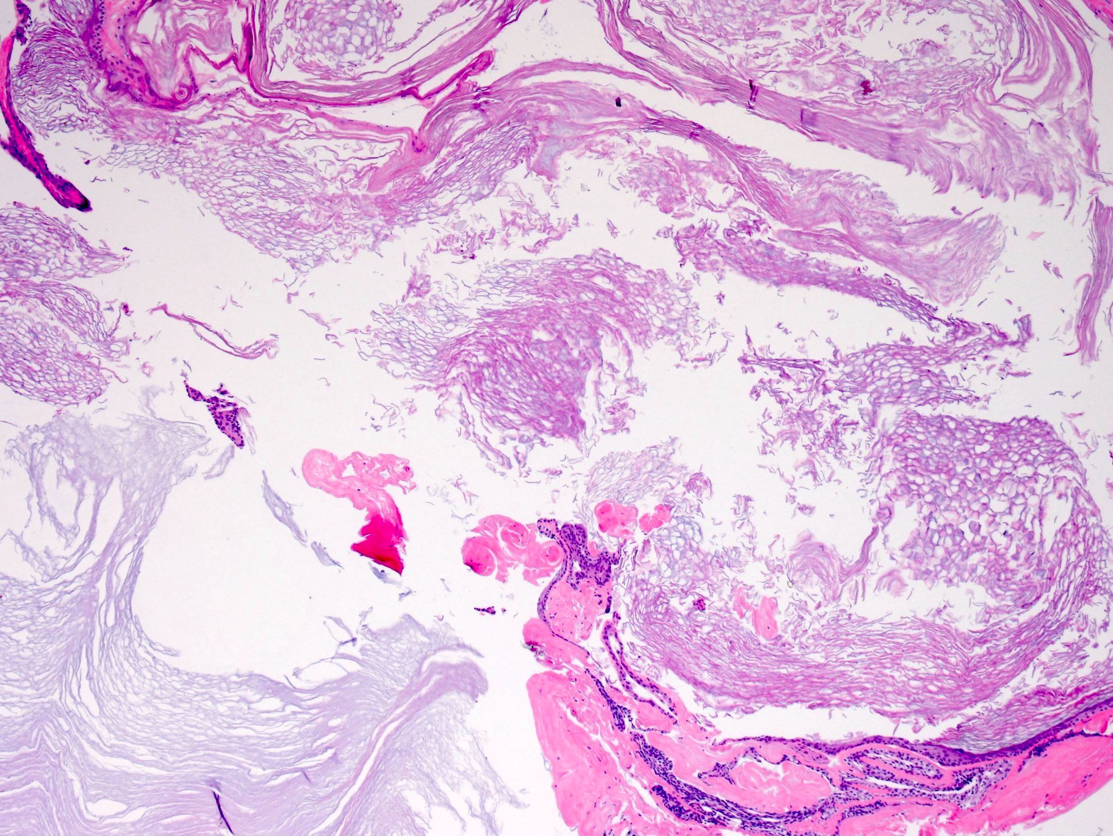
Contributed by the University of Colorado Department of Pathology, Jijgee Munkhdelger, M.D., Ph.D. and Andrey Bychkov, M.D., Ph.D.
Loose keratin flakes
Stratified squamous epithelium lining
Ruptured epidermoid cyst
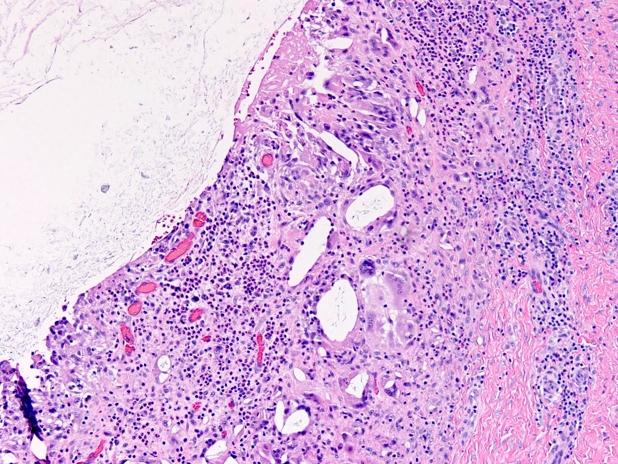
Giant cells, cholesterol clefts, mixed inflammation
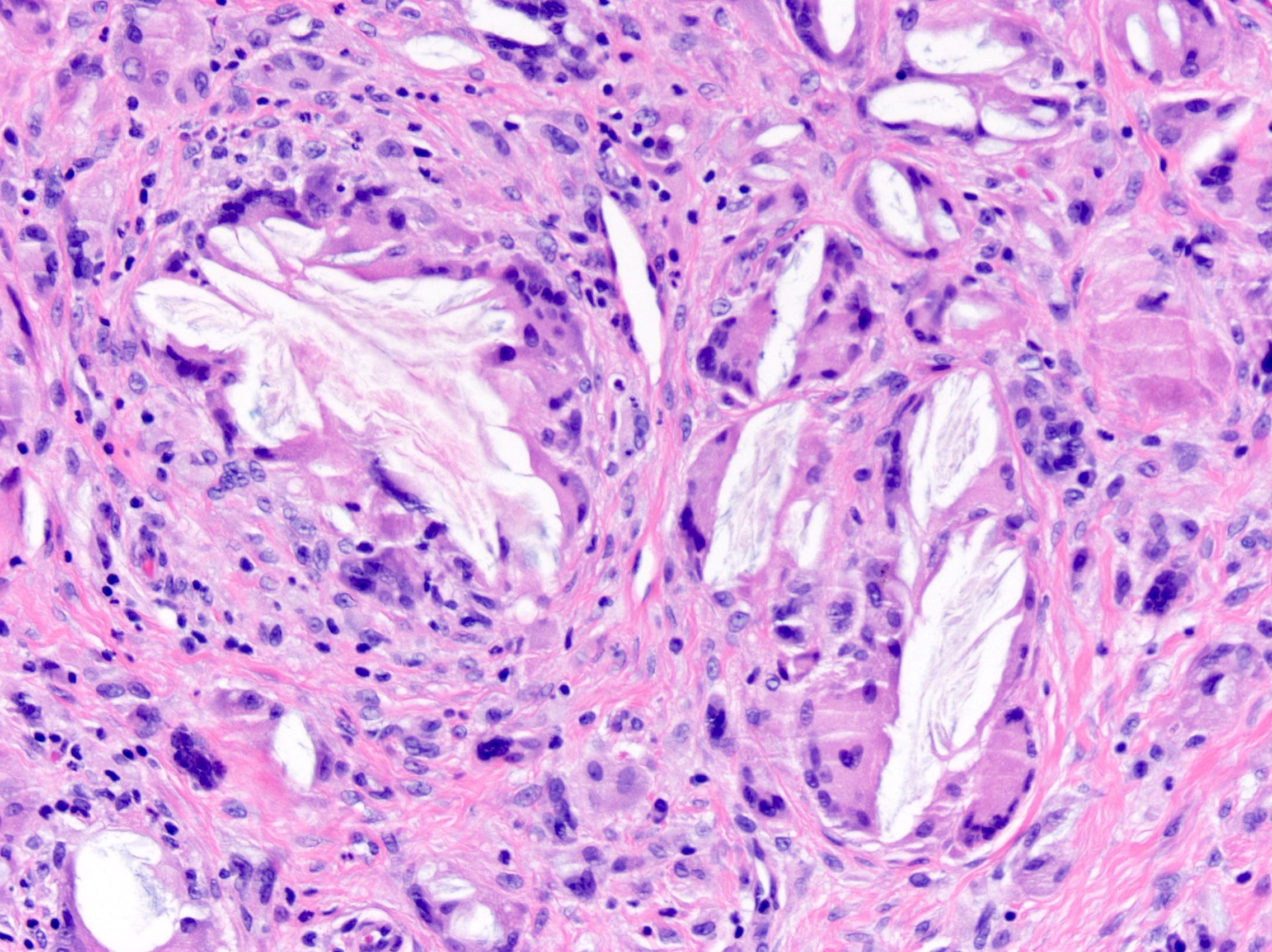
Foreign body giant cell reaction
Epidermal inclusion cyst in cerebellopontine angle

Lamellated
keratin flakes in
cranial epidermal
inclusion cyst
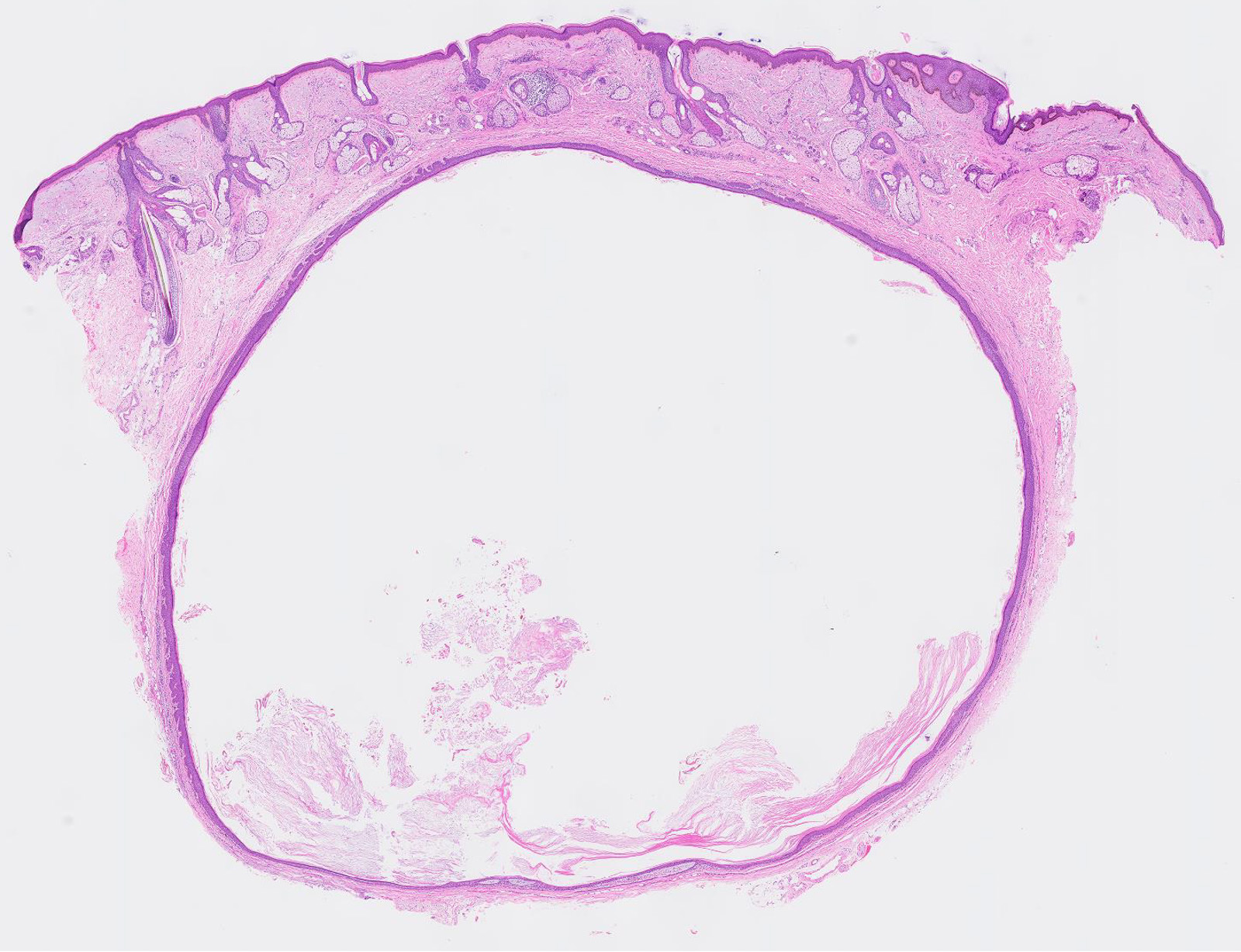
Unilocular dermal cyst
Cytology description
- Anucleate keratinizing squamous cells with some nucleated squamous cells
- Frequent erythrocytes, leukocytes, multinucleated giant cells and cholesterol crystals (J Nat Sci Biol Med 2014;5:460)
Cytology images
Contributed by the University of Colorado Department of Pathology
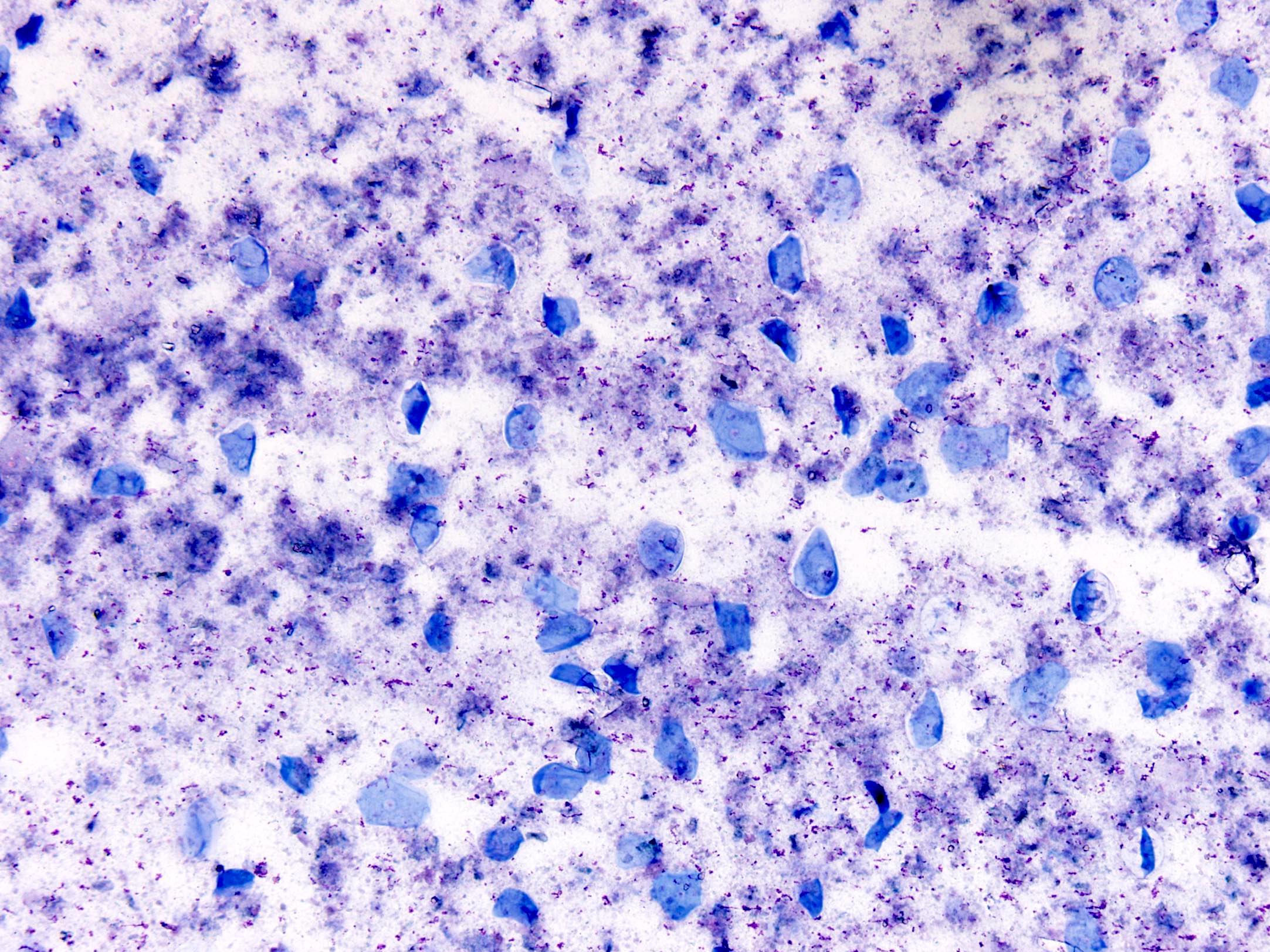
Anucleate squamous cells
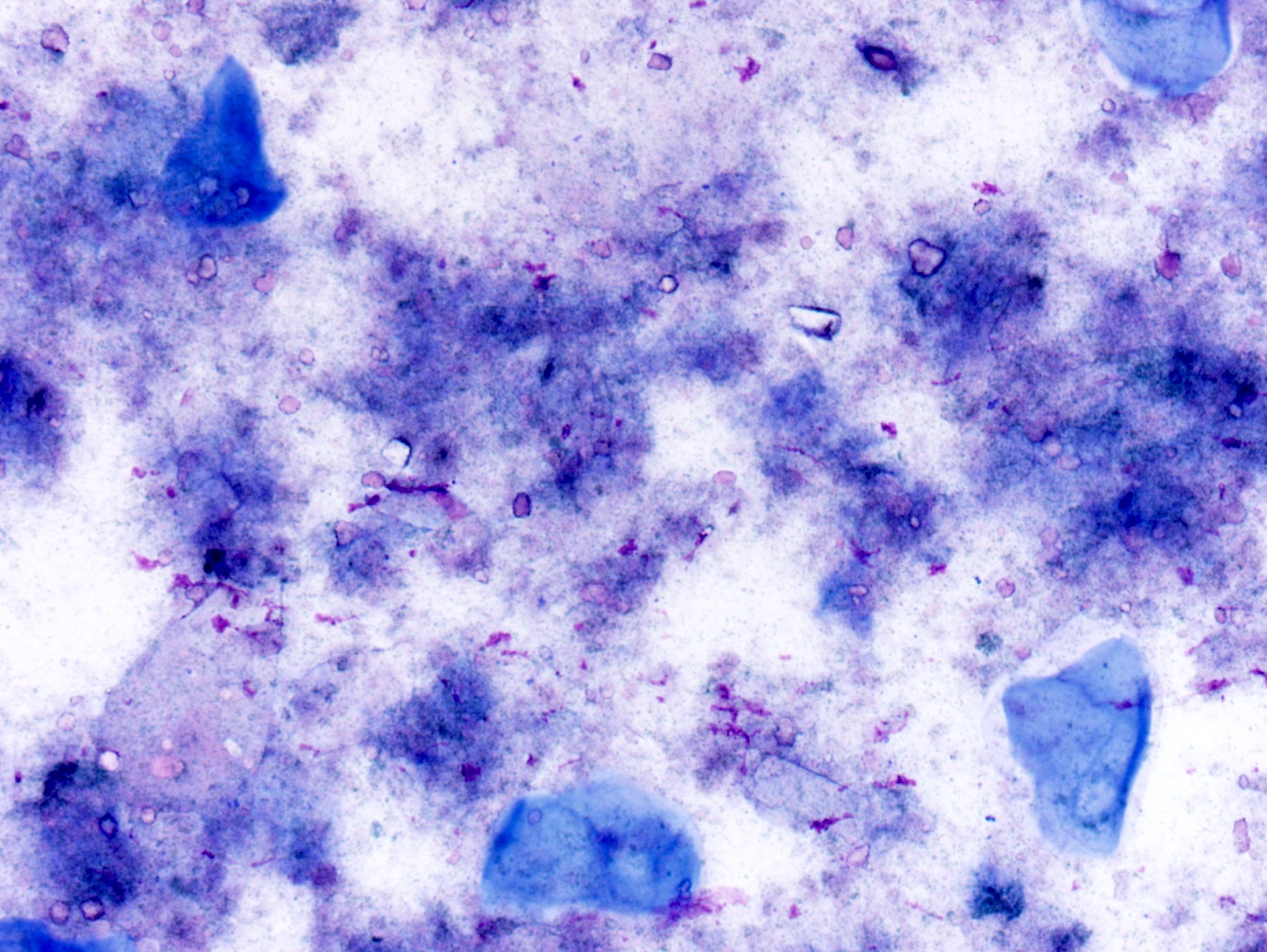
Keratinizing squamous cells
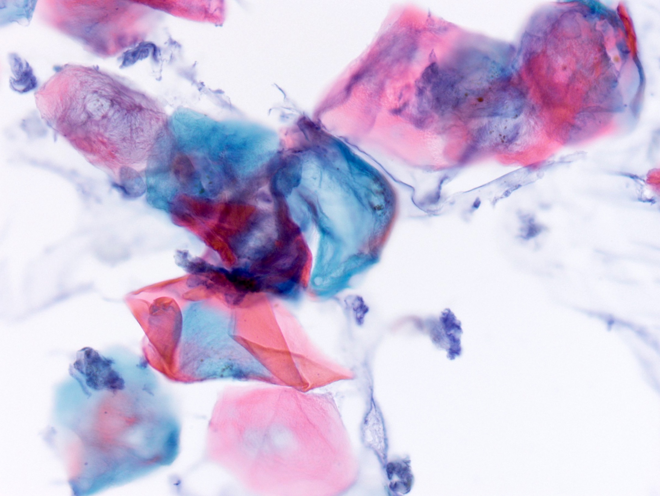
Anucleate squames and inflammatory cells
Sample pathology report
- Skin, neck, excision:
- Epidermal inclusion cyst
- Microscopic description: Cyst lined by squamous epithelium with granular layer containing lamellated keratin.
Differential diagnosis
- Proliferating epidermoid cyst:
- May show carcinomatous changes, invasion and be locally aggressive
- Trichilemmal (pilar) cyst:
- Lack a granular layer in the cyst lining
- Dense lamellated keratin cyst contents
- More likely to occur on the scalp
- Pilomatricoma:
- Eosinophilic cellular outlines of squamous cells ("ghost cells") in addition to the more basophilic matrical cells
- More likely to occur in a pediatric population
- Hybrid cyst:
- Demonstrates features of both an epidermal and trichilemmal cyst
- Dermoid cyst:
- Similar in appearance
- Has adnexal structures (ie. sebaceous glands) in cyst wall
- Arises at sites of embryonic closure such as the lateral eyebrow
- Pilonidal sinus:
- Sinus tract surrounded by epithelium and mixed inflammation
- Characteristically contains broken hair shafts
- Steatocystoma:
- Compressed sebaceous gland within the cyst wall
- Characteristic wavy eosinophilic crenulated cuticle of the lining
- Odontogenic keratocyst:
- Attenuated squamous epithelium with parakeratosis
- Retraction of epithelium with basal palisade can be a helpful finding
- Often filled with keratin debris like epidermoid cyst
Additional references
Board review style question #1
An elderly man has a soft, round subcutaneous mass on the back of the neck with a central punctum. Which of the following is the most likely diagnosis?
- Angiosarcoma
- Epidermoid cyst
- Lymphadenitis
- Pilar cyst
- Spindle cell lipoma
Board review style answer #1
Board review style question #2

The above image is a section of a subcutaneous mass removed from the axilla of a 40 year old woman. What is the most likely diagnosis?
- Dermoid cyst
- Dilated pore of Weiner
- Epidermoid cyst
- Hailey-Hailey
- Trichilemmal cyst
Board review style answer #2
