
Eye
Orbit & optic nerve
Subconjunctival herniated orbital fat
Author: Ivy John, M.D.

Last author update: 1 November 2016
Last staff update: 13 May 2021
Copyright: 2002-2024, PathologyOutlines.com, Inc.
PubMed Search: Subconjunctival herniated orbital fat
Table of Contents
Definition / general | Essential features | Case reports | Clinical images | Microscopic (histologic) description | Microscopic (histologic) images | Positive stains | Differential diagnosis | Board review style question #1 | Board review style answer #1Cite this page: John I. Subconjunctival herniated orbital fat. PathologyOutlines.com website. https://www.pathologyoutlines.com/topic/softtissueadiposeshof.html. Accessed April 19th, 2024.
Definition / general
- Rare entity; first described in path literature in 2007 (Am J Surg Pathol 2007;31:193)
- Prolapsed intraconal fat due to dehiscence of tenon capsule precipitated by age, disease, trauma or surgery
- Typically occurs in elderly obese men
- Presents as small (up to 2.5 cm), raised, convex, yellow, compressible mass in the superotemporal quadrant of the eye (Optom Vis Sci 2015;92:1021, Figure 1 & 2)
- Unilateral or bilateral
- Low recurrence rate after simple excision
Essential features
- Rare entity, caused by forward movement of intraconal fat due to dehiscence of tenon capsule precipitated by age, disease, trauma or surgery
- Typically presents in elderly obese men as a small, raised, yellow, compressible mass in the superotemporal quadrant of the eye
- Composed of an admixture of mature adipocytes, delicate fibrovascular septae, Lochkern cells and floret-like giant cells
- Principal differential diagnosis includes pleomorphic lipoma and atypical lipomatous tumor
Case reports
- An infant with a yellowish subconjunctival lesion (Eur J Pediatr 2010;169:1427)
- An elderly man presented with a 1.5 cm "orbital" mass (Case of the Week #426)
Clinical images
Images hosted on other servers:

Prolapsed orbital fat
Microscopic (histologic) description
- Composed of uniformly shaped mature adipocytes separated by a delicate fibrovascular septae
- Scattered Lochkern cells (adipocytes with enlarged nuclei containing intranuclear vacuoles) and floret-like giant cells (multinucleated giant cells with a wreath like arrangement of bland, uniform, often vacuolated nuclei)
- Varying numbers of inflammatory cells, including lymphocytes, plasma cells, histiocytes and mast cells
Microscopic (histologic) images
Contributed by Ivy John, M.D.
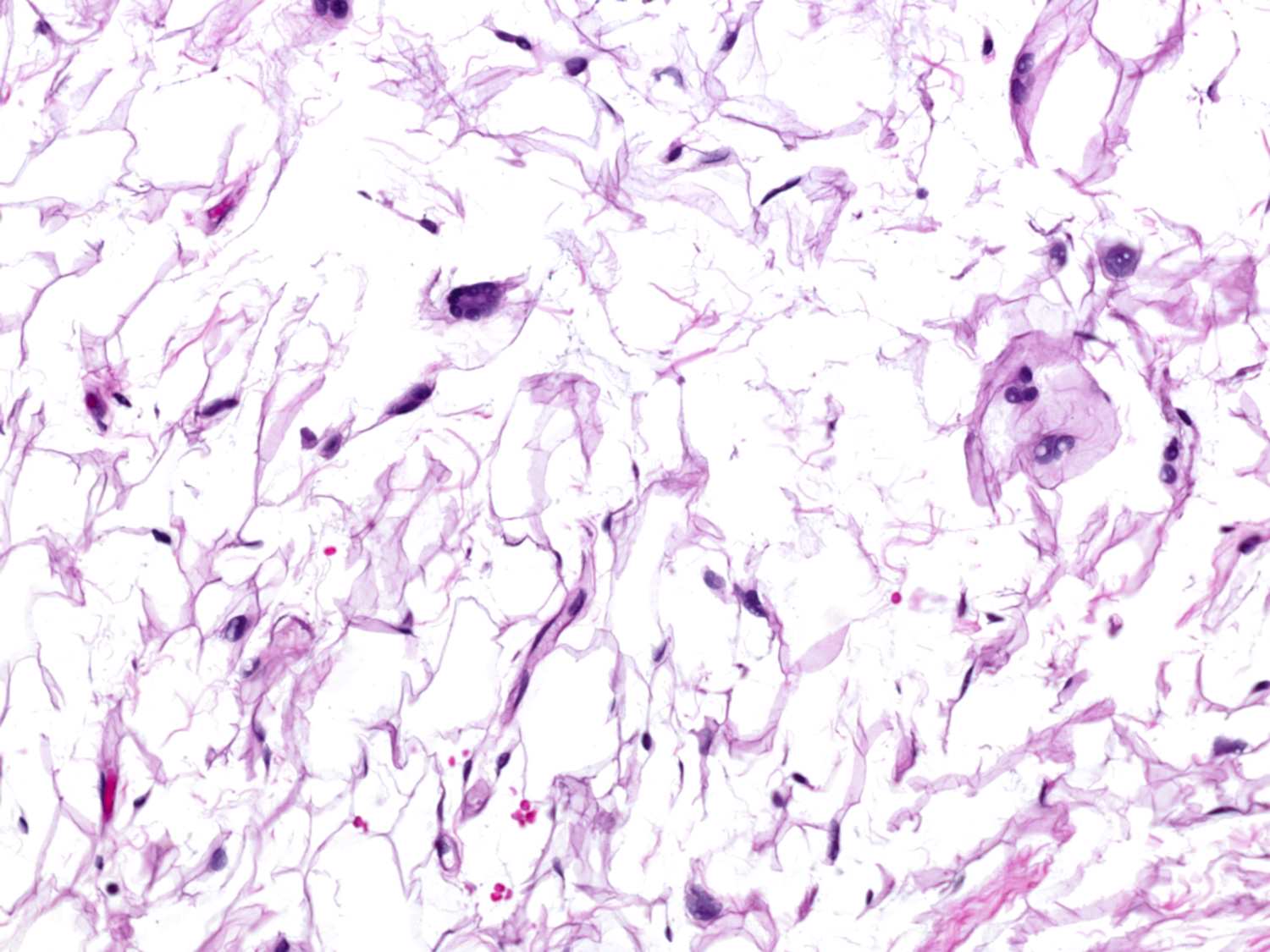

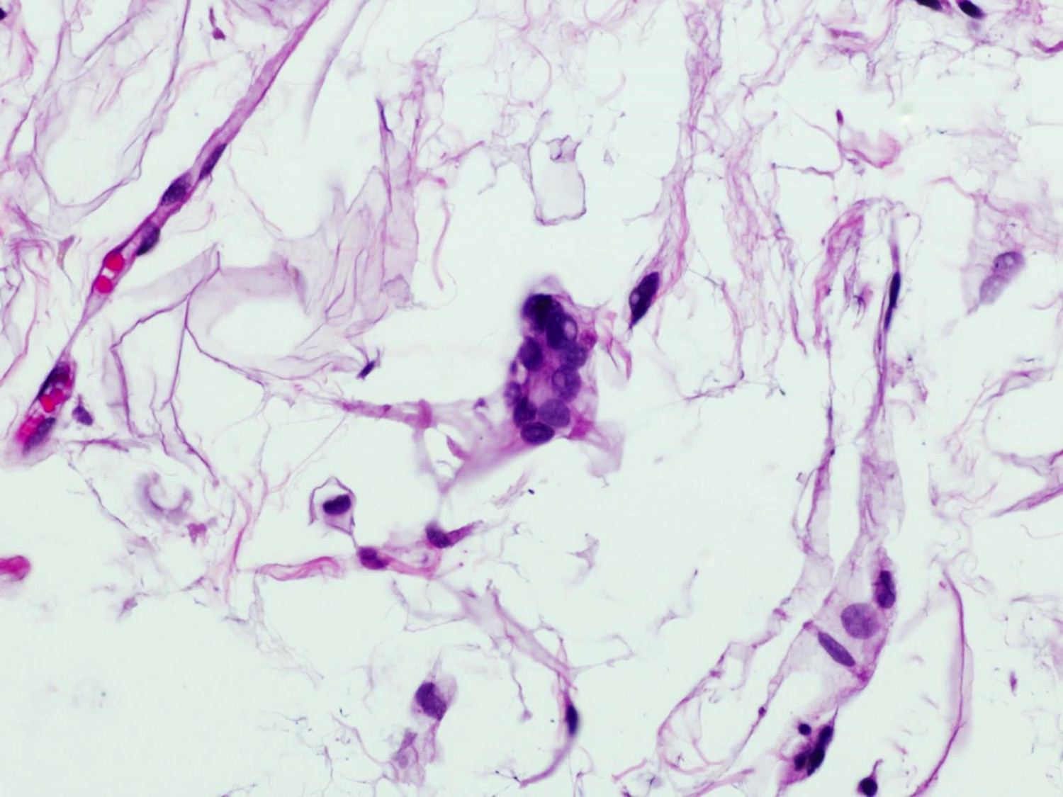
Herniated orbital fat
Differential diagnosis
- Pleomorphic lipoma:
- Different location; PL often involves soft tissues of back and shoulder of elderly men
- Areas typical of spindle cell lipomas, including variably myxoid stroma, wire-like collagen and bland spindle cell proliferation are focally present
- Floret-like giant cells show notable nuclear enlargement and hyperchromasia
- Well differentiated liposarcoma:
- May rarely involve the orbit (Ann Diagn Pathol 2001 Oct;5:255)
- Similar to other sites, will contain enlarged hyperchromatic cells within fibrous septae
- MDM2 amplification
Board review style question #1
Regarding subconjunctival herniated orbital fat, which of the following statements is true?
- Associated with MDM2 amplification
- Floret-like giant cells with nuclear hyperchromasia and pleomorphism are typically seen in SHOF
- Nuclei of Lochkern cells and floret-like giant cells are highlighted by CD34 immunohistochemistry
- Has an aggressive course and the management includes radiation therapy
Board review style answer #1
C. The nuclei of Lochkern cells and floret-like giant cells in SHOF are highlighted by CD34 immunohistochemistry (although not necessary for diagnosis)
Comment Here
Reference: Subconjunctival herniated orbital fat
Comment Here
Reference: Subconjunctival herniated orbital fat
