
Table of Contents
Definition / general | Terminology | Clinical images | Gross description | Microscopic (histologic) description | Microscopic (histologic) images | Positive stains | Negative stains | Molecular / cytogenetics descriptionCite this page: AIDS associated Kaposi sarcoma (epidemic). PathologyOutlines.com website. https://www.pathologyoutlines.com/topic/softtissuekaposiaids.html. Accessed April 18th, 2024.
Definition / general
- Historically, 40% of homosexual men with AIDS got Kaposi vs. 5% of others with AIDS
- Incidence of Kaposi has been decreasing over time (Hum Path 2001;32:649Arch Pathol Lab Med 2002;126:182)
- Early involvement of lymph nodes and gut and wide dissemination
- Usually not a direct cause of death, although 1 / 3 develop lymphoma or another second malignancy
Terminology
- Macule / patch:
- Pink purple macules of lower extremity or feet
- Superficial or mid dermal proliferation of collagen dissecting jagged capillary vessels with inconspicuous spindle cell component
- May be confluence of vessels
- Plaque:
- Dermal, dilated, jagged vascular channels that dissect collagen fibers and contain isolated or small groups of spindle cells
- Red blood cell extravasation prominent
- Also hemosiderin laden macrophages, pink hyaline globules
- Nodule / tumor:
- More distinctly neoplastic, most of lesion composed of spindle cells with intersecting fascicle like pattern in a background of inflammatory cells and red blood cells
- Small vessels and slitlike spaces with hyaline droplets and rows of red blood cells
- Mitotic figures common
- May involve lymph nodes and viscera (African and AIDS variants)
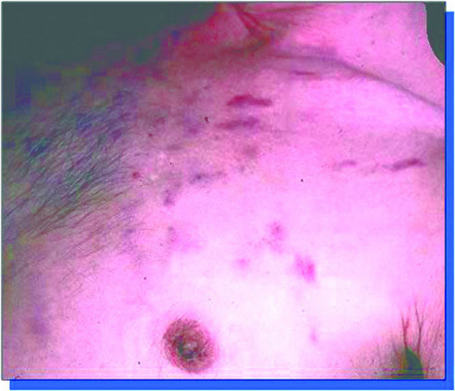
Clinical images
Images hosted on PathOut server:
Images hosted on other servers:


Ecchymosis like, courtesy of Dr. Mark R. Wick
Images hosted on other servers:
Rapidly growing nodule
Various images
Gross description
- Indolent disease has 3 stages:
- Early - macule / patch
- Intermediate - plaque
- Late - nodule / tumor
Microscopic (histologic) description
- Dilated irregular blood vessels in background of lymphocytes, plasma cells, macrophages
- Resembles granulation tissue
- Disease spreads proximally, converts to raised
Microscopic (histologic) images
Images hosted on other servers:


Various stains
Positive stains
- Smooth muscle actin, D2-40 (Mod Pathol 2002;15:434), other vascular markers
Negative stains
Molecular / cytogenetics description
- Detect HHV8 by PCR or in situ hybridization
