

Skin nonmelanocytic tumor
Neural tumors
Neurothekeoma
Author: Vijay Shankar, M.D.

Last author update: 1 November 2012
Last staff update: 9 November 2020
Copyright: 2002-2024, PathologyOutlines.com, Inc.
PubMed Search: Neurothekeoma [title] soft tissue
Table of Contents
Definition / general | Sites | Clinical features | Case reports | Treatment | Clinical images | Microscopic (histologic) description | Microscopic (histologic) images | Positive stains | Negative stains | Differential diagnosisCite this page: Shankar V. Neurothekeoma. PathologyOutlines.com website. https://www.pathologyoutlines.com/topic/softtissueneurothekeoma.html. Accessed April 24th, 2024.
Definition / general
- Superficial tumor, first described in 1980
- Originally, derivation thought to be from nerve sheath; now thought to derive from fibroblasts with ability to differentiate into myofibroblasts and to recruit histiocytes (Am J Surg Pathol 2007;31:1103)
Sites
- Usually head, upper extremities or shoulder girdle
Clinical features
- Solitary, superficial, slow growing mass up to 2 cm
- 60% women, mean age 17 years (range 2 - 85 years), 80% are < age 30 at initial diagnosis
Case reports
- 5 month old boy with thumb swelling (Indian J Dermatol 2009;54:59)
- 19 year old woman with painful subcutaneous nodule on lower back (Dermatol Online J 2009;15:3)
- 66 year old man with painless swelling in eyelid (Indian J Ophthalmol 2008;56:334)
Treatment
- Excision, may recur
Clinical images
Images hosted on other servers:

Thumb nodule

Lower back

Lower eyelid
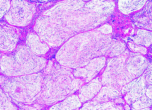
Microscopic (histologic) description
- Cellular, myxoid or mixed subtypes
- Involves dermis or subcutis
- Multinodular mass with myxoid matrix and peripheral fibrosis
- Whorled or focally fascicular patterns of spindled and epithelioid mononuclear cells with abundant cytoplasm, indistinct cell borders
- Margins usually positive; usually occasional multinucleated giant cells
- Variable nuclear atypia
- Median 4 MF / 25 HPF, may have 10+ MF / 25 HPF, may be atypical
Microscopic (histologic) images
Contributed by Mark R. Wick, M.D.
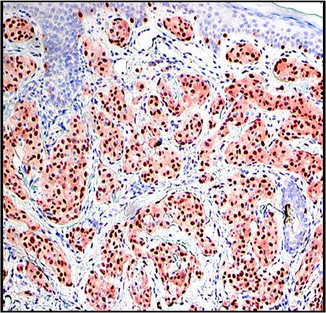
MiTF

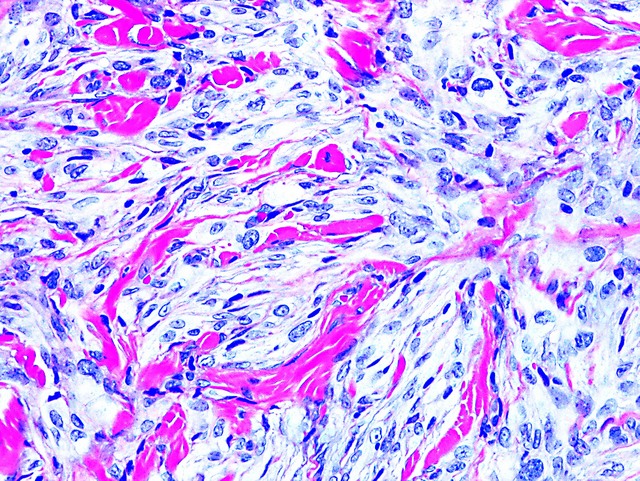

Cellular type
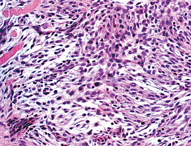
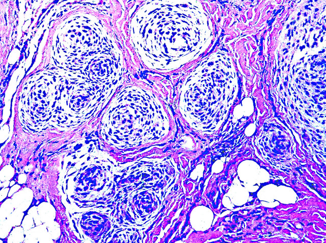
Cellular type, skin
Images hosted on other servers:


Left thumb nodule of 5 month old boy


Lower eyelid of elderly man
Differential diagnosis
