

Soft tissue
Skeletal muscle
Rhabdomyosarcoma
Spindle cell / sclerosing rhabdomyosarcoma
Editorial Board Member: Nasir Ud Din, M.B.B.S.
Deputy Editor-in-Chief: Borislav A. Alexiev, M.D.


Last author update: 6 April 2023
Last staff update: 6 April 2023
Copyright: 2002-2024, PathologyOutlines.com, Inc.
PubMed Search: Sclerosing rhabdomyosarcoma
Table of Contents
Definition / general | Essential features | Terminology | ICD coding | Epidemiology | Sites | Pathophysiology | Clinical features | Diagnosis | Radiology description | Radiology images | Prognostic factors | Case reports | Treatment | Clinical images | Gross description | Microscopic (histologic) description | Microscopic (histologic) images | Positive stains | Molecular / cytogenetics description | Sample pathology report | Differential diagnosis | Additional references | Board review style question #1 | Board review style answer #1 | Board review style question #2 | Board review style answer #2Cite this page: Kao E, Belzarena C. Spindle cell / sclerosing rhabdomyosarcoma. PathologyOutlines.com website. https://www.pathologyoutlines.com/topic/softtissuesclerosingrhabdo.html. Accessed April 20th, 2024.
Definition / general
- Spindle cell / sclerosing rhabdomyosarcoma (RMS) is a rare type of rhabdomyosarcoma characterized by fascicular spindle cell morphology
- Subclassification depends on the presence of genetic alterations associated with prognosis
- Predilection for the head and neck / extremities
- 5 - 10% of RMS (Arch Pathol Lab Med 2015;139:1281)
Essential features
- Rhabdomyosarcoma with prominent fascicular spindle cell or sclerosing features
- Relatively similar morphology but a genetically and clinically heterogeneous disease, especially in the pediatric population
- Tumors with VGLL2, NCOA2 and CITED2 gene rearrangements typically arise in infants and have a favorable prognosis (Am J Surg Pathol 2016;40:224)
- Tumors with MYOD1 mutations usually arise in adolescents and adults and have an unfavorable prognosis (Mod Pathol 2019;32:27)
- Tumors with TFCP2::NCOA2 gene rearrangements can be intraosseous (Mod Pathol 2020;33:404)
Terminology
- Spindle cell / sclerosing are considered the same diagnostic variant of rhabdomyosarcoma
ICD coding
- ICD-O: 8912/3 - spindle cell rhabdomyosarcoma
- ICD-11: 2B55.Z & XH7NM2 - rhabdomyosarcoma, unspecified primary site & spindle cell rhabdomyosarcoma
Epidemiology
- M > F
Sites
- Sclerosing is more common in adults in the extremities and head and neck regions (Am J Surg Pathol 2002;26:1175)
- Spindle cell is most common in the paratestis, followed by head and neck
- More common in the paratesticular region in the pediatric population (Arch Pathol Lab Med 2015;139:1281)
Pathophysiology
- Multiple genetic abnormalities have been identified
- Congenital / infantile spindle cell type: VGLL2, SRF, TEAD1, NCOA2, CITED2 gene fusions (Am J Surg Pathol 2016;40:224)
- Spindle cell / sclerosing type in adolescents / adults: MYOD1 mutation (J Pathol 2014;232:300)
- Intraosseous spindle cell / epithelioid type: EWSR1 or FUS fused with TFCP2 or MEIS1::NCOA2 gene fusions (Mod Pathol 2020;33:404, Am J Surg Pathol 2019;43:695)
Clinical features
- Infantile spindle cell RMS with VGLL2::NCOA2 associated gene fusions has a very good prognosis (Am J Surg Pathol 2016;40:224)
- Spindle cell / sclerosing RMS with no identified genetic abnormality has a good prognosis (Am J Surg Pathol 2016;40:224)
- MYOD1 mutant and TFCP2 associated gene fusion RMS have a poor prognosis (Mod Pathol 2019;32:27, Mod Pathol 2020;33:404)
Diagnosis
- Cellular spindle cell fascicles or tumor cells in sclerotic / collagenous background with positivity for desmin, myogenin and MyoD1 (Ann Diagn Pathol 2018;36:50)
- Demonstration of a MYOD1 mutation or various gene rearrangements may be helpful for subclassification (Oral Surg Oral Med Oral Pathol Oral Radiol 2022;134:354)
Radiology description
- Hypointense to isointense on T1 weighted images and hypointense to hyperintense on T2 weighted images (Pathol Res Pract 2019;215:152399)
- Heterogeneously enhancing soft tissue mass (J Oral Maxillofac Pathol 2022;26:S103)
Radiology images
Images hosted on other servers:

Large masticator space mass in neck

Cranial MRI

CT scan

MRI
Prognostic factors
- In adults, recurrence and metastasis rate is 40 - 50% (Arch Pathol Lab Med 2015;139:1281)
- Congenital / infantile spindle cell variant with gene fusions has a favorable prognosis (Am J Surg Pathol 2016;40:224)
- MYOD1 mutant variants have a poor prognosis (Mod Pathol 2019;32:27)
Case reports
- 7 year old girl with a spindle cell rhabdomyosarcoma in the parotid gland (Ann Pathol 2021;41:123)
- 15 year old girl with aggressive head tumor and involvement of the optic nerve (Cureus 2022;14:e21062)
- 16 year old girl presented with aggressive intraosseous tumor (Cold Spring Harb Mol Case Stud 2022;8:a006209)
- 27 year old woman with facial spindle cell rhabdomyosarcoma (J Oral Maxillofac Pathol 2022;26:S103)
- 29 year old man presented with tongue tumor (J Maxillofac Oral Surg 2021;20:464)
Treatment
- No consensus for optimal treatment
- In localized congenital type, complete surgical resection was associated with favorable prognosis (Eur J Cancer 2022;168:56)
- Most cases are treated with combination of surgery, radiotherapy and chemotherapy (J Maxillofac Oral Surg 2021;20:464)
- Recurrence and progression are common (J Maxillofac Oral Surg 2021;20:464)
Clinical images
Images hosted on other servers:

Cystic mass
Gross description
- Variably circumscribed tumor with a tan, sometimes whorled cut surface
Microscopic (histologic) description
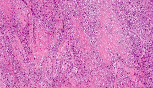
- Fascicles of spindle cells with a herringbone growth pattern resembling leiomyosarcoma or fibrosarcoma
- Primitive undifferentiated areas with round cells and hyperchromatic nuclei may focally be present
- Tadpole or strap cells and rhabdomyoblasts can be seen in some cases
- Sclerosing RMS shows prominent hyalinization / sclerosis; tumor cells in cords, nests or trabeculae in a pseudovascular growth pattern
- In bone, there can be epithelioid cells in sheets and fascicles in addition to the typical spindle cell morphology (Histopathology 2018;73:514)
Microscopic (histologic) images
Contributed by Erica Kao, M.D. and Borislav A. Alexiev, M.D.
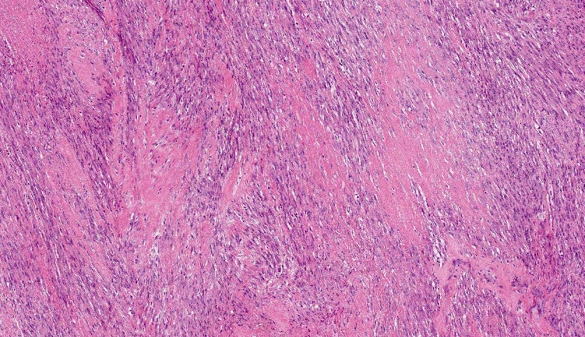
Sclerotic and collagenous stroma
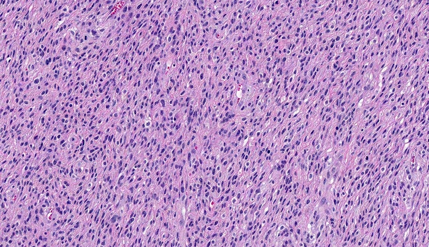
Fascicular growth
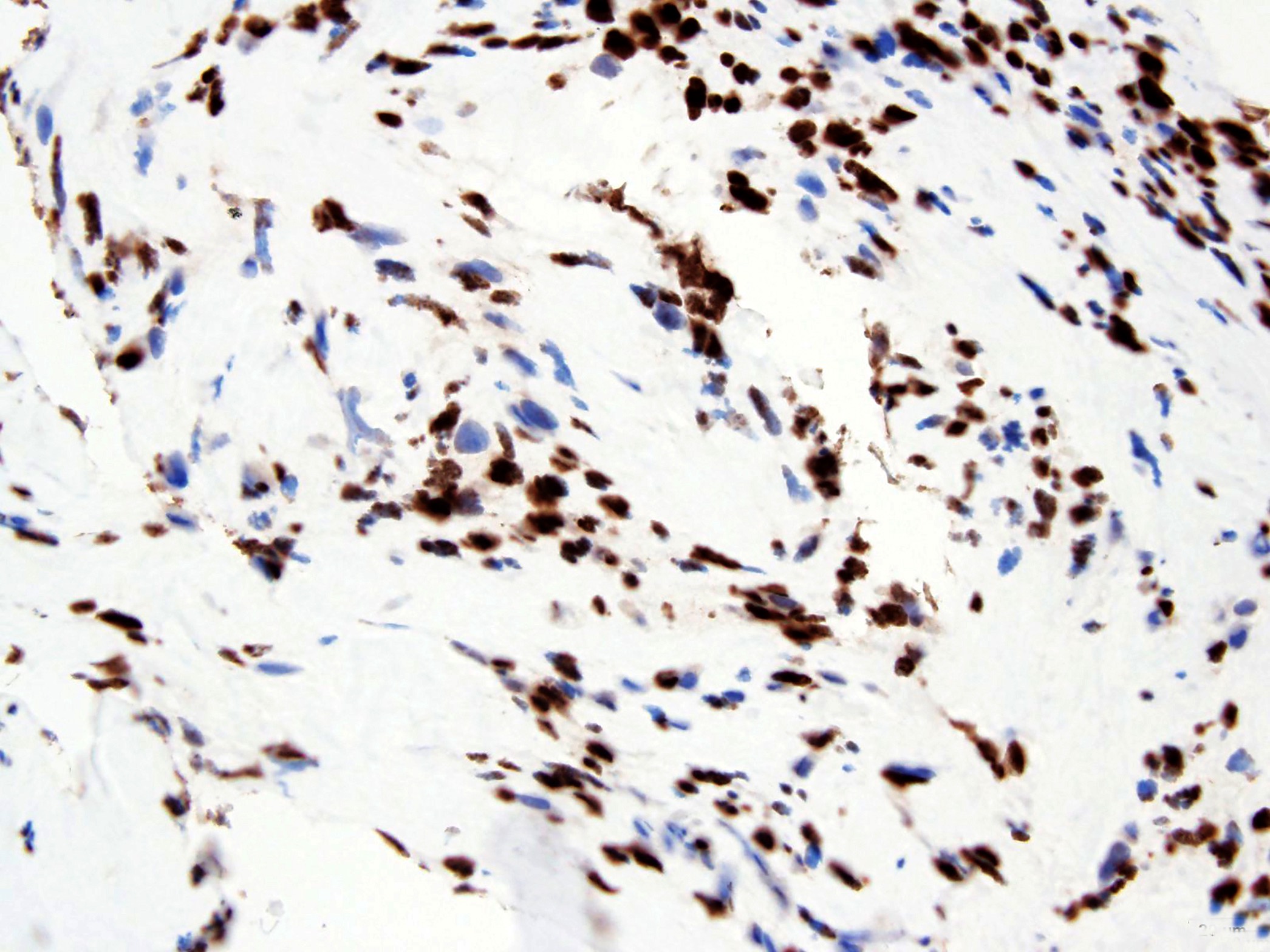
MyoD1
Positive stains
Molecular / cytogenetics description
- Should be PAX fusion negative
- Congenital / infantile spindle cell type: VGLL2, SRF, TEAD1, NCOA2, CITED2 gene fusions (Am J Surg Pathol 2016;40:224)
- Spindle cell / sclerosing type in adolescents / adults: MYOD1 mutation
- Intraosseous spindle cell / epithelioid type: EWSR1::FUS fused with TFCP2 or MEIS1::NCOA2 gene fusions (Mod Pathol 2020;33:404, Am J Surg Pathol 2019;43:695)
Sample pathology report
- Soft tissue, neck, biopsy:
- Rhabdomyosarcoma with spindle cell features (see comment)
- Comment: The biopsy contains cellular fascicles of tumor cells that are strongly positive for desmin, myogenin and MyoD1. Molecular testing will be performed for further subtyping and reported in an addendum.
Differential diagnosis
- Alveolar rhabdomyosarcoma:
- Different morphology; has PAX3 and PAX7::FOXO1 fusion transcripts (Pediatr Dev Pathol 2004;7:583)
- Myogenin is generally seen as weak, focal and nuclear
- Angiosarcoma:
- Mesenchymal chondrosarcoma:
- Characterized by a distinctive biphasic histology composed of round to spindled tumor cells admixed with areas of hyaline cartilage
- Spindle cell areas may even show limited expression of desmin, myogenin and MyoD1 (Hum Pathol 2018;77:28)
- Metastatic carcinoma:
- Positive expression of cytokeratin is seen in metastatic carcinoma
- Desmin and MyoD1 are negative
- Osteosarcoma:
- Has osteoid associated with cytologically malignant spindled cells
- Sclerosing epithelioid fibrosarcoma:
- Composed of epithelioid cells arranged in nests and cords and deposited in a densely hyalinized collagenous matrix
- However, in almost all cases the tumor also shows foci of spindle shaped sarcoma similar to conventional fibrosarcoma and exhibits EMA or MUC4 positivity (Head Neck Pathol 2015;9:147)
- Also synovial sarcoma, leiomyosarcoma, spindle cell carcinoma, spindle cell melanoma, fibrosarcoma, malignant peripheral nerve sheath tumor and malignant triton tumor
Additional references
Board review style question #1
Which soft tissue tumor is more commonly found in the head and neck region?
- Angiolipoma
- Dermatofibrosarcoma protuberans
- Liposarcoma
- Spindle cell / sclerosing rhabdomyosarcoma
Board review style answer #1
D. Spindle cell / sclerosing rhabdomyosarcoma
Comment Here
Reference: Spindle cell / sclerosing rhabdomyosarcoma
Comment Here
Reference: Spindle cell / sclerosing rhabdomyosarcoma
Board review style question #2
Which soft tissue tumor is shown in the image above and is associated with fascicular spindle cell morphology?
- Alveolar rhabdomyosarcoma
- Embryonal rhabdomyosarcoma
- Pleomorphic rhabdomyosarcoma
- Spindle cell / sclerosing rhabdomyosarcoma
Board review style answer #2
D. Spindle cell / sclerosing rhabdomyosarcoma. Fascicular spindle cell morphology is one of the essential features of spindle cell / sclerosing
rhabdomyosarcoma.
Comment Here
Reference: Spindle cell / sclerosing rhabdomyosarcoma
Comment Here
Reference: Spindle cell / sclerosing rhabdomyosarcoma
