

Soft tissue
General
Histology-skeletal muscle
Author: Norbert Sule, M.D., Ph.D.
Editorial Board Member: Borislav A. Alexiev, M.D.
Editor-in-Chief: Debra L. Zynger, M.D.

Last author update: 11 January 2021
Last staff update: 21 April 2021
Copyright: 2002-2024, PathologyOutlines.com, Inc.
PubMed Search: Skeletal muscle[TI] soft tissue[TIAB] pathology
Table of Contents
Definition / general | Essential features | Embryogenesis | Terminology | Physiology | Diagrams / tables | Microscopic (histologic) description | Microscopic (histologic) images | Special and histochemical stains | Positive stains | Negative stains | Electron microscopy description | Electron microscopy images | Additional references | Board review style question #1 | Board review style answer #1 | Board review style question #2 | Board review style answer #2Cite this page: Sule N. Histology-skeletal muscle. PathologyOutlines.com website. https://www.pathologyoutlines.com/topic/softtissueskeletalnormal.html. Accessed April 16th, 2024.
Definition / general
- Specialized contractile tissue responsible for the voluntary movements
Essential features
- Contraction of skeletal muscle is controlled by large motor nerve fibers of the alpha neuron located in the anterior horn of the spinal cord and motor nuclei of the brain
- Neurotransmitter of the neuromuscular junction is acetylcholine, which binds to the nicotinic receptors
- Skeletal muscle fiber is an elongated cylindrical, unbranched, multinucleated contractile unit formed by fusion of single cells resulting in multinucleated syncytia
- Sarcoplasm is filled with myofibrils, a protein structure composed of contractile proteins and organized in a repeating pattern resulting in the light microscopic and electron microscopic appearance of cross striation
- Biochemically, there are 2 types of fibers: type I (aerobic, slow, red) and type II (anaerobic, fast, white)
Embryogenesis
- Paraxial mesoderm / somite → dermatomyotome → myoblast → myotubes → muscle fiber
- Normal skeletal muscle is formed from the primitive mesenchymal element of the paraxial mesoderm (somite) which divides into ventral dorsolateral somite or dermatomyotome
- Mesenchymal cells of the dermatome differentiate into dermal fibroblast and myotomes differentiate into myoblast (portion of skin and group of skeletal muscles derived from the same segment of dermatomyotome is supplied by a specific segmental spinal nerve)
- Myoblast proliferation and fusion results in formation of multinucleated cells called myotubes
- After formation of myotubes, synthesis of fibrillary protein starts in the center of the cells and causes peripheral displacement of the nuclei
- This process is mostly complete at the time of birth
- Only a few residual satellite myoblasts are present in the mature muscle that are able to play a role in the repair and regeneration
- Reference: J Anat 2015;227:673
Terminology
- Synonyms: striated muscle, voluntary muscle
- Its basic cellular unit is the muscle fiber (also called myofibers or myocytes)
- Group of muscle fibers controlled by the same nerve fibers is called the motor unit
- Excitation of the nerve fiber results in simultaneous contraction of all the muscle fibers in the same motor unit
- Structural organization of skeletal muscle:
- Muscle fiber = cellular unit (enwrapped by endomysium)
- Fasciculi = bundle of muscle fibers (enwrapped by perimysium)
- Muscle = collection of fasciculi (enwrapped by epimysium)
- Special terminology to describe the highly specialized cellular components of muscle fibers:
- Plasma membrane = sarcolemma / plasmalemma
- Endoplasmic reticulum = sarcoplasmic reticulum
- Mitochondria = sarcosomes
- Specialized intramuscular stretch receptors are called neuromuscular spindles
Physiology
- Contraction of skeletal muscle is controlled by large motor nerve fibers of the alpha neuron located in the anterior horn of the spinal cord and motor nuclei of the brain
- Intercellular junction between the nerve ending and the muscle fiber is called the neuromuscular junction
- Stimulation by the motor nerve causes simultaneous contraction
- Neurotransmitter of the neuromuscular junction is acetylcholine, which binds to the nicotinic receptors
- Excitation of the muscle cell membrane results in the release of Ca2+ from the sarcoplasmic reticulum
- Ca2+ binds to troponin tropomyosin complex, unmasking the cross bridge site on the actin molecules
- Contraction is the result of a sliding mechanism of the thick (myosin) and thin (actin) fibers
- ATP hydrolysis activates sites on the myosin fibers connecting to actin molecules and forming cross bridges
- This connection causes configurational changes of the myosin fiber, shortening the sarcomere and resulting in movement of the muscle
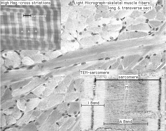
Diagrams / tables
Images hosted on other servers:

Organization from
gross muscle fibers to
contractile filaments

T tubules and sarcoplasmic reticulum
Microscopic (histologic) description
- Skeletal muscle
- Organization of skeletal muscle:
- Muscle fiber enwrapped by endomysium, loose connective tissue with reticulin and collagen fibers
- Fasciculi enwrapped by perimysium, loose connective tissue with elastin and collagen fibers
- Muscle enwrapped by epimysium, dense collagenous sheath
- Organization of skeletal muscle:
- Muscle fiber
- Elongated cylindrical, unbranched, multinucleated contractile unit formed by fusion of single cells resulting in multinucleated syncytia
- Size: diameter varies 10 - 100 micrometers; length: up to 35 cm
- Flattened nuclei are pushed to periphery of the muscle fiber (just underneath the sarcolemma)
- Sarcoplasm is filled with myofibrils, a protein structure composed of contractile proteins and organized in a repeating pattern resulting in the light microscopic and electron microscopic appearance of cross striation
- Myofibrils composed of thin (actin) and thick (myosin) filaments
- Biochemically there are 2 types of fibers
- Type I (aerobic, slow, red):
- Small cross section
- Abundant mitochondria
- Large amount of myoglobin (oxygen storage)
- Type II (anaerobic, fast, white): intense, short, rapid but sporadic movements
- Large cross section
- Few mitochondria
- Little myoglobin
- Rich in glycogen and glycolytic enzymes
- Type I (aerobic, slow, red):
- Neuromuscular spindles
- Encapsulated, cigar shaped structures (~6 micrometer) embedded in endomysium and perimysium
- Composed of modified muscle fibers (intrafusal fibers) and associated sensory myelinated and nonmyelinated nerve endings, positioned parallel to the muscle fibers
- Reference: Ageing Res Rev 2018;47:123
Microscopic (histologic) images
Contributed by Norbert Sule, M.D., Ph.D.
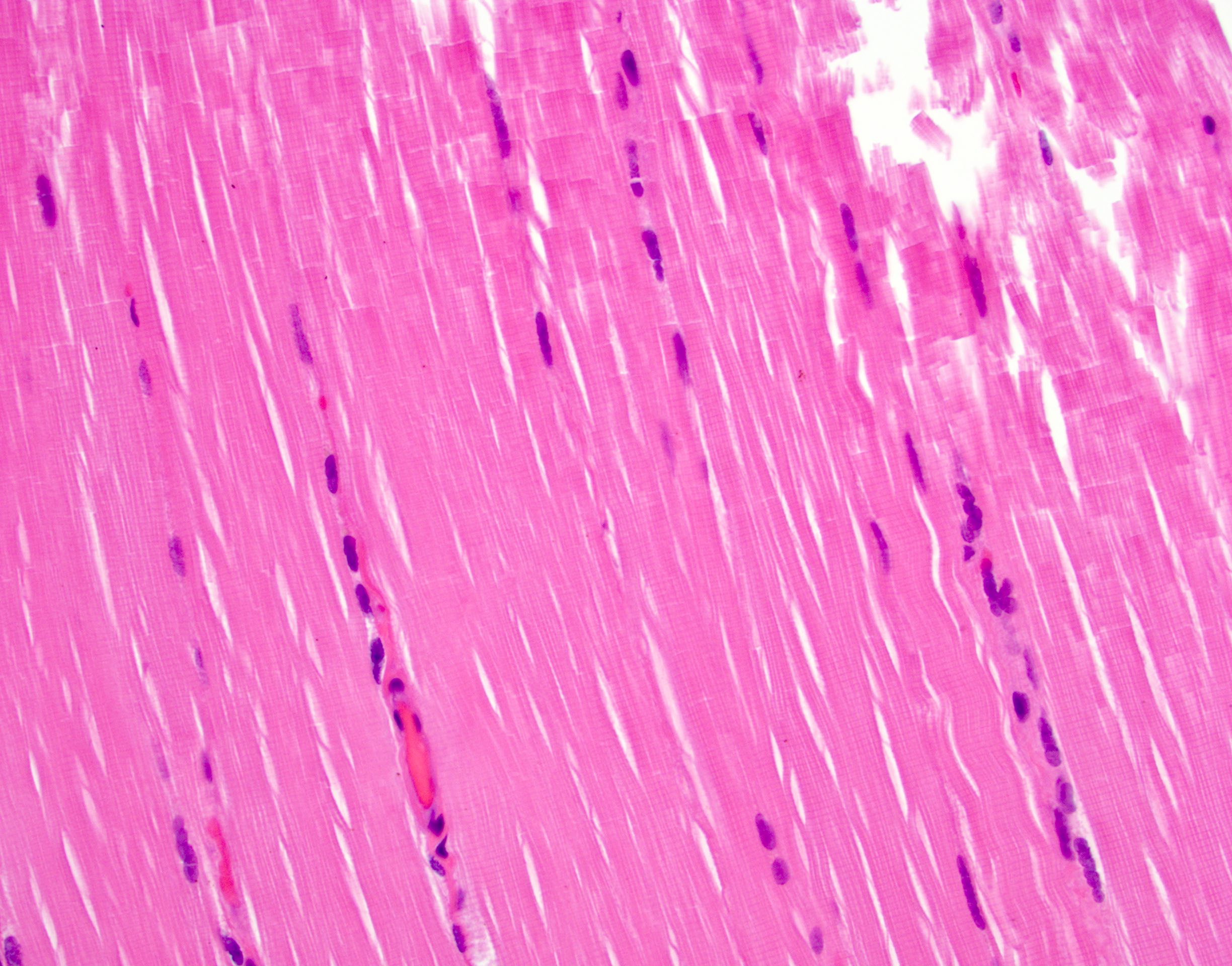
Cross striation
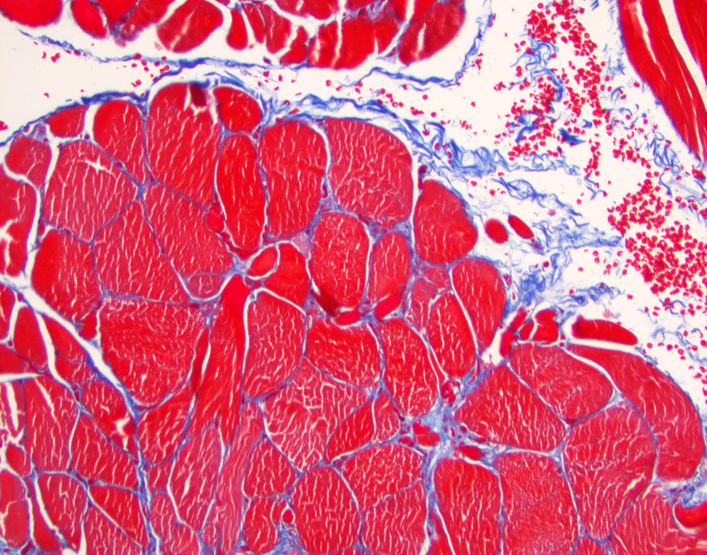
Masson trichrome
Special and histochemical stains
- Masson trichrome: epi, peri and endomysium
- Iron hematoxylin: cross striation
- PAS - glycogen content:
- Type 1 / red fibers: lighter
- Type 2 / white fibers: darker
- Histochemical stains (to reveal the metabolic properties of red and white fibers):
- Succinate dehydrogenase - Kreb cycle enzyme:
- Type 1 / red fibers: darker
- Type 2 / white fibers: lighter
- ATPase pH 4.3 - pattern depends on pH:
- Type 1 / red fibers: darker
- Type 2 / white fibers: lighter
- ATPase pH 9.4 - pattern depends on pH:
- Type 1 / red fibers: lighter
- Type 2 / white fibers: darker
- Succinate dehydrogenase - Kreb cycle enzyme:
Positive stains
- Vimentin (nonspecific)
- Desmin
- Muscle specific actin
- Myogenic regulatory protein (MyoD1) (Am J Pathol 1994;144:693)
- Myogenin (Am J Surg Pathol 2001;25:1150)
- Myoglobin (Am J Pathol 2009;175:201, J Exp Biol 2010;213:2741)
Electron microscopy description
- Electron microscopy reveals structural components (Curr Diagn Pathol 2002;8:225):
- I (isotropic) band: thin filaments only
- A (anisotropic) band: overlapping thin and thick filaments
- H band: thick filaments only
- Z line: divides center of I band; serves as attachment site for the sarcomere, the repeating individual unit of the muscle fiber
- T tubule:
- Tubular plasma membrane system / extension at the level of A and I band junction surrounding the myofibril
- Lumen is continuous with the extracellular space
- Sarcoplasmic reticulum:
- Smooth endoplasmic reticulum derived membrane system located between the T tubules with sac-like dilatation called terminal cisternae
- Calcium ion concentrated in the lumens
- Triad: a complex composed of T tubule and 2 terminal cisternae
Electron microscopy images
Images hosted on other servers:
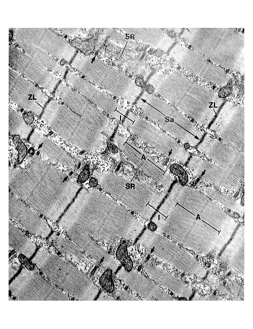
Contractile filament within muscle fiber resulting in band-like arrangement
Additional references
Board review style question #1
Which of the following statements regarding the connective tissue of skeletal muscle is correct?
- Endomysium consists of fine reticular fibers surrounding the muscle fiber
- Epimysium separates each muscle fascicle from the others
- Perimysium envelopes the whole skeletal muscle
- Perimysium enwraps the individual muscle fibers within a fascicle
Board review style answer #1
A. Endomysium consists of fine reticular fibers surrounding the muscle fiber. Skeletal muscle is enwrapped in a dense collagenous sheath called epimysium. The muscle represents a collection of muscle fasciculi separated by perimysium, which is a loose connective tissue of elastin and collagen fibers. The individual skeletal muscle fiber enwrapped by loose connective tissue is called endomysium, which is composed of reticulin and collagen fibers.
Comment Here
Reference: Histology - skeletal muscle
Comment Here
Reference: Histology - skeletal muscle
Board review style question #2
A 66 year old woman undergoes resection of a thigh mass composed of large pleomorphic epithelioid cells with eosinophilic cytoplasm, necrosis and increased mitotic activity. The cells are diffusely positive for vimentin, desmin and MyoD1 (subset) and negative for CD45, S100, MelanA, pancytokeratin and MDM2. What differentiation do the pleomorphic cells show?
- Smooth muscle
- Adipocytic
- Epithelial
- Skeletal muscle
Board review style answer #2
D. Skeletal muscle. The histology describes a pleomorphic epithelioid cell neoplasm. Based on the morphology alone, no definite differentiation is evident. Considering the age, the differential diagnosis is wide and includes dedifferentiated liposarcoma, pleomorphic leiomyosarcoma, melanoma, malignant peripheral nerve sheath tumor and rhabdomyosarcoma. The performed immunohistochemical stains revealed positive reaction with vimentin and desmin. Considering the morphology and the immunohistochemical profile, the findings are most consistent with muscular differentiation. The positive reaction of MyoD1 is compatible skeletal muscle differentiation (vimentin+, desmin+ and MyoD1+; pleomorphic rhabdomyosarcoma) and rules out smooth muscle differentiation (vimentin+, desmin+ and MyoD1-; pleomorphic leiomyosarcoma). The lack of expression of S100 and MDM2 rules out melanocytic (S100+; melanoma), adipocytic (S100+, MDM2+; pleomorphic liposarcoma) and neural differentiation (S100+ focal; malignant peripheral nerve sheath tumor). The negativity with CD45 and pancytokeratin excludes leukocytic and epithelial differentiation, respectively.
Comment Here
Reference: Histology - skeletal muscle
Comment Here
Reference: Histology - skeletal muscle
