
Lymph nodes & spleen, nonlymphoma
Spleen-vascular tumors
Sclerosing angiomatoid nodular transformation
Author: Valentina Sangiorgio, M.D.
Editorial Board Member: Elizabeth Courville, M.D.
Deputy Editor-in-Chief: Patricia Tsang, M.D., M.B.A.

Last author update: 6 April 2023
Last staff update: 6 April 2023
Copyright: 2003-2024, PathologyOutlines.com, Inc.
PubMed Search: Sclerosing angiomatoid nodular transformation of the spleen
Table of Contents
Definition / general | Essential features | Terminology | ICD coding | Epidemiology | Sites | Etiology | Clinical features | Diagnosis | Laboratory | Radiology description | Radiology images | Prognostic factors | Case reports | Treatment | Clinical images | Gross description | Gross images | Microscopic (histologic) description | Microscopic (histologic) images | Positive stains | Negative stains | Electron microscopy description | Sample pathology report | Differential diagnosis | Board review style question #1 | Board review style answer #1 | Board review style question #2 | Board review style answer #2Cite this page: Sangiorgio V. Sclerosing angiomatoid nodular transformation. PathologyOutlines.com website. https://www.pathologyoutlines.com/topic/spleensclerosingangiomatoid.html. Accessed April 25th, 2024.
Definition / general
- Rare, benign splenic lesion of unknown etiology, characterized by confluent angiomatoid nodules surrounded by concentric collagen fibers
Essential features
- Nonneoplastic condition that affects the spleen only; not described in other sites
- Microscopically characterized by multiple angiomatoid nodules in a fibrosclerotic stroma
- Surgery is curative in all the cases and recurrence has not been reported
Terminology
- Multinodular hemangioma (obsolete)
ICD coding
- ICD-10: D18.03 - hemangioma of intra-abdominal structures
Epidemiology
- Female predilection (F:M = 2:1)
- Mean age of 48 years
- Range is 30 - 60 years (Int J Surg Case Rep 2012;3:492)
Sites
- Only described in the spleen
- Confined to the splenic parenchyma, with no extrasplenic involvement
Etiology
- Etiology and pathogenesis unknown
- Exaggerated sclerotic and neoangiogenic splenic reaction could be secondary to different pre-existing conditions, including vascular lesions undergoing thrombosis, infarcts and even hamartoma (Int J Surg Case Rep 2012;3:492, Am J Surg Pathol 2004;28:1268)
Clinical features
- Asymptomatic in the majority of the cases; incidentally discovered by imaging studies or at time of surgery for unrelated processes (Am J Surg Pathol 2004;28:1268)
- Symptomatic patients may experience nonspecific symptoms, including abdominal discomfort, abdominal fullness, nausea, vomiting and undesired weight loss
Diagnosis
- Clinically and radiologically indistinguishable from other lesions of the spleen, including hemangioma, hamartoma and rarely, malignancies
- Final diagnosis is achieved only with histopathologic examination and immunohistochemistry of resected spleen specimens
- Splenectomy serves both diagnostic and treatment functions
- References: Am J Surg Pathol 2004;28:1268, Semin Diagn Pathol 2021;38:159
Laboratory
- Laboratory values are mostly unaltered
- Occasional reports of nonspecific findings, including raised erythrocyte sedimentation rate, leukocytosis and polyclonal hypergammaglobulinemia (Am J Surg Pathol 2004;28:1268)
Radiology description
- No characteristic and unique radiologic findings of this entity
- Abdominal ultrasound, CT scan and MRI studies usually reveal an isolated, multinodular solid splenic mass
- References: J Comput Assist Tomogr 2019;43:863, Diagn Interv Imaging 2021;102:389
Radiology images
Images hosted on other servers:

CT findings

Expansible low density mass

5 cm tumor in hilus

Hypointensity of tumor

Splenic lesion at medial aspect

Lesion in lower spleen

Hypoenhancing mass
Prognostic factors
- Benign clinical behavior with favorable prognosis and no reported recurrences after splenectomy (Am J Surg Pathol 2004;28:1268)
Case reports
- 21 year old woman with mild thrombocytopenia and a hypervascular splenic mass (Radiol Case Rep 2018;14:521)
- 26 year old man and 65 year old woman presenting with undesired weight loss and a lasting feeling of abdominal fullness (World J Clin Cases 2020;8:103)
- 37 year old woman with a 5 cm hypoechoic mass in the spleen with low standardized uptake value (SUV) at FDG PET / CT scan (Acta Radiol Open 2016;5:2058460116649799)
- 49 year old woman with a newly diagnosed invasive clear cell carcinoma of the uterus and a 4 cm isolated splenic nodule suspicious for metastatic involvement (J Med Case Rep 2018;12:377)
- 67 year old man with an isolated 55 mm splenic lesion suspicious for an abscess (Int J Surg Case Rep 2019;56:1)
Treatment
- Splenectomy performed at discovery of the splenic mass is curative in all the cases
Clinical images
Images hosted on other servers:

Macroscopic findings

Laparoscopic view
Gross description
- Single unencapsulated, well circumscribed solid mass with a striking multinodular quality and variable fibrotic strands (Semin Diagn Pathol 2021;38:159)
Gross images
Contributed by Shafinaz Hussein, M.D. and Huifei Liu, M.D., Ph.D. (Case #364)
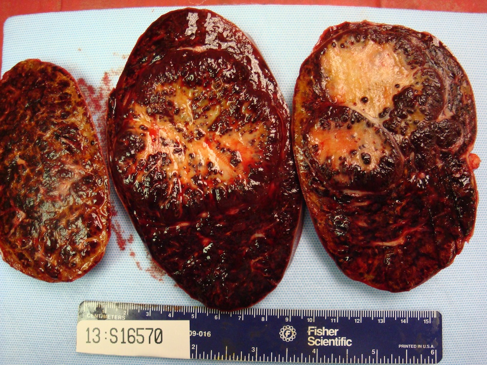
Circumscribed, bosselated, firm, tan colored mass
Images hosted on other servers:

Well demarcated, unencapsulated lesion

Splenic resection

Well defined, nonencapsulated lesion

Red-brown, multinodular lesions
Microscopic (histologic) description
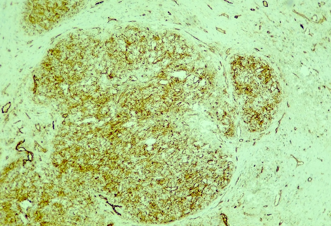
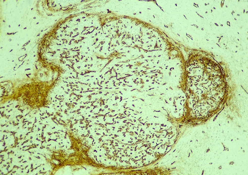
- Multiple, well circumscribed and confluent angiomatoid nodules surrounded by a variable fibrosclerotic stroma and occasionally, by a fibrinoid rim imparting a granuloma-like appearance at low magnification (Arch Pathol Lab Med 2007;131:974, Semin Diagn Pathol 2021;38:159)
- Nodules contain a mixture of slit-like, round or irregular vascular spaces lined by cytologically bland, sometimes plump endothelial cells; lacking atypia, necrosis and mitotic figures
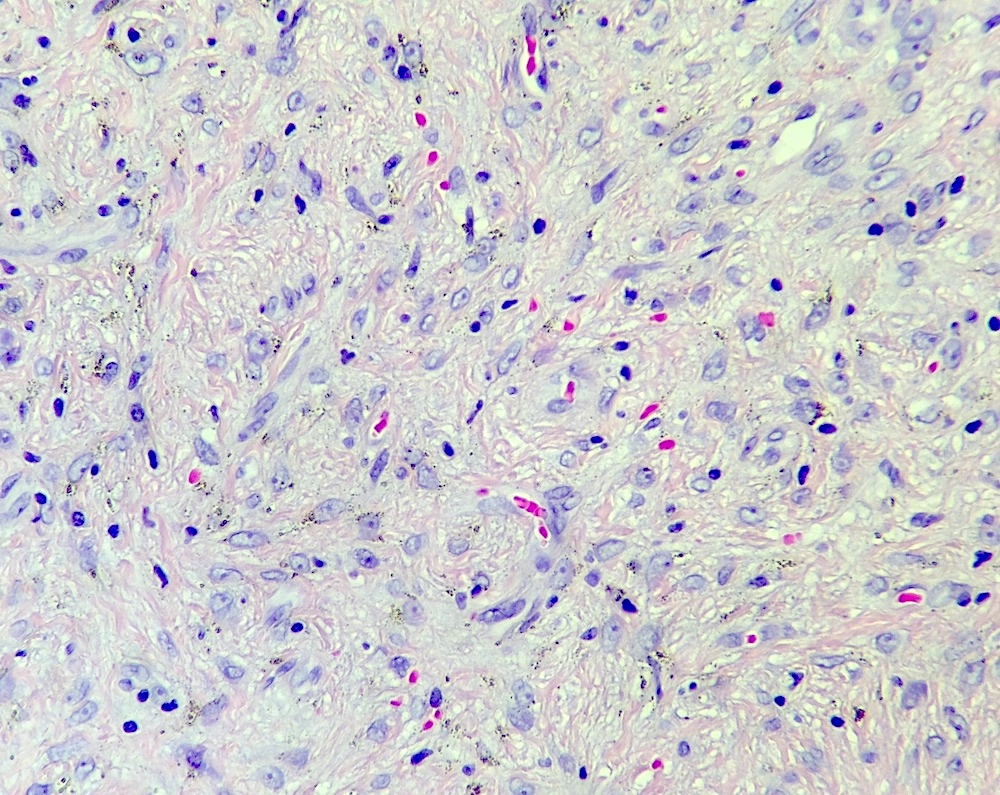
- 3 different types of vessels within the nodules can be highlighted by immunohistochemistry (see Positive stains), which recapitulate the normal red pulp composition: capillaries, sinusoids and small veins
- Stroma separating the nodules is fibrous to fibromyxoid with bland fibroblasts and myofibroblasts; contains abundant inflammatory cells, including lymphocytes, hemosiderin laden macrophages and polytypic plasma cells (Semin Diagn Pathol 2021;38:159)
- IgG4 positive plasma cells occasionally numerous with IgG4:IgG ratio increased; however, serum IgG4 concentration not elevated (Pathol Int 2009;59:844)
- Adjacent splenic tissue is unremarkable
Microscopic (histologic) images
Contributed by Valentina Sangiorgio, M.D., Shafinaz Hussein, M.D. and Huifei Liu, M.D., Ph.D. (Case #364)
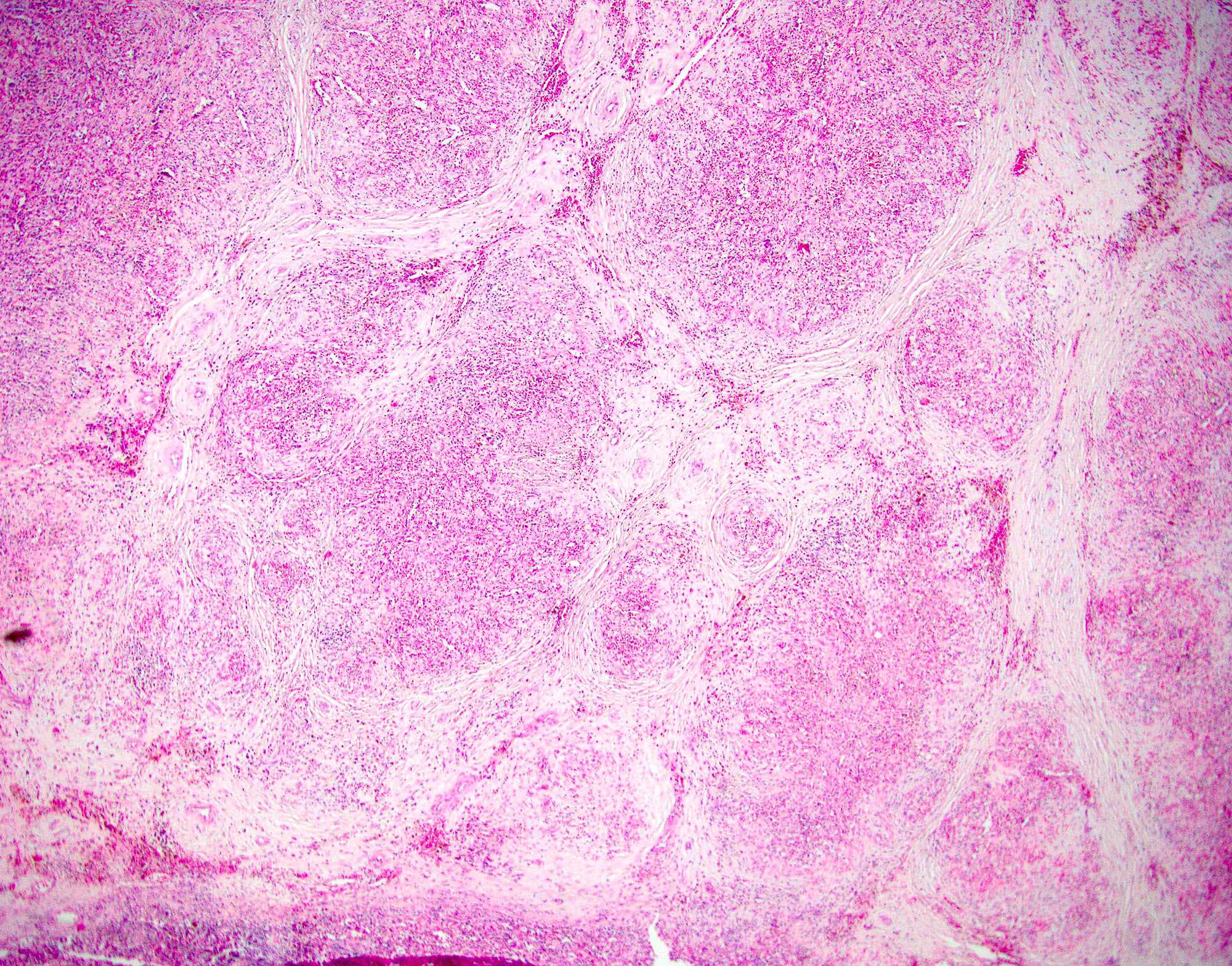
Multiple vascular nodules
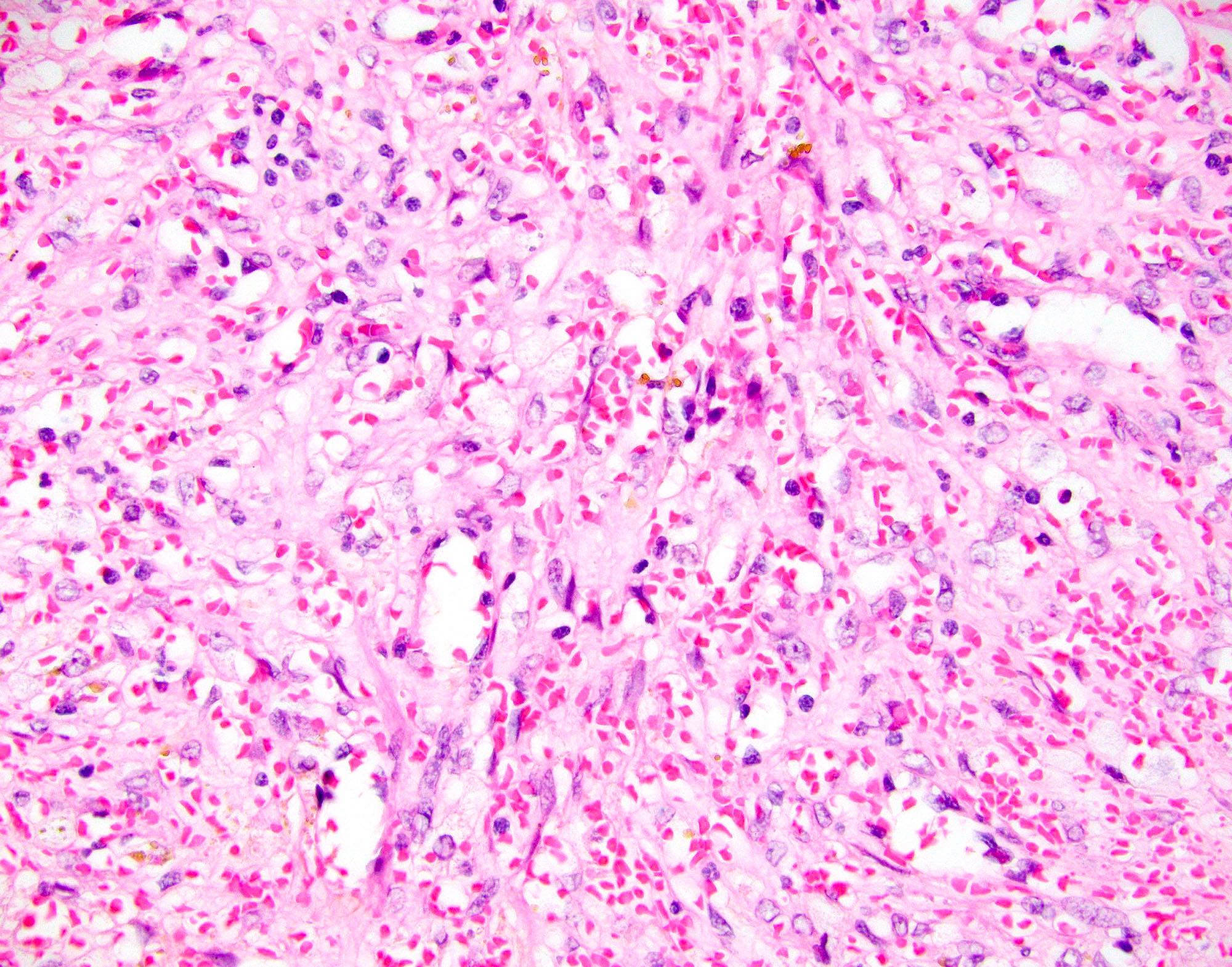
Predominantly small capillaries
Bland endothelial cells
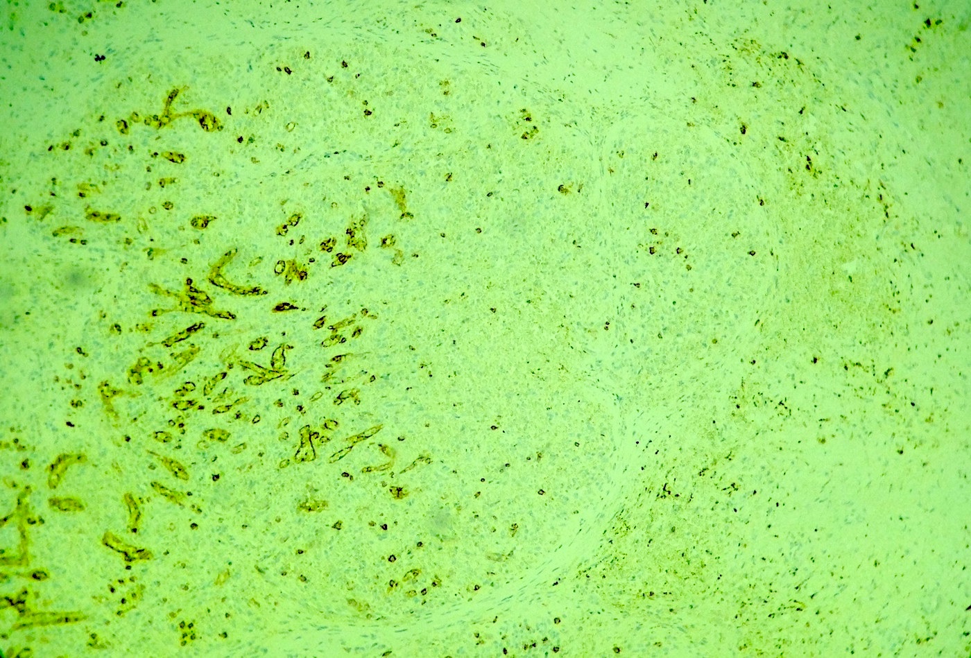
CD8
CD31
CD34
Positive stains
- 3 types of vessels (as seen in the normal splenic red pulp):
- SMA positive in myofibroblasts in the nodules
- CD68 positive in histiocytes / macrophages
- Polytypic expression of kappa and lambda light chains within plasma cells
- References: Am J Surg Pathol 2004;28:1268, Arch Pathol Lab Med 2013;137:1309
Negative stains
- D2-40
- Epstein Barr virus (EBV) in situ hybridization
Electron microscopy description
- Small vascular spaces lined by endothelial cells with pinocytotic vesicles but no Weibel-Palade bodies (Am J Surg Pathol 2004;28:1268)
Sample pathology report
- Spleen, splenectomy:
- Sclerosing angiomatoid nodular transformation (SANT) of the spleen
Differential diagnosis
- Hemangioma:
- Often of cavernous type
- Cystically dilated, blood filled spaces lined by a layer of bland endothelial cells
- Vascular spaces separated by thin fibrous septa, without the striking angiomatoid nodular appearance of sclerosing angiomatoid nodular transformation (Semin Diagn Pathol 2021;38:154)
- Littoral cell angioma:
- Macroscopically enlarged spleen overrun by multiple hemorrhagic nodules with a vague lobular configuration and a spongy appearance; uncommonly solitary nodules
- Nodules composed of pleomorphic vascular spaces filled with papillary projections
- Papillary stalks lined by plump endothelial cells
- Characteristic immunophenotype of splenic littoral cells with dual histiocytic and endothelial differentiation lacking CD34
- Hemangioendothelioma and angiosarcoma:
- Ill defined vascular spaces lined by variably atypical, malignant endothelial cells, with necrosis and increased mitotic activity (Semin Diagn Pathol 2021;38:154)
- Hamartoma:
- Structurally disorganized red pulp tissue, with no Malpighian follicles and only occasional fibrous trabeculae
- Compared with sclerosing angiomatoid nodular transformation, the borders are poorly defined and it lacks the angiomatoid nodular appearance (Semin Diagn Pathol 2021;38:154)
Board review style question #1
What does this image from a sclerosing angiomatoid nodular transformation of the spleen show?
- Areas of geographic necrosis
- Atypical endothelial cells with prominent nucleoli
- Easily identifiable mitotic figures (≥ 1 per 10 high power fields)
- Small capillaries admixed with scattered inflammatory cells, including small lymphocytes, histiocytes and plasma cells
Board review style answer #1
D. Small capillaries admixed with scattered inflammatory cells, including small lymphocytes, histiocytes and plasma cells. Necrosis, atypia and increased mitotic activity (≥ 1 per 10 high power fields) are not seen in this image. They are not seen in otherwise conventional sclerosing angiomatoid nodular transformation and their identification should prompt alternative diagnoses, including hemangioendothelioma or angiosarcoma.
Comment Here
Reference: Sclerosing angiomatoid nodular transformation
Comment Here
Reference: Sclerosing angiomatoid nodular transformation
Board review style question #2
Which of the following statements best describes sclerosing angiomatoid nodular transformation of the spleen?
- Benign condition that affects the spleen only, is cured with splenectomy and does not manifest any metastatic potential
- Benign neoplasm that can affect parenchymatous organs (mostly spleen, liver and lung) and does not manifest any metastatic potential
- Neoplasm of uncertain malignant potential as it can recur after resection in sites other than spleen
- Neoplasm of uncertain malignant potential as occasional cases transform into overt angiosarcoma
Board review style answer #2
A. Benign condition that affects the spleen only, is cured with splenectomy and does not manifest any metastatic potential. Sclerosing angiomatoid nodular transformation is a nonneoplastic lesion which affects the spleen only, with no description in other anatomic sites. It is cured by splenectomy and never recurs thereafter.
Comment Here
Reference: Sclerosing angiomatoid nodular transformation
Comment Here
Reference: Sclerosing angiomatoid nodular transformation
