
Stains & CD markers
IMP3

Copyright: 2020-2024, PathologyOutlines.com, Inc.
PubMed Search: IMP3[TI] free full text[SB]
IMP3
Author: David Wachter, M.D.
Editor-in-Chief: Debra L. Zynger, M.D.
Last author update: 2 June 2020
Last staff update: 2 July 2021
Copyright: 2020-2024, PathologyOutlines.com, Inc.
PubMed Search: IMP3[TI] free full text[SB]

Table of Contents
Definition / general | Essential features | Terminology | Pathophysiology | Interpretation | Uses by pathologists | Prognostic factors | Microscopic (histologic) description | Microscopic (histologic) images | Positive staining - normal | Positive staining - disease | Negative stainingCite this page: Wachter D. IMP3. PathologyOutlines.com website. https://www.pathologyoutlines.com/topic/stainsIMP3.html. Accessed April 23rd, 2024.
Definition / general
- Insulin-like growth factor II messenger ribonucleic acid (mRNA) binding protein 3 (IMP3) is an oncofetal protein
- Belongs to the IMP family, which consists of IMP1, IMP2 and IMP3 (Scand J Clin Lab Invest Suppl 2001;234:93)
- Plays ubiquitous role in embryogenesis, including development of the intestine, thymus, pancreas and kidneys (Mech Dev 1999;88:95)
- Promising marker for malignancies (Histopathology 2012;60:278)
Essential features
- Cytoplasmic marker with expression in many malignancies (Histopathology 2012;60:278)
- Tendency of higher expression in more aggressive neoplasms (Onco Targets Ther 2017;10:2849)
- Marker not specific for any malignancy
- Could be used as independent prognostic factor in the future
Terminology
- KH domain containing protein overexpressed in cancer (KOC) (Mech Dev 1999;88:95)
Pathophysiology
- IMPs are thought to enter the nucleus, bind to target mRNA and are then exported to the cytoplasm where they possibly influence posttranscriptional expression of the bound mRNA (Biochem J 2003;376:383)
- Functional analyses of IMP3 suggested its potential influence on tumor cell adhesion and invasive growth in cancer (J Biol Chem 2005;280:18517, EMBO J 2006;25:1456)
Interpretation
- Cytoplasmic stain
Uses by pathologists
- Differentiation between reactive epithelial changes and high grade dysplasia / carcinoma in different organ systems (bile ducts and pancreas, ampulla of Vater, gastric epithelium, colonic epithelium)
- Differentiation between benign and malignant tumors in different organ systems (liver, breast, testis)
Prognostic factors
- Several studies have shown immunohistochemical IMP3 expression to be an independent negative prognostic factor for different malignancies:
- Clear cell carcinoma and high grade serous carcinoma of the ovary (Front Oncol 2020;9:1570, Ann Diagn Pathol 2019;40:30)
- Triple negative carcinoma of the breast (Medicine (Baltimore) 2020;99:e19091)
- Esophageal squamous cell carcinoma (J Thorac Dis 2019;11:3776)
- Nonmuscle invasive urothelial carcinoma (Medicine (Baltimore) 2019;98:e16009)
- Malignant peritoneal mesothelioma (Hum Pathol 2018;81:138)
- Colorectal carcinoma (Oncol Lett 2017;14:7304)
- Uterine leiomyosarcoma (Histopathology 2018;72:739)
- Hepatocellular carcinoma (Histopathology 2012;60:278)
- Testicular teratoma in postpubertal patients (Virchows Arch 2020 Mar 6 [Epub ahead of print])
- Bile duct carcinoma (Hum Pathol 2009;40:1377)
Microscopic (histologic) description
- Positivity most often defined as moderate to strong expression in more than 10% of tumor cells
- Interpretation limited by use of different cut offs and different scoring systems in the literature
Microscopic (histologic) images
Contributed by David Wachter, M.D.
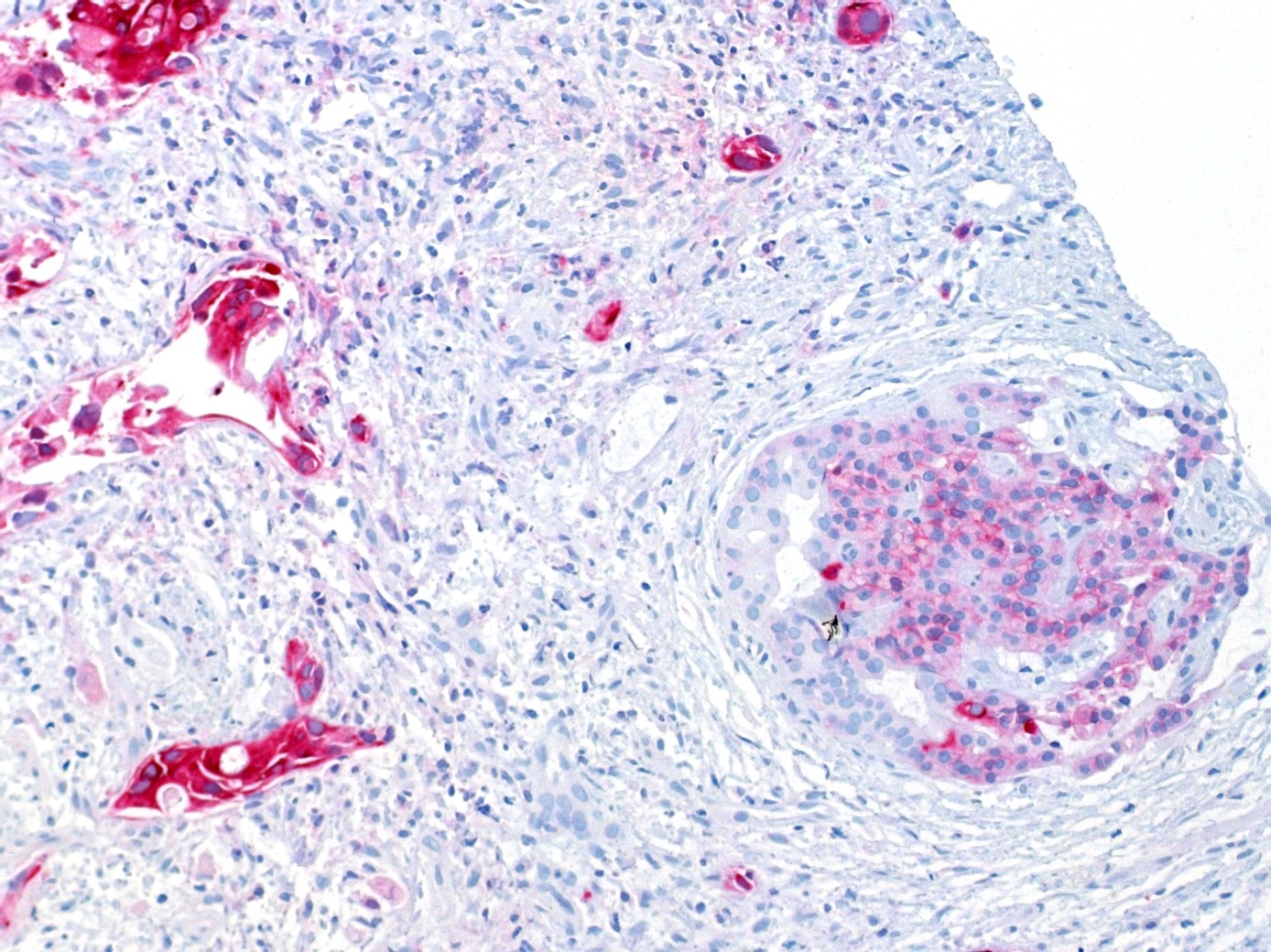
Ductal adenocarcinoma of pancreas and pancreatic isle
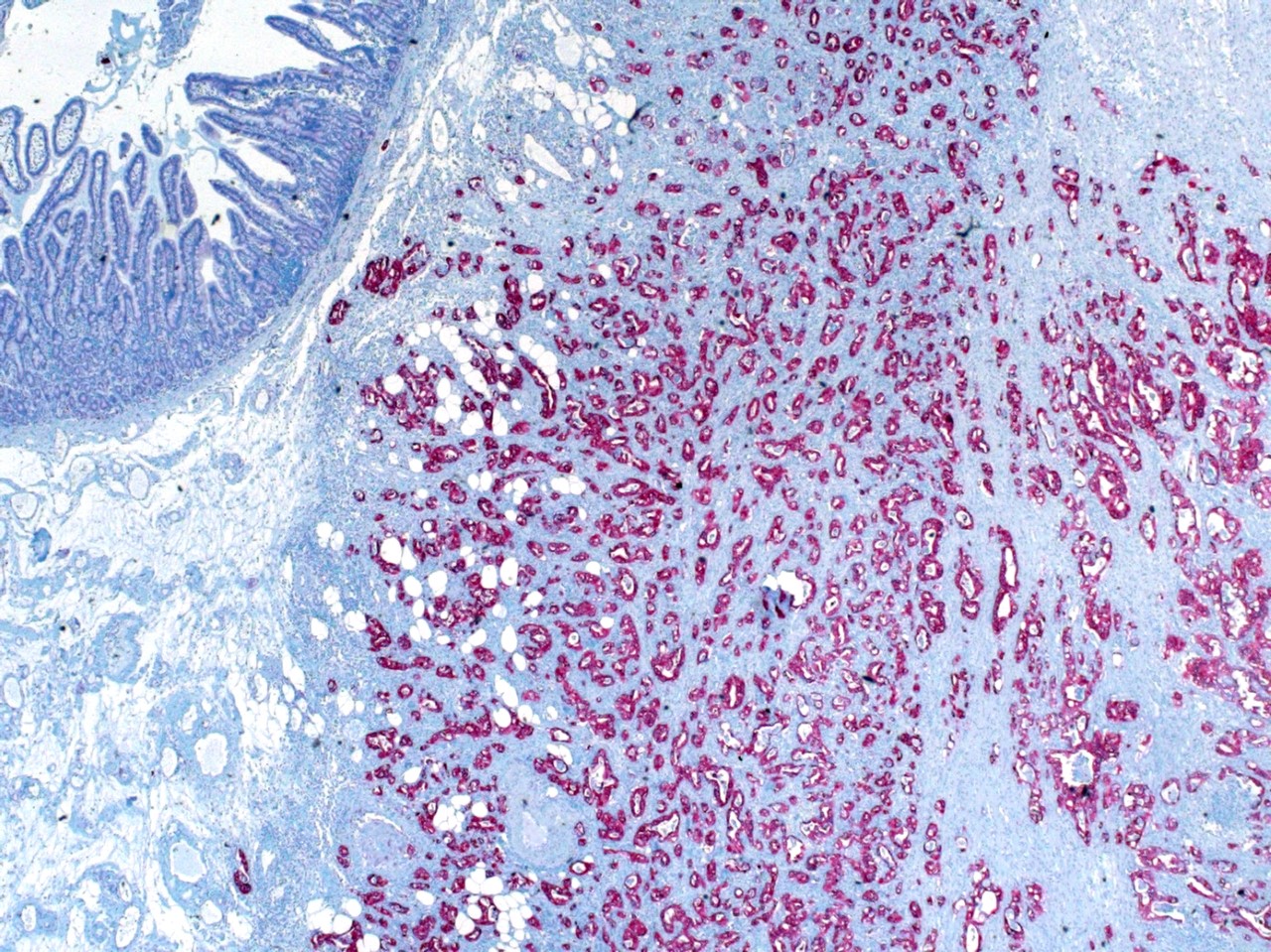
Ductal adenocarcinoma of pancreas and duodenum
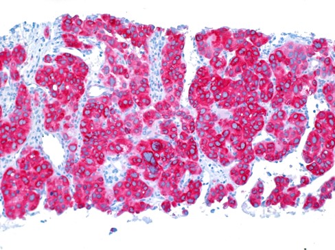
Hepatocellular carcinoma
Positive staining - normal
- TMA study analyzed IMP3 expression in normal tissues (Oncol Rep 2018;39:3)
- Weakly positive: amnion, chorion cells, decidua, thymocytes, crypt cells of rectal mucosa, ciliated cells of bronchial mucosa, secretory cells of cervix, cells of the adenohypophysis of the anterior lobe of your the pituitary gland
- Moderately positive: mesenchymal cells of placenta, mucous cells of submandibular and sublingual and bronchial glands, spermatogonia of testis
- Strongly positive: syncytiotrophoblast, cytotrophoblast, lymph follicles in lymph nodes and tonsils with lymphoblasts, lymphocytes, absorptive cells of ileum, ciliated cells of fallopian tube
Positive staining - disease
- Ductal adenocarcinoma of the pancreas (88.4%) (Am J Surg Pathol 2011;35:873)
- High grade dysplasia of the bile ducts (Hum Pathol 2009;40:1377)
- While using a scoring system including p53 expression, high grade dysplasia (83%) and carcinoma (65%) of the gastric mucosa (Int J Clin Exp Pathol 2014;7:2091)
- Colorectal carcinoma (70.6%) (Hum Pathol 2017;64:137)
- Adenocarcinoma of the ampulla of Vater (87.4%) (Anticancer Res 2019;39:4947)
Negative staining
- Hepatocellular carcinoma (negative 81.6%) (Histopathology 2012;60:278)
- Normal or inflamed exocrine pancreatic tissue (negative 72.3%, weakly positive 23.1%) (Am J Surg Pathol 2011;35:873)
- Normal and nontumorous liver tissue and benign liver tumors (Histopathology 2012;60:278)
- No or weak expression in normal, inflamed and low grade dysplastic bile duct epithelium (Hum Pathol 2009;40:1377)
- While using a scoring system including p53 expression, 96% of nonneoplastic lesions of the gastric mucosa (Int J Clin Exp Pathol 2014;7:2091)
- Normal colonic mucosa and hyperplastic polyps, majority of colonic adenomas (97.2%) (Hum Pathol 2017;64:137)

