
Stains & CD markers
PAS (Periodic acid-Schiff)


Copyright: 2002-2024, PathologyOutlines.com, Inc.
PubMed Search: PAS
PAS (Periodic acid-Schiff)
Authors: Monika Vyas, M.D., Jonathan E. Zuckerman, M.D., Ph.D., Nicole K. Andeen, M.D., Patricia Tsang, M.D., M.B.A.
Editorial Board Member: Christian M. Schürch, M.D., Ph.D.
Last author update: 15 July 2022
Last staff update: 15 July 2022
Copyright: 2002-2024, PathologyOutlines.com, Inc.
PubMed Search: PAS

Table of Contents
Definition / general | Essential features | Terminology | Etiology | Uses by pathologists | Microscopic (histologic) images | Cytology images | Positive staining - normal | Positive staining - disease | Negative staining | Sample pathology report | Board review style question #1 | Board review style answer #1 | Board review style question #2 | Board review style answer #2Cite this page: Vyas M, Zuckerman JE, Andeen NK, Tsang P. PAS (Periodic acid-Schiff). PathologyOutlines.com website. https://www.pathologyoutlines.com/topic/stainsPAS.html. Accessed April 25th, 2024.
Definition / general
- A special stain, not an immunostain
Essential features
- Substances with nearby glycol groups or their amino or alkylamino derivatives are oxidized by periodic acid to form dialdehydes, which combine with Schiff reagent to form an insoluble magenta compound
- Stains basement membrane (normal and in tumors), glycogen, some mucins (see below) and mucopolysaccharides
- PAS stains neutral and acid simple nonsulfated and acid complex sulfated mucins
Terminology
- PASD: PAS with predigestion with diastase
Etiology
- Substances with nearby glycol groups or their amino or alkylamino derivatives are oxidized by periodic acid to form dialdehydes, which combine with Schiff reagent to form an insoluble magenta compound
- Used for formalin fixed tissue and enzyme cytochemistry; can be used for frozen sections with modifications (Eur J Gynaecol Oncol 1998;19:482, Am J Surg Pathol 1992;16:87)
- Stains basement membrane (normal and in tumors), glycogen, some mucins (see below) and mucopolysaccharides
- Routine stain in brain (with Luxol fast blue), cornea, kidney, liver (glycogen stains strongly for PAS without diastase) and skeletal muscle specimens for nontumor pathology
- Some mucins (see below) are PASD (PAS with predigestion with diastase) positive (i.e., stain is present after diastase predigestion; also called diastase resistant); glycogen is PASD negative (also called diastase sensitive because diastase removes PAS staining)
- PAS stains neutral and acid simple nonsulfated and acid complex sulfated mucins
- PAS does not stain acid simple mesenchymal mucins and acid complex connective tissue mucins
- Also stains various inclusions, bodies, granules and secretions composed of mucopolysaccharides or mucins
Uses by pathologists
- Breast cytology: PASD positive cells with internal structure and producing nuclear indentation, particularly in dissociated or atypical cells, correlate with malignant histology (J Clin Pathol 2001;54:146)
- Microorganisms:
- Stains polysaccharide laden fungal cell walls
- PAS+ granule at anterior end of mature spores is diagnostic of microsporidia (BMC Clin Pathol 2006;6:6)
- Stains Whipple disease: foamy macrophages containing PAS+ (PASD+) intracytoplasmic granules (Tropheryma whippelii bacteria)
- PAS+ (PASD+) bacteria include Bacillus cereus, Corynebacterium diphtheriae, Propionibacterium acnes, Klebsiella pneumoniae and Micrococcus luteus (Arch Dermatol 1991;127:543)
- Hematopathology (Arch Pathol Lab Med 1991;115:346, Haematologica 2002;87:148):
- Stains lymphoblasts in acute lymphoblastic leukemia (ALL)
- Most types of acute myeloid leukemia (AML) are negative for PAS but exceptions are pronormoblasts in pure erythroid leukemia, M5a, M6 (60%), M7
- Stains Pautrier microabscesses in mycosis fungoides
- Kidney:
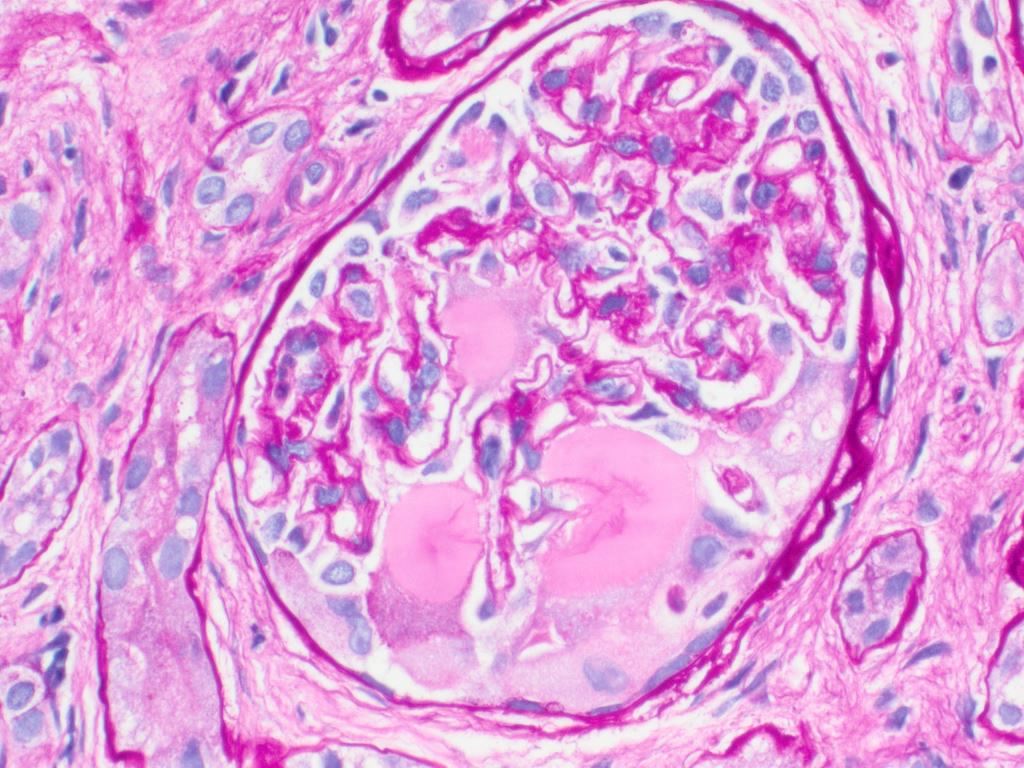
- Recommended for routine evaluation of medical renal biopsies due to basement membrane staining
- Particularly helpful for distinguishing tubulitis (in allograft), diabetic Kimmelstiel-Wilson nodules (PAS positive), amyloid (PAS negative) and atypical casts in light chain cast nephropathy (PAS negative)
- Liver:
- Routine stain for hepatocytes (PAS without diastase)
- Glycogenic hepatopathy (PAS+ diastase sensitive)
- Differential diagnosis of eosinophilic inclusions in liver
- Alpha-1 antitrypsin (PAS+ diastase resistant)
- Polyglucosan-like inclusions (PAS+ diastase sensitive) (Lafora disease, cyanamide poisoning, medication) (Gastroenterology 2006;131:713)
- Type IV glycogenosis (PAS+ diastase sensitive) (Mol Genet Metab Rep 2020;24:100601)
- Cystic fibrosis (PAS+ diastase sensitive) (Am J Clin Pathol 2016;146:22)
- Parenteral nutrition (PAS+ diastase sensitive) (Pediatr Dev Pathol 2009;12:79)
- Lung: stains amorphous or granular globules in bronchoalveolar lavage (BAL) fluid in pulmonary alveolar proteinosis (J Clin Pathol 1997;50:981)
- Muscle biopsies: routine stain to demonstrate glycogen
- Pancreas: acinar cell carcinoma (PASD+)
- Parotid glands: zymogen granules are PAS+
- Prostate: Cowper glands are PASD+ (Am J Surg Pathol 1997;21:550)
- Skin: eosinophilic globoid bodies (Kamino bodies) in Spitz nevus are PASD+
- Esophagus:
- Glycogenic acanthosis (PAS+ diastase sensitive)
- Candida esophagitis
- Small intestine:
- Stains Whipple disease bacteria (Am J Clin Pathol 2002;118:742, Hum Pathol 2003;34:589)
- Strong cytoplasmic staining present in microvillous inclusion disease versus linear brush border staining in normals (Am J Surg Pathol 2002;26:902)
- Testis: stains germ cell neoplasia in situ (GCNIS) and seminoma (PAS+, PASD negative) but not normal seminiferous tubules (Am J Surg Pathol 1994;18:947)
- Tumors: adenocarcinoma of various sites (mucin is PASD+), alveolar soft parts sarcoma (PASD+ crystalline structures), apocrine carcinomas, basement membrane containing tumors (cylindroma, eccrine spiradenoma), clear cell tumors (stains glycogen), glycogen rich carcinomas, glycogen rich / balloon cell melanoma, granular cell tumor (cytoplasmic granules), hyaline globules in renal tumors, mucinous tumors, Paget disease of breast (Am J Surg Pathol 2001;25:823, Arch Pathol Lab Med 1998;122:353, Hum Pathol 1997;28:400)
- Other: stains malakoplakia
- Enzyme cytochemistry: coarse granular staining
Microscopic (histologic) images
Contributed by Andrey Bychkov, M.D., Ph.D.
Thyroid:

Graves disease
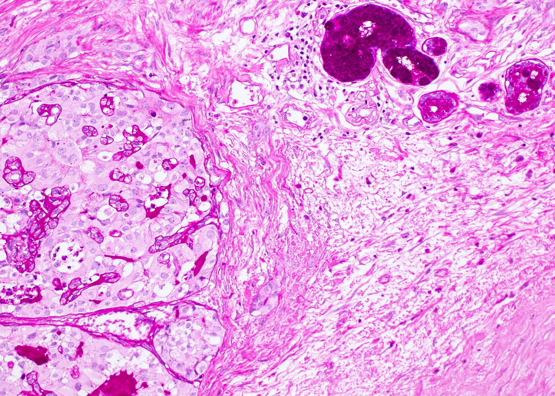
PASD staining
of PTC with
laryngeal invasion
PAS staining of thyroid
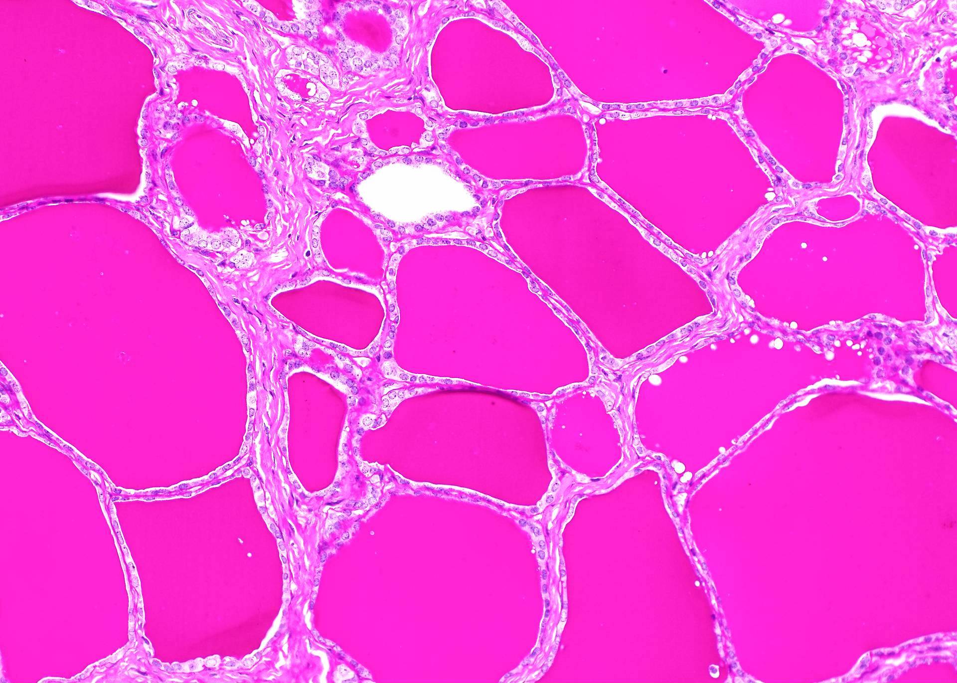
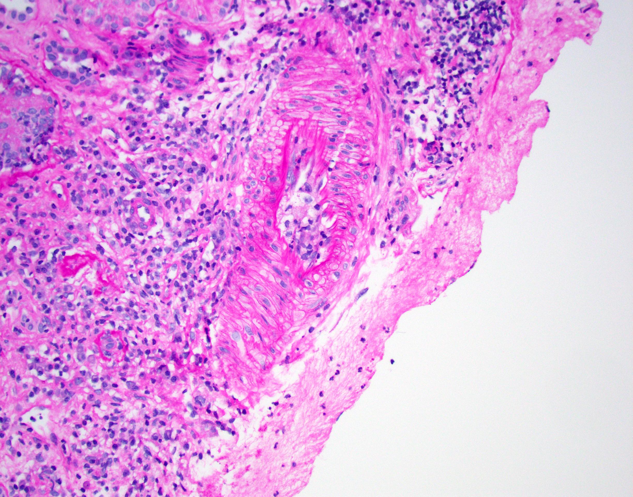
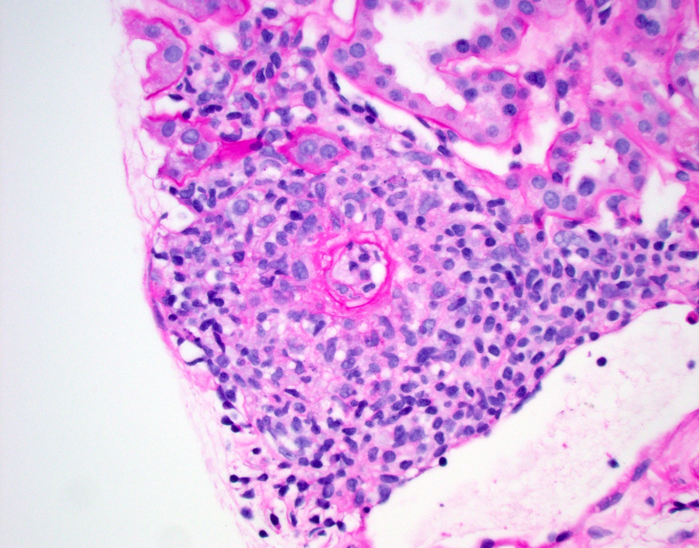
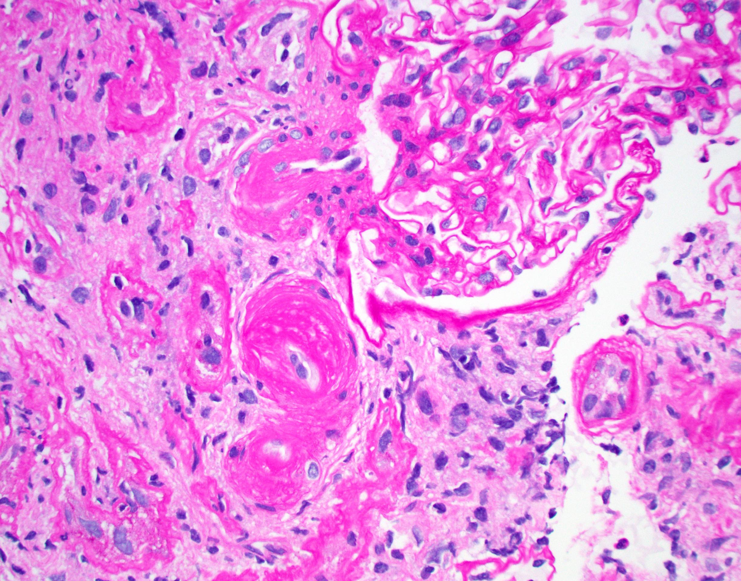
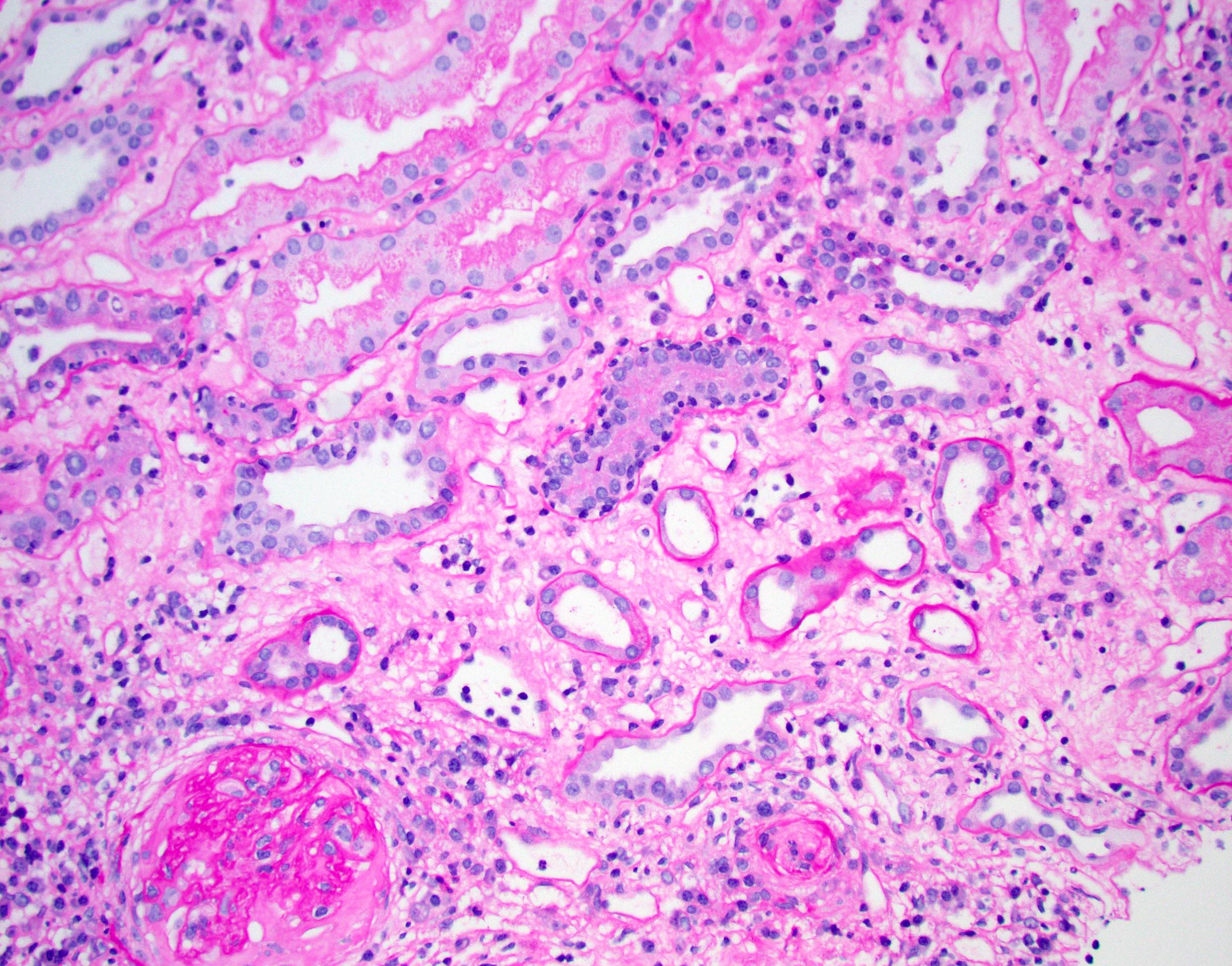
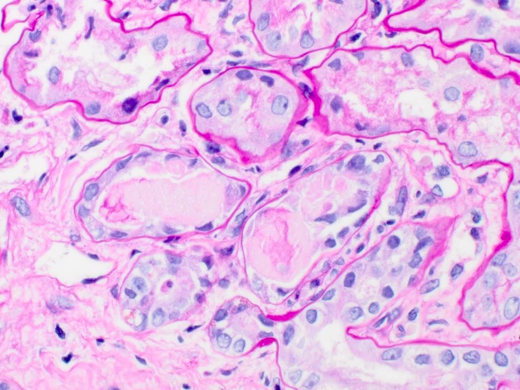
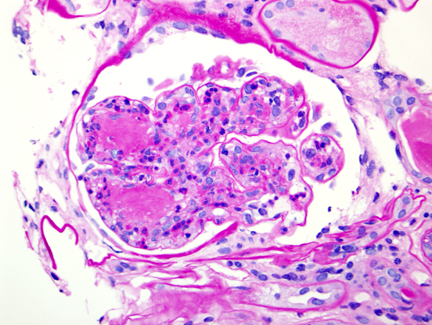
Contributed by Jonathan E. Zuckerman, M.D., Ph.D., Nicole K. Andeen, M.D. and cases #30 and #51
Kidney:
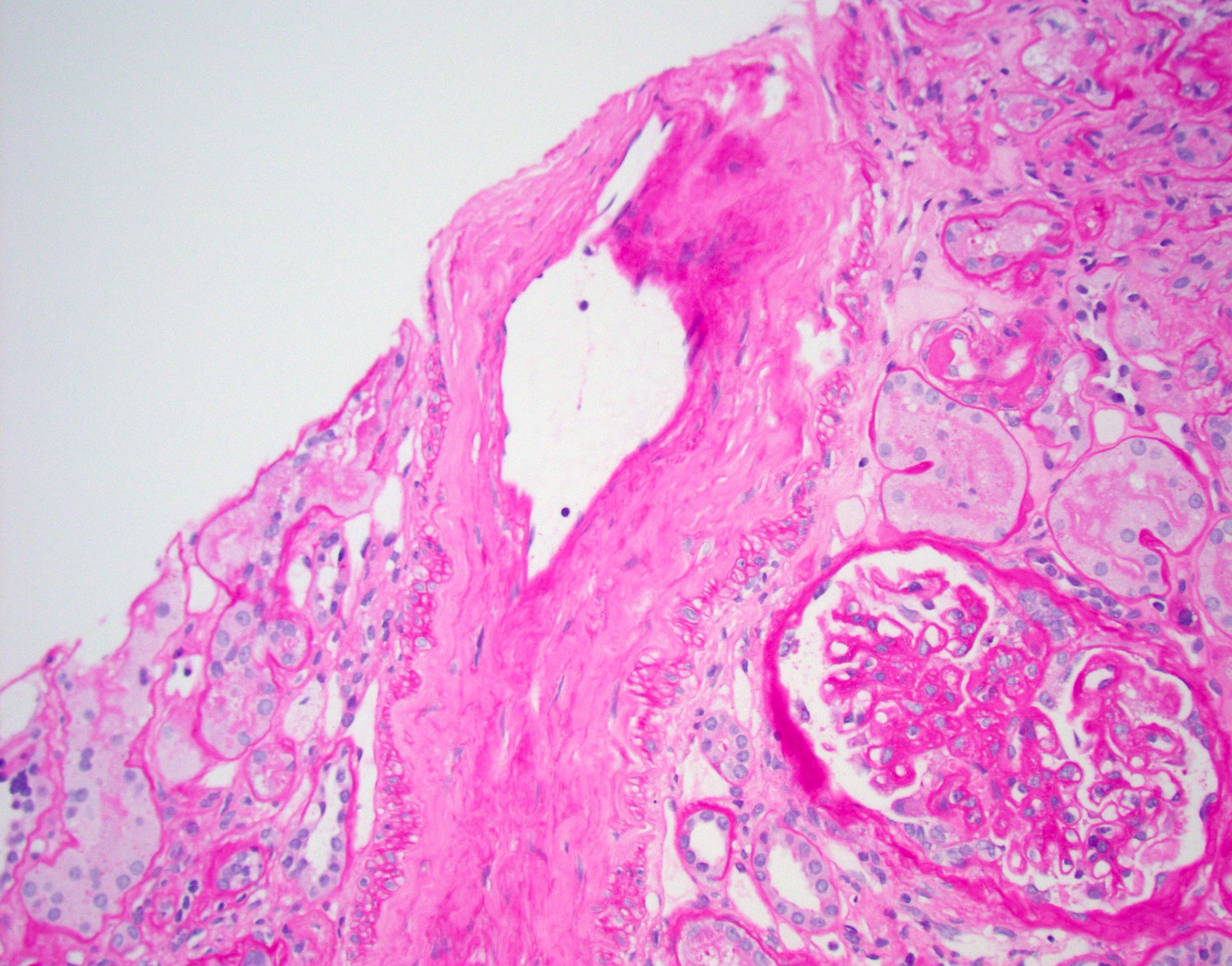
Arteriosclerosis
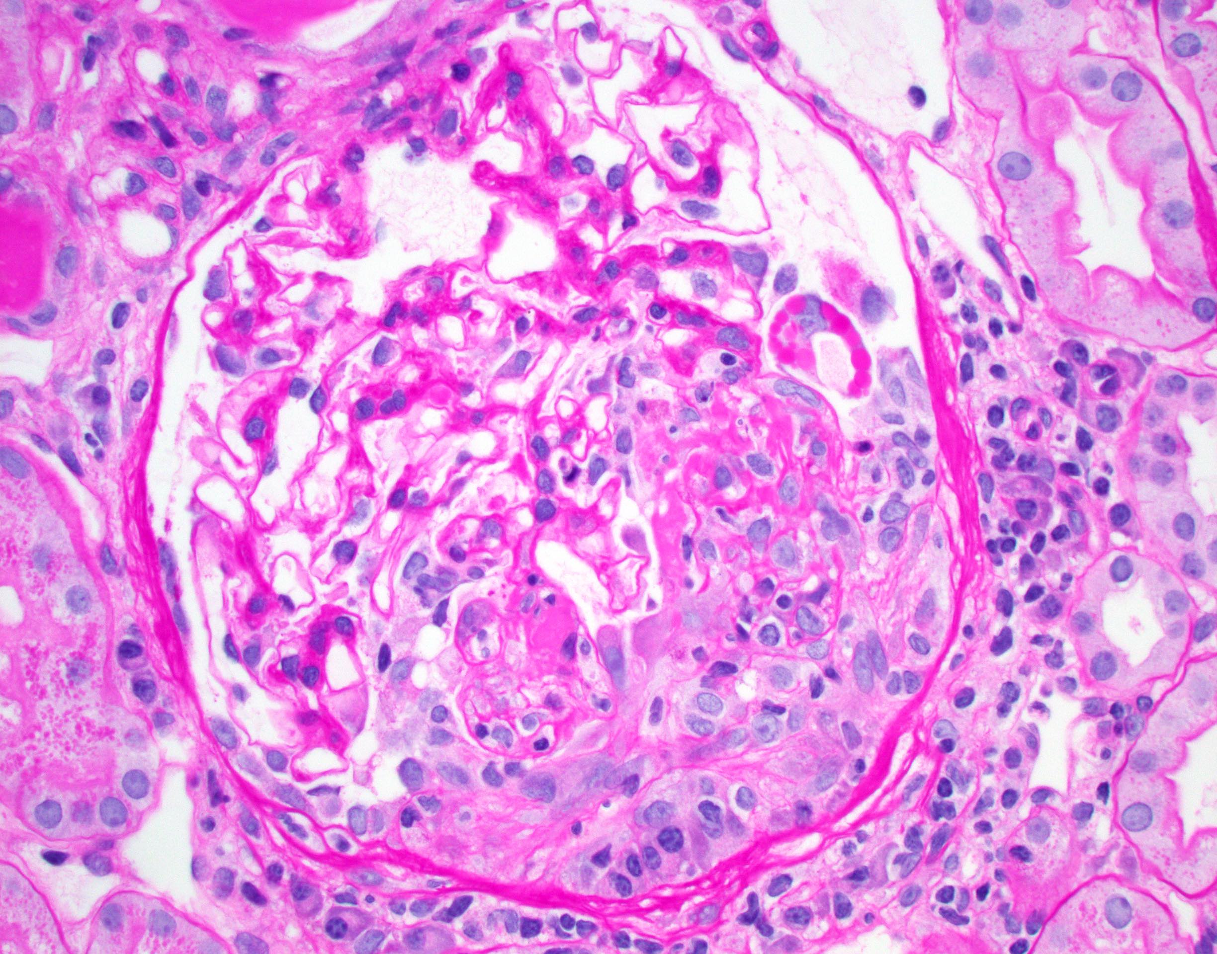
Cellular crescent
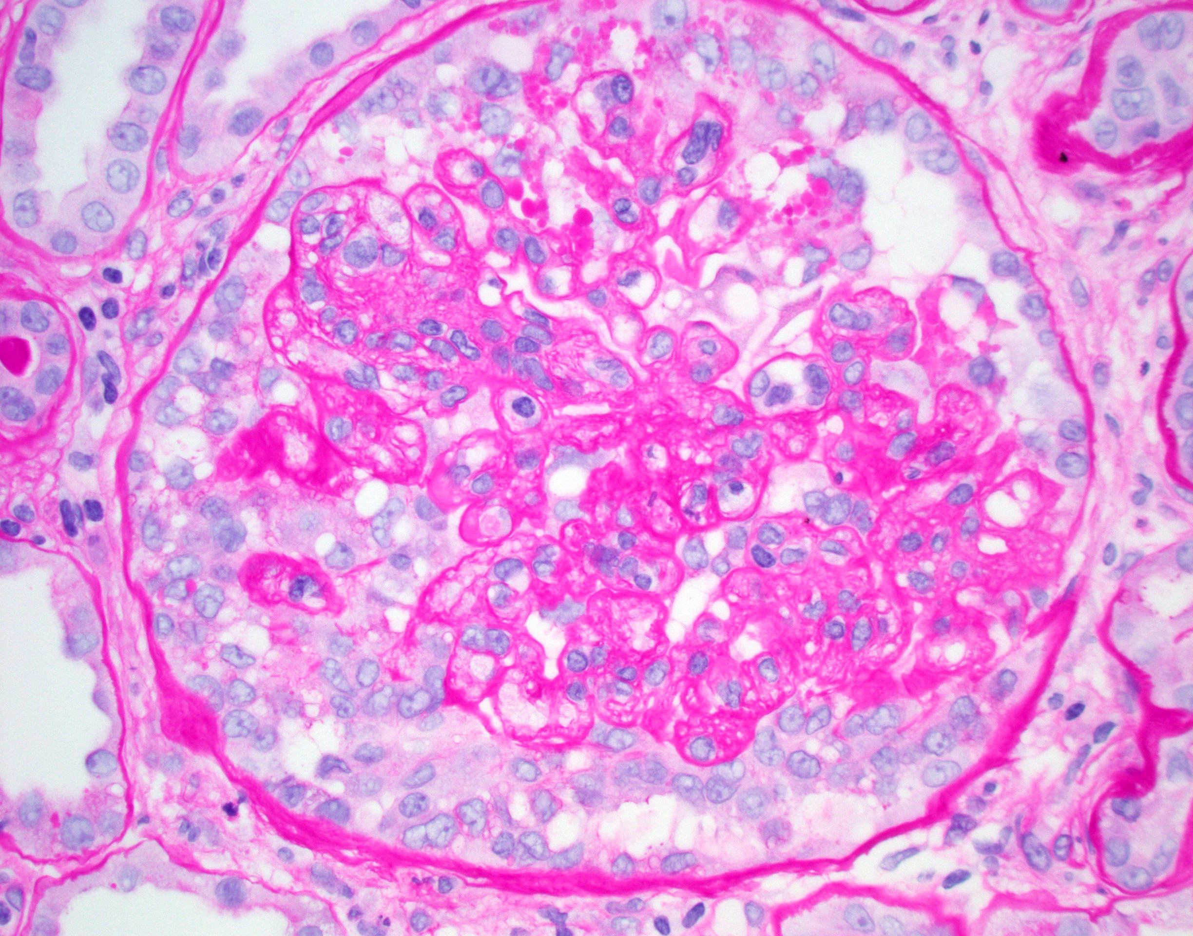
Collapsing glomerulopathy
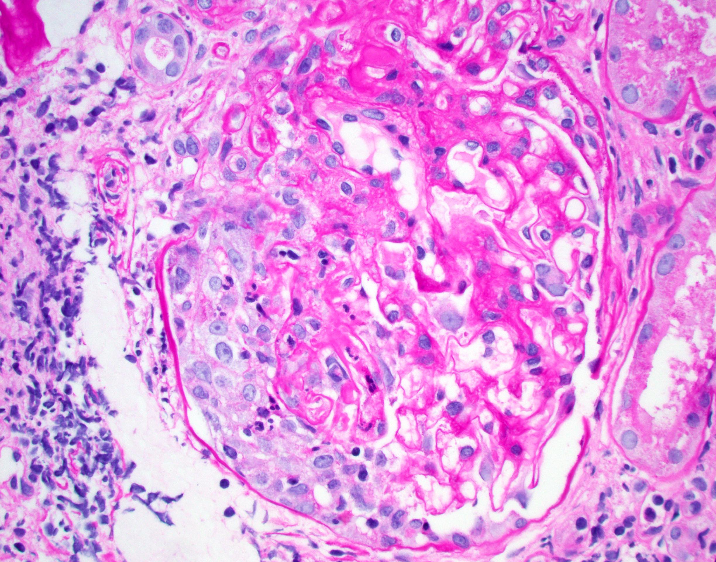
Crescent
Endotheliitis
Necrotizing arteriolitis

PAS- light chain casts
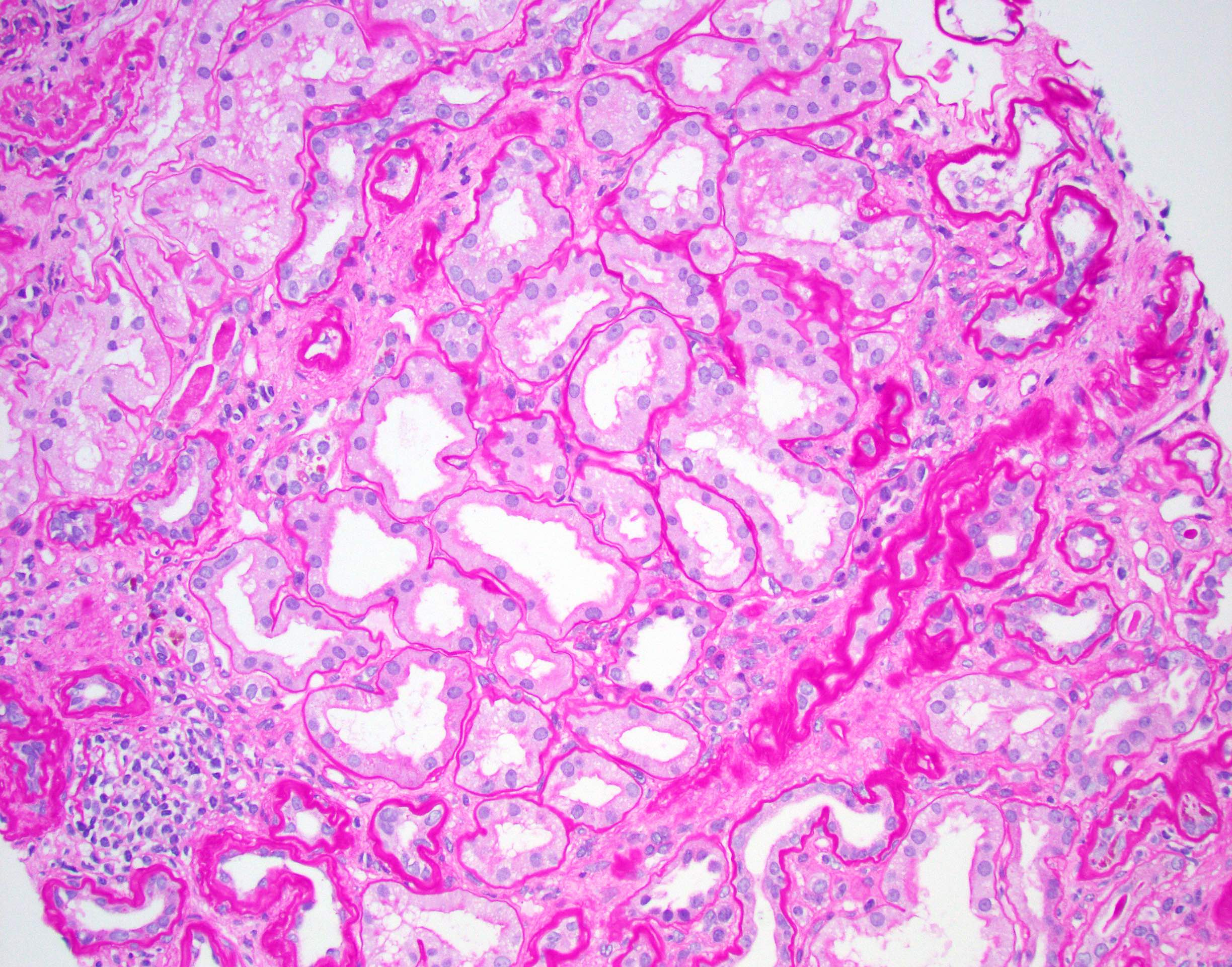
PAS+ thickened tubular basement membranes
PAS+ arteriolar hyalinosis
Tubulitis
PAS- amyloid in glomerulus
PAS- casts of light chain cast nephropathy
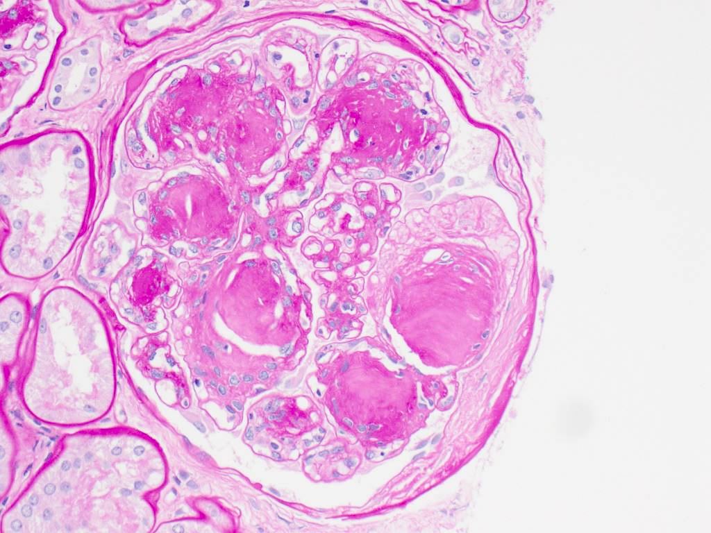
PAS+ nodular
mesangial expansion
in diabetic
nephropathy
Nodular
glomerulosclerosis
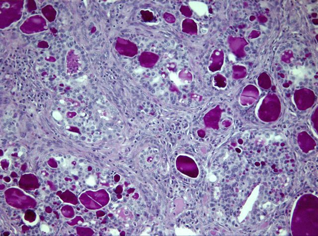
Tubulocystic carcinoma, kidney (tumor)
Cytology images
Contributed by Andrey Bychkov, M.D., Ph.D. and Patricia Tsang, M.D., M.B.A.
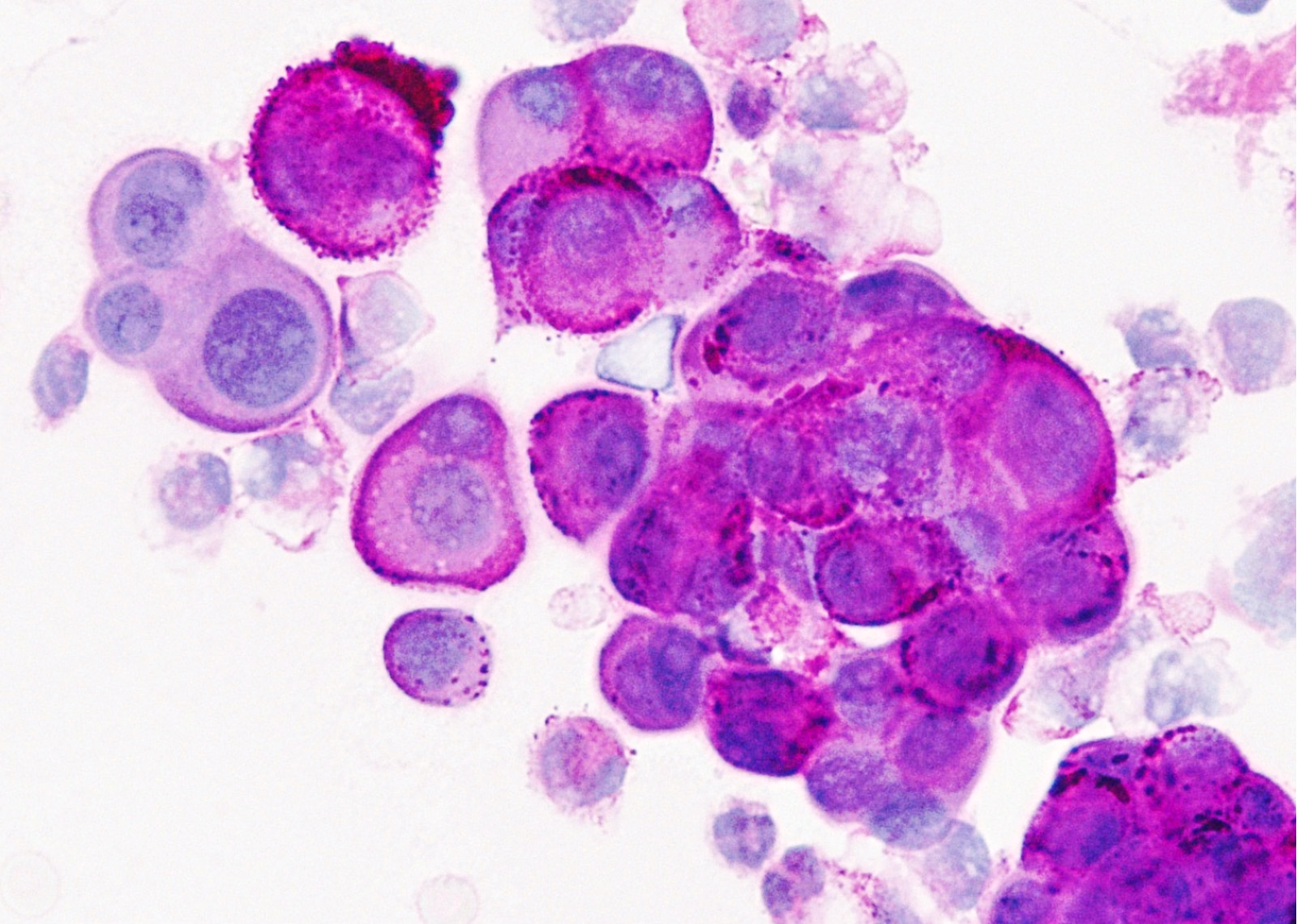
Glycogen granules
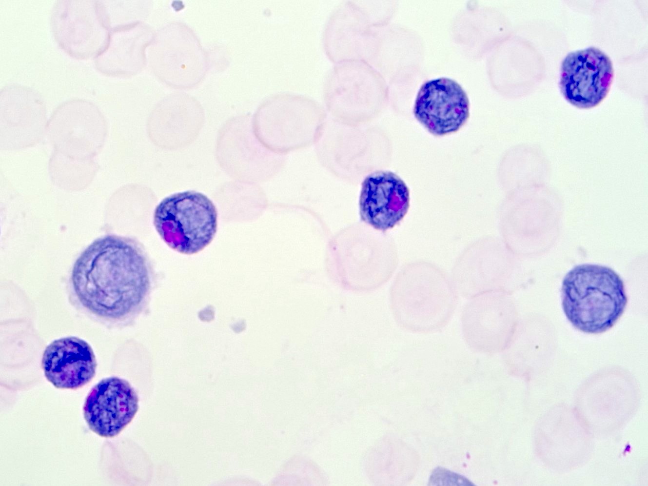
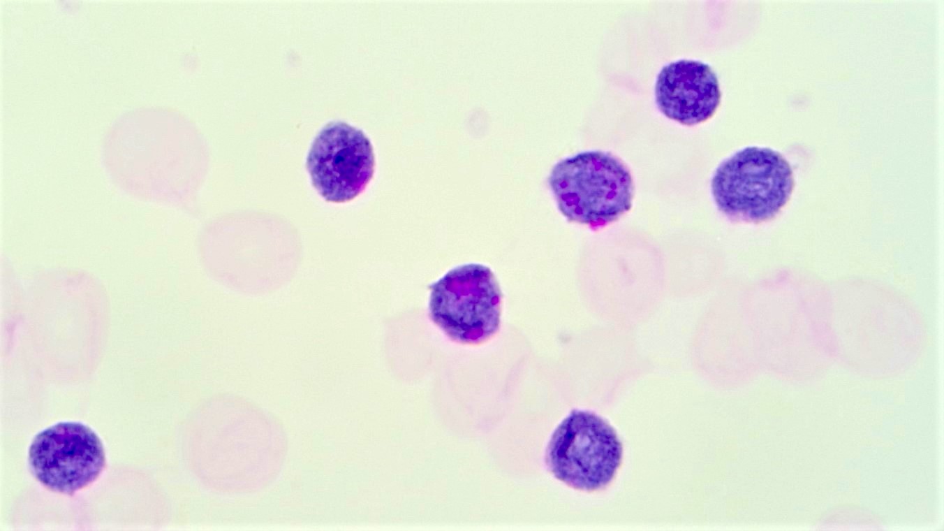
B lymphoblastic leukemia
Positive staining - normal
- Basement membrane, fungi, glycogen (removed after diastase or amylase predigestion), mucins (neutral and acid simple nonsulfated and acid complex sulfated types), surfactant
Positive staining - disease
- Acute lymphoblastic leukemia (75%, block staining), alpha-1-antitrypsin inclusions, alveolar soft part sarcoma (intracytoplasmic crystals), acute myeloid leukemia M5a, M6 (60%), M7, basement membrane containing tumors (cylindroma), clear cell tumors, malakoplakia, renal cell carcinoma (PAS+ glycogen removed with diastase), parasites, signet ring cell carcinoma of stomach, Whipple disease (J Histochem Cytochem 2006;54:615, Exp Mol Pathol 2014;96:274)
Negative staining
- Mucins (acid simple mesenchymal and acid complex connective tissue types)
Sample pathology report
- Left kidney, needle biopsy:
- Changes consistent with diabetic nephropathy, including PAS+ afferent and efferent arteriolar hyalinosis
Board review style question #1
Which of the following is typical of PAS staining?
- Stains acid simple mesenchymal mucins
- Stains basement membranes
- Stains glycogen after diastase or amylase predigestion
- Stains seminiferous tubules
Board review style answer #1
B. Stains basement membranes. PAS stains glycogen but not after diastase or amylase predigestion. It stains only some mucins - neutral and acid simple nonsulfated and acid complex sulfated types but not acid simple mesenchymal and acid complex connective tissue types. It does not typically stain normal seminiferous tubules.
Comment Here
Reference: PAS (Periodic acid-Schiff)
Comment Here
Reference: PAS (Periodic acid-Schiff)
Board review style question #2
Which of the following statements about PAS staining in this patient with diabetes is true?
- Highlights amyloid in blood vessels
- Highlights atypical casts in light chain cast nephropathy
- Highlights Kimmelstiel-Wilson nodules
- Recommended for routine evaluation of medical renal biopsies due to nuclear staining
Board review style answer #2
C. Highlights Kimmelstiel-Wilson nodules. PAS staining is particularly helpful for distinguishing tubulitis (in allograft), diabetic Kimmelstiel-Wilson nodules (PAS positive), amyloid (PAS negative) and atypical casts in light chain cast nephropathy (PAS negative). It is a standard stain for medical renal biopsies but it is a membranous stain and not a nuclear stain.
Comment Here
Reference: PAS (Periodic acid-Schiff)
Comment Here
Reference: PAS (Periodic acid-Schiff)

