
Stains & CD markers
Cytokeratin 5/6 and CK5


Copyright: 2019-2024, PathologyOutlines.com, Inc.
PubMed Search: CK5/6
Cytokeratin 5/6 and CK5
Editorial Board Member: Brandon Umphress, M.D.
Editor-in-Chief: Debra L. Zynger, M.D.
Last author update: 9 June 2023
Last staff update: 9 June 2023
Copyright: 2019-2024, PathologyOutlines.com, Inc.
PubMed Search: CK5/6

Table of Contents
Definition / general | Essential features | Terminology | Pathophysiology | Clinical features | Interpretation | Uses by pathologists | Prognostic factors | Microscopic (histologic) images | Positive staining - normal | Positive staining - disease | Negative staining | Sample pathology report | Board review style question #1 | Board review style answer #1 | Board review style question #2 | Board review style answer #2Cite this page: Terlević R, Vranić S. Cytokeratin 5/6 and CK5. PathologyOutlines.com website. https://www.pathologyoutlines.com/topic/stainsck5and6.html. Accessed April 25th, 2024.
Definition / general
- Basic (type II) cytokeratins of molecular weight 58 kDa (CK5) and 56 kDa (CK6) (Cell 1982;31:11)
- Common antibodies are directed against both cytokeratin 5 and 6
- Major partner of CK5 is CK14, while CK6 pairs with CK16/17 (Exp Cell Res 2007;313:2244, Eur J Cell Biol 2004;83:735)
- CK5 is also detected by antibodies against high molecular weight cytokeratins (such as 34 beta E12, also known as K903) and pan-CK markers (such as CK AE1 / AE3)
Essential features
- Stains basal cells of prostate and basal / myoepithelial cells of breast (can be used to rule out invasion)
- Useful for detecting benign breast proliferations (mosaic-like pattern) versus ductal carcinoma in situ (DCIS) (negative or rarely diffusely positive) (Pathology 2009;41:68)
- Together with p63, used to detect squamous cell origin in poorly differentiated carcinomas (Am J Clin Pathol 2001;116:823)
Terminology
- K5, K6, keratin 5, keratin 6, basal cytokeratins, intermediate molecular weight cytokeratins
Pathophysiology
- Cytoskeletal constituents of the intermediate filament family of proteins, which provide the cell with a structural framework stretching from around the nucleus to desmosomes and hemidesmosomes (Science 1998;279:514)
- In stratified epithelia, CK5 preferentially stains basal layer, while CK6 stains suprabasal layer (Virchows Arch 2022;480:433)
- CK5 and its partner CK14 constitute up to 25% of total cell protein in basal cells of epidermis; expression in skin correlates with mitotic activity (Science 1998;279:514, J Biol Chem 1995;270:21362, Development 1994;120:2369)
- CK6 is expressed in wound healing and hyperproliferative cancer cells (J Biol Chem 1995;270:21362)
- CK5/6 expression may be useful in subclassifying epidermolysis bullosa cases (Appl Immunohistochem Mol Morphol 2018;26:586)
- CK5 is a marker of breast luminal progenitor and stem cells (Breast Cancer Res Treat 2011;128:45)
- CK5 positive cells may represent a chemoresistant population in breast and ovarian cancer (Breast Cancer Res Treat 2011;128:45, Int J Gynecol Cancer 2015;25:1565)
Clinical features
- CK5 and CK14 mutations may cause epidermolysis bullosa simplex and Dowling-Degos disease (Hum Mutat 2006;27:719, Am J Hum Genet 2006;78:510)
- Important in tooth enamel formation (J Biol Chem 2003;278:20293)
- CK6 mutations involved in pachyonychia congenita (Nat Genet 1995;10:363)
Interpretation
- Diffuse cytoplasmic staining with perinuclear enhancement
Uses by pathologists
- Identify breast basal / myoepithelial cells and prostate basal cells
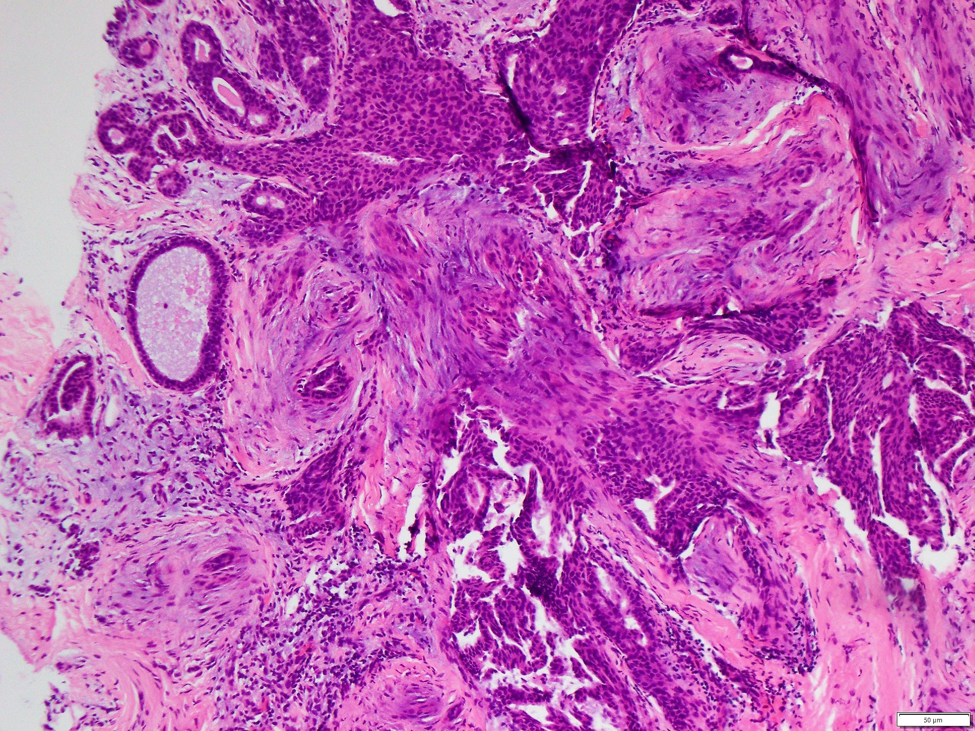
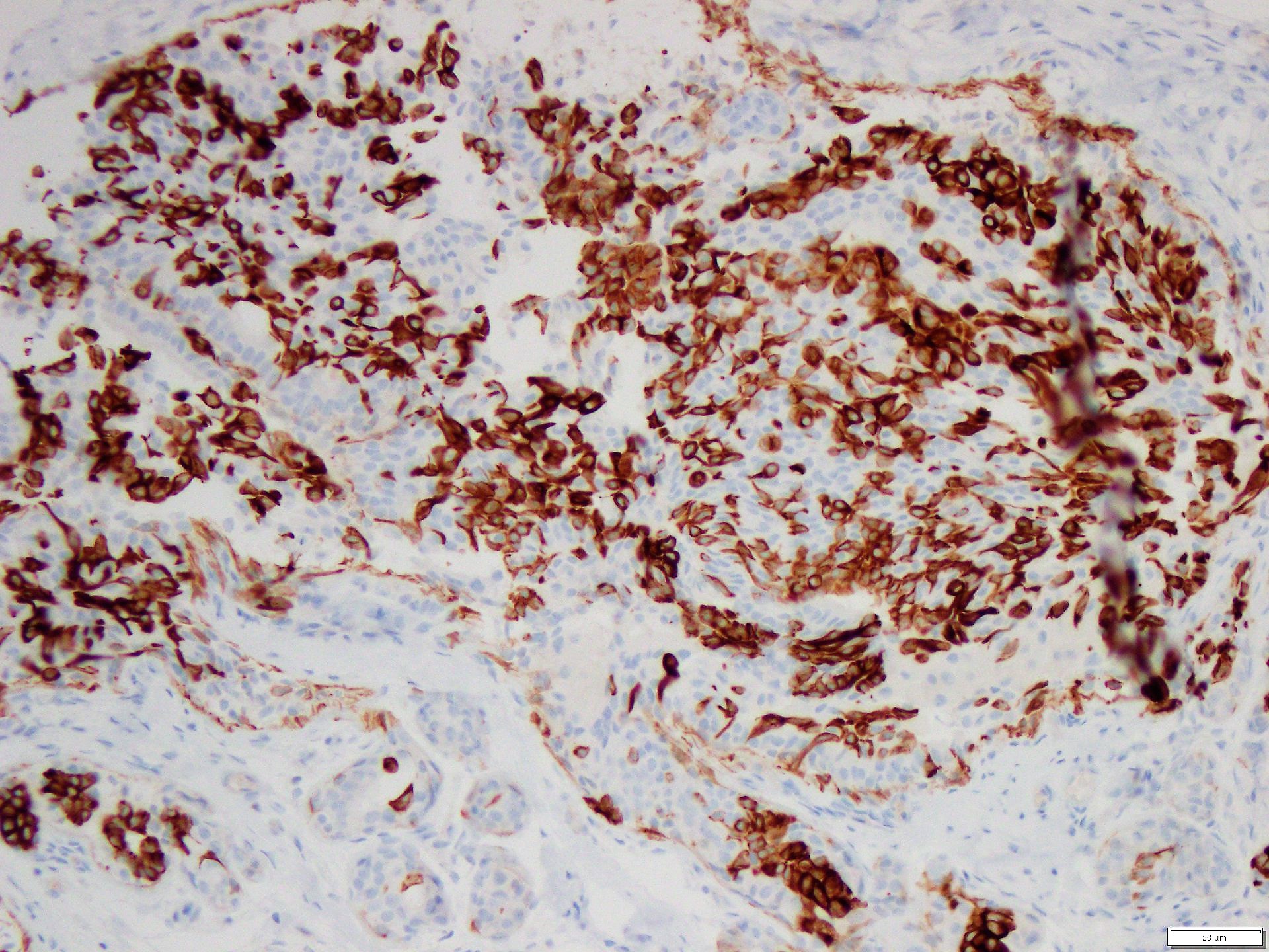
- Distinguish breast usual ductal hyperplasia (UDH) and papillary lesions (mosaic-like pattern) from DCIS (usually negative, rarely diffusely positive) (Pathology 2009;41:68)
- Distinguish epithelioid mesothelioma (CK5/6+ in 83%) from lung adenocarcinoma (CK5/6- in 85%), including in pleural effusions (CK5/6+ in 90% mesothelioma, 0% adenocarcinoma) (Histopathology 2006;48:223, Diagn Cytopathol 2006;34:801)
- Distinguish cutaneous spindle squamous cell carcinoma (CK5/6+ in 100%) from superficial epithelioid sarcoma (rare focal positivity) (J Cutan Pathol 2003;30:114)
- Together with p63+, identify squamous origin in poorly differentiated metastatic carcinomas (e.g., cervical) (Am J Clin Pathol 2001;116:823)
- CK5 preferred to K903 in antibody cocktails for the detection of prostate carcinoma (Diagn Pathol 2012;7:81)
- As part of panel to discriminate between undifferentiated sinonasal carcinoma (CK5/6-) and other poorly differentiated head and neck tumors (poorly differentiated squamous cell carcinoma, olfactory neuroblastoma and nasopharyngeal carcinoma) (Ann Diagn Pathol 2014;18:261)
- CK5 used as part of panel to distinguish cutaneous metastasis of breast cancer from sweat gland carcinoma (Arch Pathol Lab Med 2011;135:975)
- CK5/6 full thickness positivity specific for bladder condyloma acuminatum versus basal / patchy staining in papillary noninvasive urothelial carcinoma (Int J Surg Pathol 2022;30:260)
Prognostic factors
- CK5/6+ / p63+ breast intraductal papillomas show lower risk of subsequent invasive carcinoma (Pathol Int 2015;65:81)
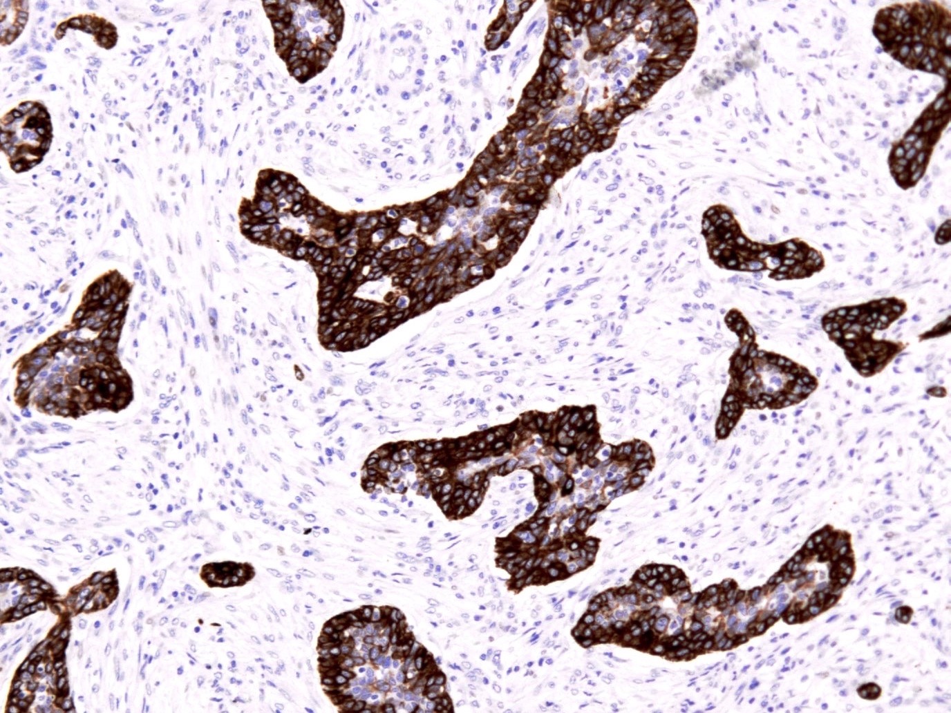
- Diffuse positivity in DCIS defines basal-like subtype with poorer prognosis (Hum Pathol 2007;38:197)
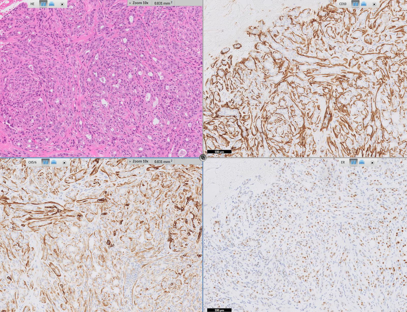
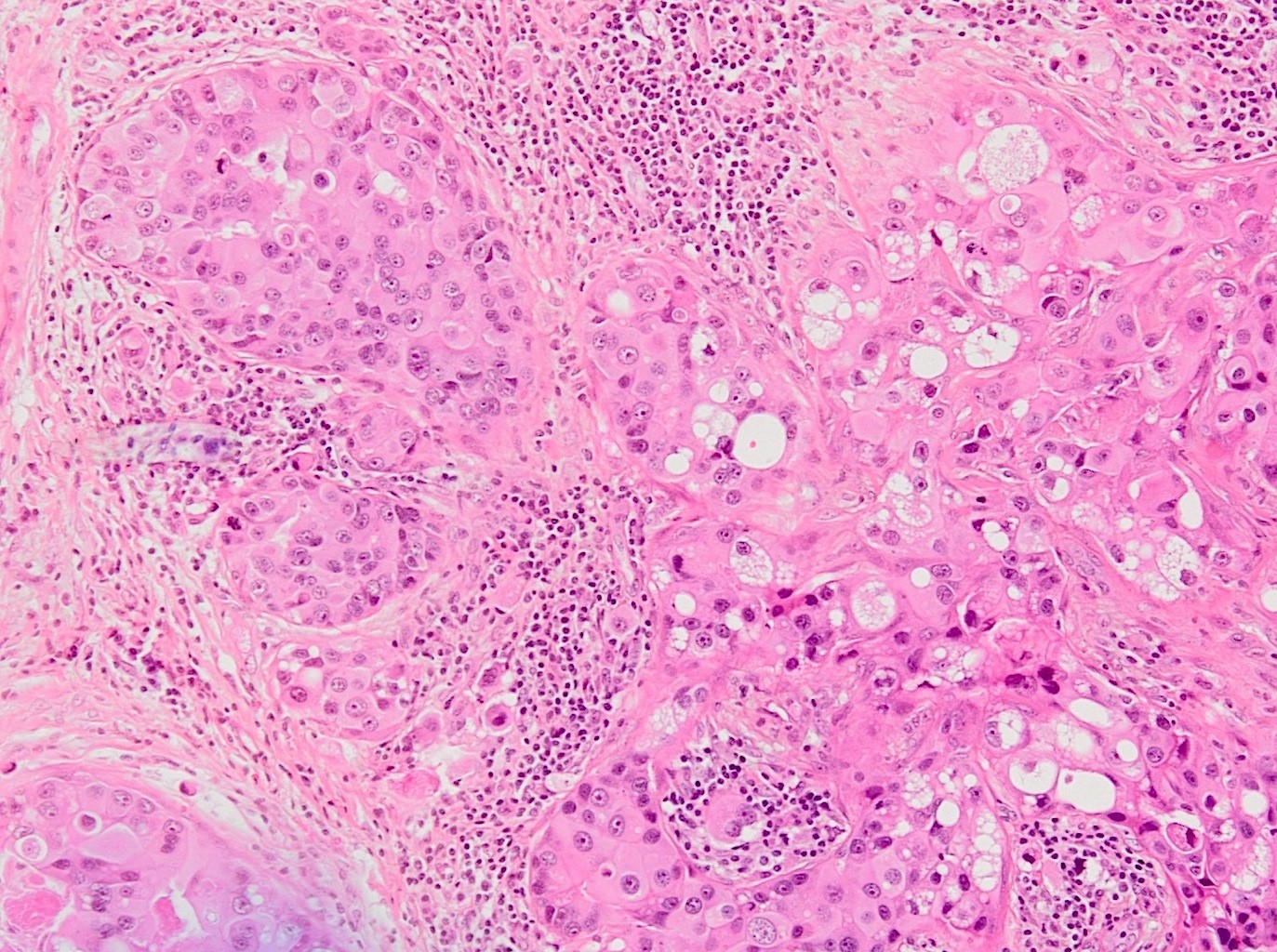
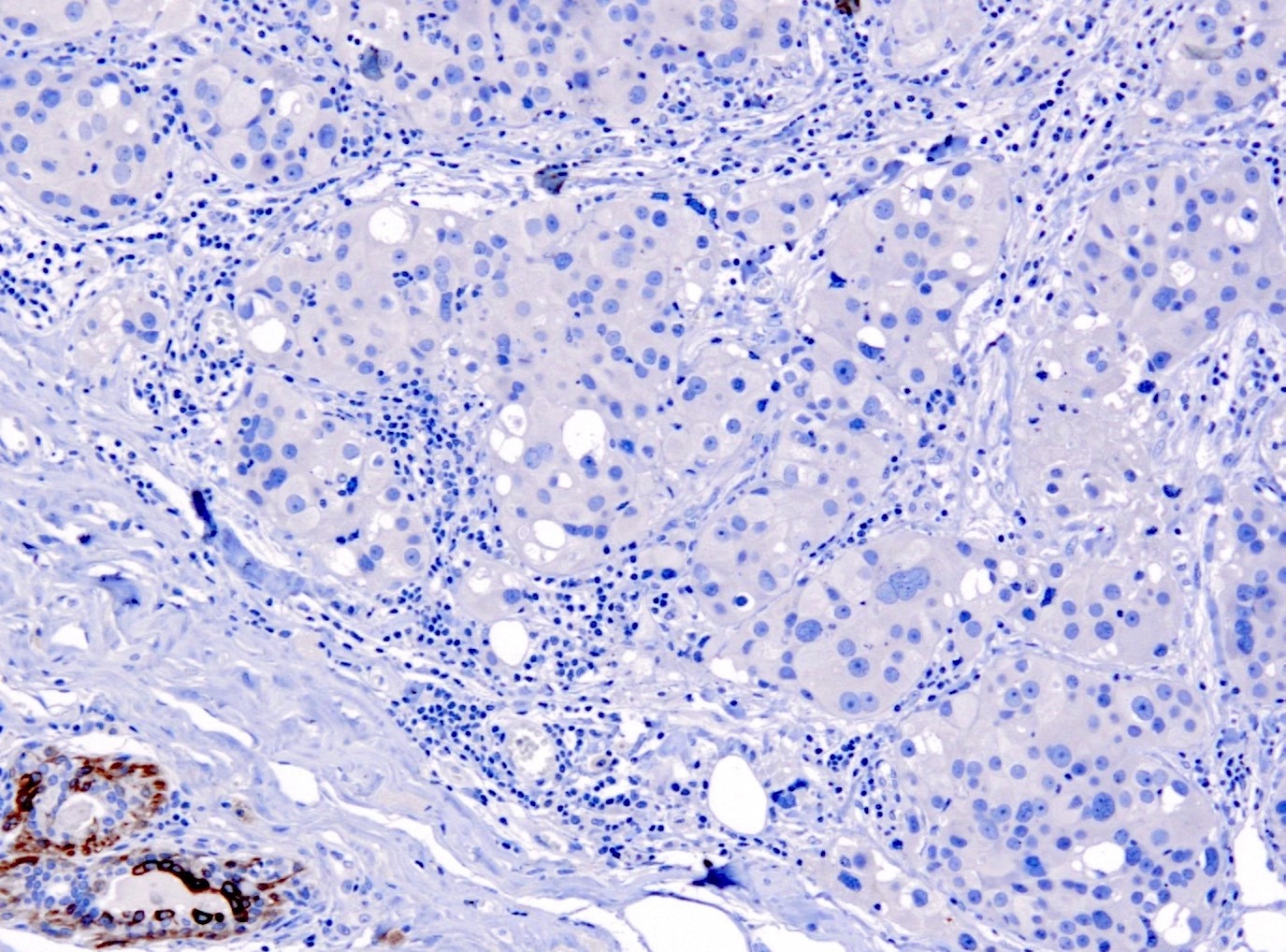
- CK5/6 positivity helps define a basal-like subtype of triple negative breast carcinoma (TNBC) with poorer prognosis; associated with premenopausal African American women, BRCA1 mutations and higher propensity for brain metastases (Transl Cancer Res 2021;10:1193, Clin Cancer Res 2006;12:1533, JAMA 2006;295:2492, Mod Pathol 2005;18:1321, J Natl Cancer Inst 2003;95:1482, Am J Surg Pathol 2006;30:1097)
- CK5/6+ muscle invasive urothelial carcinoma associated with poor survival (Int J Mol Sci 2021;22:628)
- CK5+ together with CD44+ defines invasive and noninvasive basal-like urothelial carcinoma subtype associated with more aggressive behavior (Pathol Res Pract 2015;211:610, Appl Immunohistochem Mol Morphol 2016;24:575, Sci Rep 2021;11:21186)
- CK5/6-, CD44-, CK20+ nonmuscle invasive papillary upper tract urothelial carcinoma associated with worse outcome (Histopathology 2019;74:483)
Microscopic (histologic) images
Contributed by Jijgee Munkhdelger, M.D., Ph.D., Andrey Bychkov, M.D., Ph.D. and Semir Vranić, M.D., Ph.D.
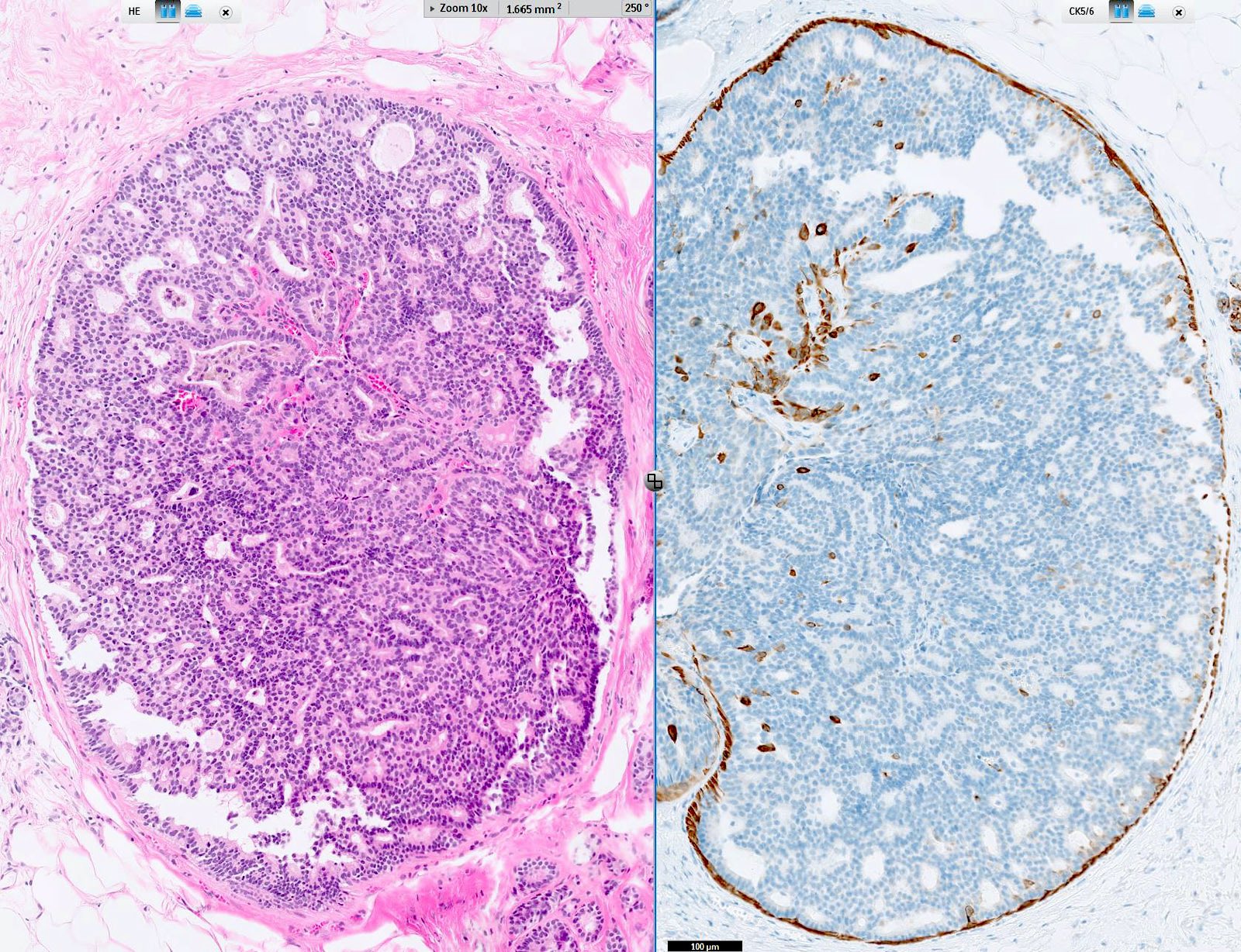
Breast tubular adenoma immunoprofile
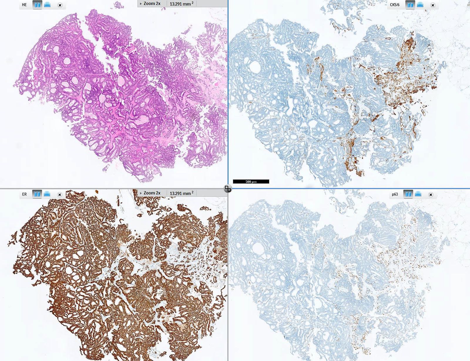
Intraductal papilloma with DCIS immunoprofile
Intraductal papilloma with DCIS, CK5/6
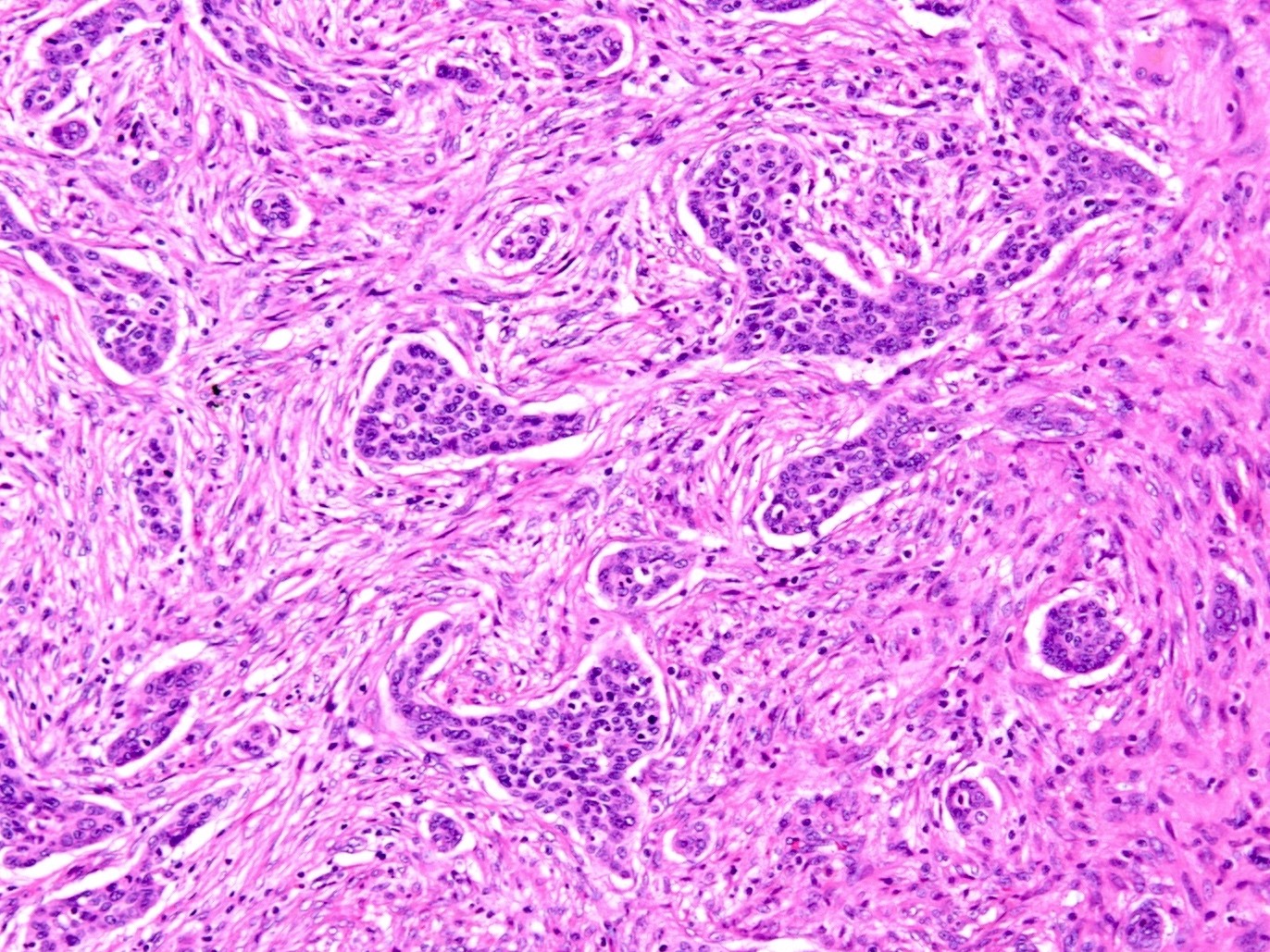
Basal-like triple negative breast cancer

Apocrine metaplasia of the breast
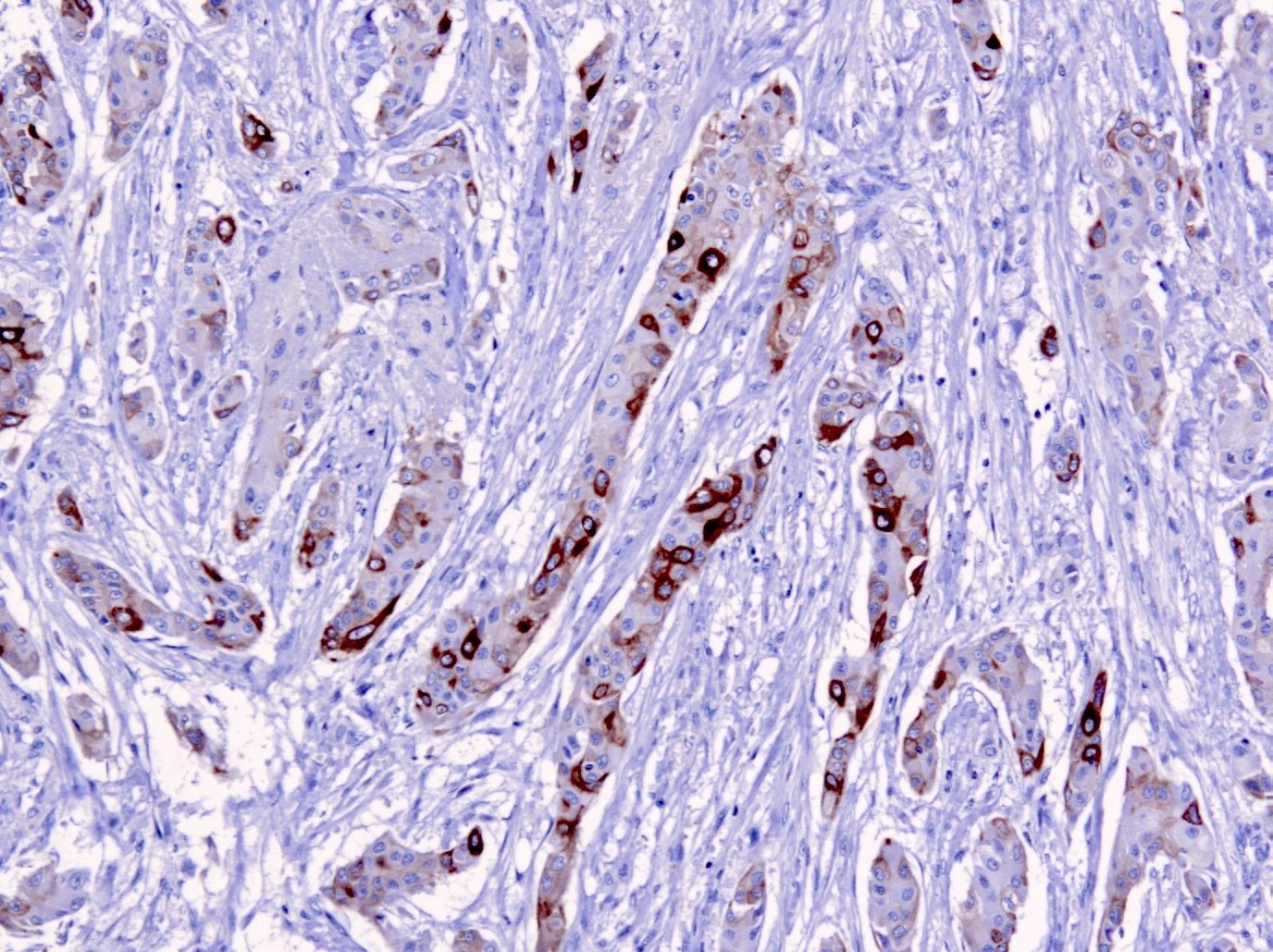
Metastatic squamous cell carcinoma of the cervix
Apocrine carcinoma of the breast
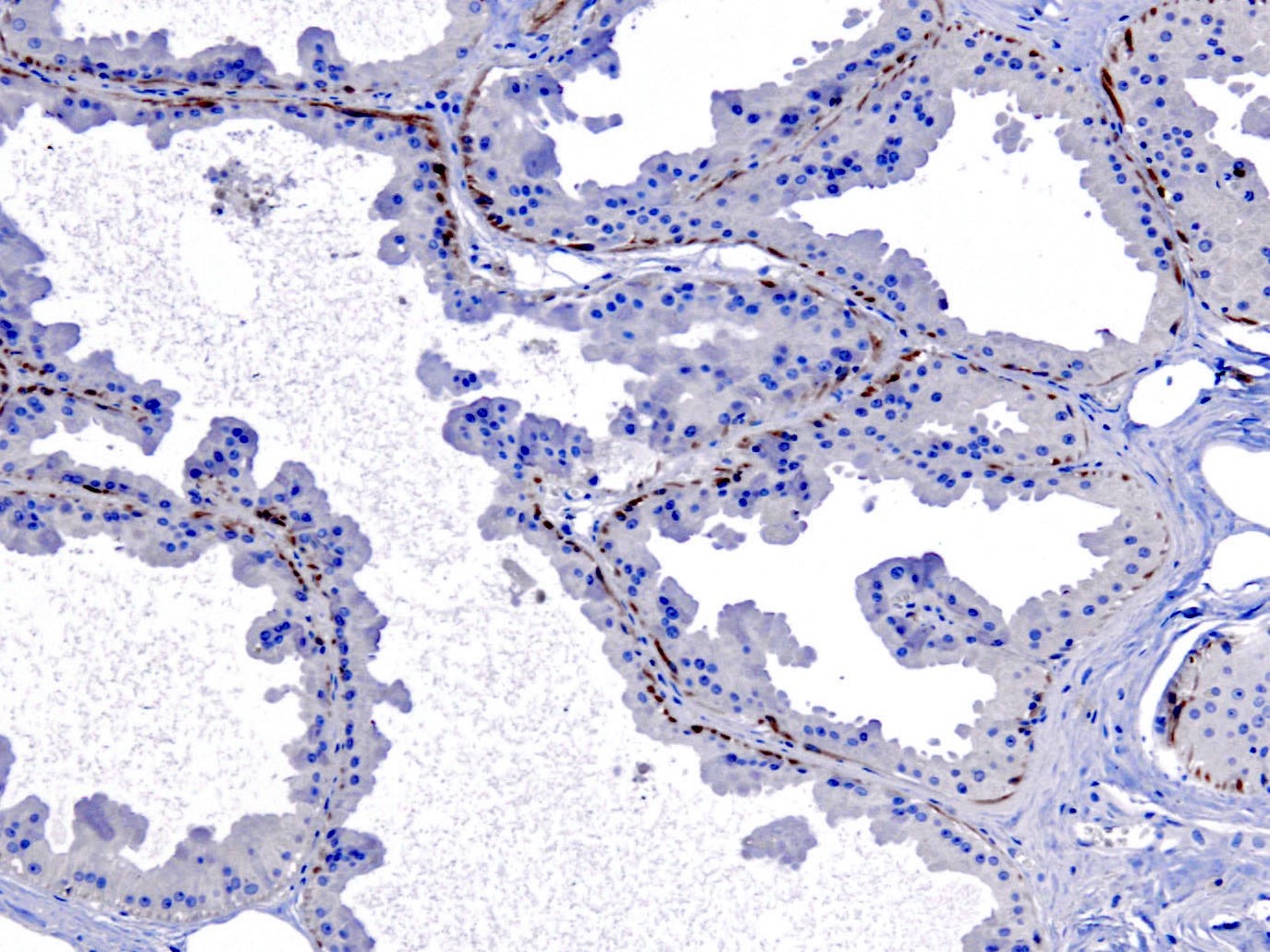
Complex sclerosing lesion of the breast
Positive staining - normal
- CK5 is the major keratin of basal cells in the epidermis, while CK6 preferentially stains the suprabasal layer (Exp Cell Res 2007;313:2244, Virchows Arch 2022;480:433)
- Also expressed in basal cells of respiratory epithelium, male genital tract (efferent ducts, epididymis, deferent duct and seminal vesicle), breast basal / myoepithelial cells, mesothelium, prostate basal cells, cornea, endocervical squamous metaplasia, spermatogenic cells, basal cells of urinary tract transitional epithelium (J Pathol 2001;195:563, Ann N Y Acad Sci 1985;455:282, Mol Reprod Dev 2002;61:1, J Histochem Cytochem 1985;33:415, Mol Cell Proteomics 2002;1:269)
- CK5 stains thyroid solid cell nests (Endocr Pathol 2016;27:83)
- CK5 and CK6 are both expressed in palmar and plantar epidermis; basal cells of eccrine and apocrine glands; hair follicle; myoepithelial cells of salivary gland acini; basal cells of excretory ducts; mucosal stratified epithelia of the oral cavity, esophagus, female genital tract and glans penis; thymus epithelial cells (J Biol Chem 1995;270:21362, Ann N Y Acad Sci 1985;455:282)
Positive staining - disease
- Tumors derived from stratified epithelia (skin and mucosa) are positive (Mod Pathol 2002;15:6)
- Some adenocarcinomas may show focal staining (pancreas, breast, lung, uterine endometrioid, cholangiocarcinoma and hepatocellular carcinoma, lung) (Mod Pathol 2002;15:6, Hum Pathol 2019;84:221)
- Poorly differentiated squamous cell carcinoma identified by both CK5/6+ and p63+ with 77% sensitivity and 96% specificity (Am J Clin Pathol 2001;116:823)
- Urothelial carcinoma (63%) (Mod Pathol 2002;15:6)
- Breast usual ductal hyperplasia and papillary lesions (mosaic-like pattern) (Pathology 2009;41:68)
- Basal-like breast DCIS (Hum Pathol 2007;38:197)
- In breast and salivary gland adenoid cystic carcinoma, CK5/6 stains cells lining true lumina (Virchows Arch 2016;469:213, Int J Clin Exp Pathol 2011;4:336)
- Metaplastic breast cancer (87%) (Appl Immunohistochem Mol Morphol 2021;29:265)
- High grade serous ovarian carcinoma (focal or diffuse pattern in 69%) (Hum Pathol 2017;67:30)
- Epithelioid mesothelioma (CK5/6+ in 83%) (Histopathology 2006;48:223)
- Stains both glandular and myoepithelial components of salivary gland tumors (Mod Pathol 2002;15:6)
- Thymoma (100%, 8/8 cases) (Mod Pathol 2002;15:6)
- Nasopharyngeal carcinoma (90%) and poorly differentiated squamous cell carcinoma of the nasal tract (100%) (Ann Diagn Pathol 2014;18:261)
- Cutaneous adnexal neoplasms (97%) (Am J Dermatopathol 2004;26:447)
- Primary cutaneous SMARCB1 deficient carcinoma (100%) (J Cutan Pathol 2021;48:1051)
- Amyloid deposits in cutaneous macular amyloidosis (50%) (Cureus 2020;12:e7606)
- Adenocarcinoma of the rete testis (80%) (Am J Surg Pathol 2019;43:670)
Negative staining
- Adenocarcinoma (colon, stomach), renal cell carcinoma, germ cell tumors, thyroid carcinomas, neuroendocrine carcinoma (lung, skin and gastrointestinal), epithelioid sarcoma, synovial sarcoma (Mod Pathol 2002;15:6)
- Sinonasal undifferentiated carcinoma (Ann Diagn Pathol 2014;18:261)
- SMARCA4 deficient sinonasal carcinoma (0%) (Am J Surg Pathol 2020;44:703)
- Well differentiated neuroendocrine tumors of skin (0%) (J Cutan Pathol 2017;44:557)
Sample pathology report
- Breast, core biopsy:
- Benign breast tissue with usual ductal hyperplasia (see comment)
- Comment: Sections show mildly distended breast gland lumina filled with a mixed population of cells showing a mosaic pattern expression of CK5/6, consistent with usual ductal hyperplasia.
Board review style question #1
- Which of the following is an important use of the CK5/6 immunostain?
- Confirm a diagnosis of epithelioid sarcoma
- Confirm a diagnosis of well differentiated neuroendocrine tumors of skin
- Identify a basal-like subtype of triple negative breast carcinoma (TNBC) with poorer prognosis
- Identify sinonasal undifferentiated carcinoma
Board review style answer #1
C. Identify a basal-like subtype of triple negative breast carcinoma with poorer prognosis (shown in the image above). Answers A, B and D are incorrect because CK5/6 is typically negative in epithelioid sarcoma, well differentiated neuroendocrine tumors of skin and sinonasal undifferentiated carcinoma.
Comment Here
Reference: Cytokeratin 5/6 and CK5
Comment Here
Reference: Cytokeratin 5/6 and CK5
Board review style question #2
- What is the typical immunohistochemical staining pattern in usual ductal hyperplasia?
- Diffuse CK5 negativity
- Diffuse CK5 positivity
- Diffuse ER negativity
- Diffuse ER positivity
- Mosaic pattern CK5
Board review style answer #2
E. Mosaic pattern CK5. Mosaic pattern of CK5 refers to partial staining of the intraductal myoepithelial cell population. Answers A, B, C and D are incorrect because diffusely negative or positive CK5 or ER would indicate a clonal (neoplastic epithelial) cell population. Rarely, a neoplastic cell population can be CK5 positive, therefore use of a panel (CK5, p63, ER) is usually recommended.
Comment Here
Reference: Cytokeratin 5/6 and CK5
Comment Here
Reference: Cytokeratin 5/6 and CK5

