

Table of Contents
Definition / general | Essential features | Pathophysiology | Clinical features | Interpretation | Uses by pathologists | Microscopic (histologic) images | Positive staining - normal | Positive staining - tumors | Negative staining - tumors | Molecular / cytogenetics description | Board review style question #1 | Board review style answer #1 | Board review style question #2 | Board review style answer #2Cite this page: Alikhan M. SDHB (succinate dehydrogenase). PathologyOutlines.com website. https://www.pathologyoutlines.com/topic/stainsdhb.html. Accessed April 18th, 2024.
Definition / general
- Succinate dehydrogenase (SDH) is a mitochondrial enzyme complex involved in the Krebs (or tricarboxylic acid) cycle and the electron transport chain, essential for normal aerobic respiration (Nat Rev Cancer 2011;11:325)
- Comprised of four subunits: SDHA, SDHB, SDHC and SDHD and an assembly factor, SDHAF2
- Bi-allelic inactivation of any of the five units results in instability of SDHB, cytoplasmic transport and rapid degradation
- Loss of SDHB may be indicative of syndromic disease; this is either with germline inactivation of any subunit or SDHC hypermethylation (epimutation); the latter is most often observed in a subset of gastrointestinal stromal tumors
- Immunohistochemistry for SDHB is a reliable and inexpensive surrogate for assessment of syndromic loss of succinate dehydrogenase (Lancet Oncol 2009;10:764)
- Loss of SDHB is seen in about 15% of pheochromocytomas / paragangliomas, 3% of gastrointestinal stromal tumors, < 1% of renal cell carcinomas and < 1% of pituitary adenomas (Histopathology 2018;72:106)
Essential features
- Succinate dehydrogenase is a mitochondrial enzyme of the tricarboxylic acid (Krebs) cycle and electron transport chain
- Inactivating mutations leads to increased HIF1-α levels and this promotes tumorigenesis, angiogenesis and inhibition of apoptosis
- Loss of succinate dehydrogenase is seen in a subset of pheochromocytomas / paragangliomas, gastrointestinal stromal tumors, renal cell carcinomas and rarely pituitary adenomas; succinate dehydrogenase deficient gastrointestinal stromal tumors may be associated with the Carney dyad or triad
- Loss of any succinate dehydrogenase subunit leads to degradation of SDHB, which can be demonstrated by immunohistochemistry
- It is recommended that all pheochromocytomas / paragangliomas be assessed for loss of SDHB; gastrointestinal stromal tumors with epithelioid morphology and positive staining for KIT and DOG1 are suspicious for succinate dehydrogenase deficiency and should also be assessed (Histopathology 2018 Jan;72:106)
Pathophysiology
- Succinate dehydrogenase facilitates conversion of succinate to fumarate by oxidation through flavin adenine dinucleotide
- Occurs at the SDHA subunit
- Resultant FADH2 molecule transfers electrons through the other subunits and eventually to ubiquinone
- Abnormal succinate dehydrogenase function leads to accumulation of succinate
- Increase inhibits an enzyme (prolyl hydroxylase) involved in the deactivation of hypoxia
inducible factor, HIF1-α (Cancer Cell 2005;8:155)
- Increased hypoxia inducible factor is involved in many cellular processes that lead to tumorigenesis such as vascular endothelial growth factor and erythropoietin (Cancer Cell 2005;7:77)
- Inhibition of prolyl hydroxylase may also inhibit apoptosis of paraganglioma cells
- Increase inhibits an enzyme (prolyl hydroxylase) involved in the deactivation of hypoxia
inducible factor, HIF1-α (Cancer Cell 2005;8:155)
Clinical features
- Germline mutations in any of the subunits are associated with a subset of paragangliomas, renal cell carcinomas or gastrointestinal stromal tumors
- Familial pheochromocytoma paraganglioma syndrome and familial gastrointestinal stromal tumors are a feature of SDH mutations (Clin Cancer Res 2017;23:e68)
- Some studies suggest SDH mutations may be associated with pathogenesis of other tumors such as neuroblastoma (Cold Spring Harb Mol Case Stud 2018;4:a002584)
- Hypermethylation of SDHC is associated with gastrointestinal stromal tumors and is the molecular signature for Carney triad (gastrointestinal stromal tumors, paraganglioma and pulmonary chondroma)
- Hypermethylation leads to subsequent inactivation of the SDHC subunit, which causes SDHB to localize to the cytoplasm, where it is rapidly degraded; this causes loss of the SDHB protein and negative immunohistochemical staining
- Succinate dehydrogenase deficient tumors are more likely to be multicentric and metastasize; succinate dehydrogenase deficient gastrointestinal stromal tumors are nonmutated for KIT and PDGFRA and thus do not respond to tyrosine kinase inhibitors; they often show poor response to chemotherapy as well (J Clin Pathol 2018;71:95)
- Rare clinical syndromes are also associated with SDHA mutation: subacute necrotizing encephalopathy - Leigh syndrome and neonatal dilated cardiomyopathy (Hum Genet 2000;106:236, Eur J Hum Genet 2010;18:1160)
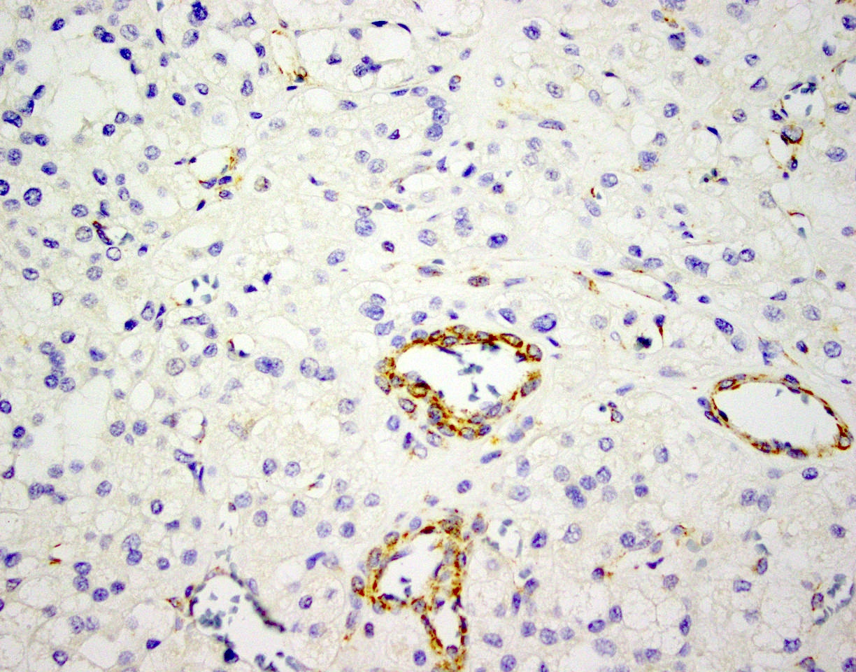
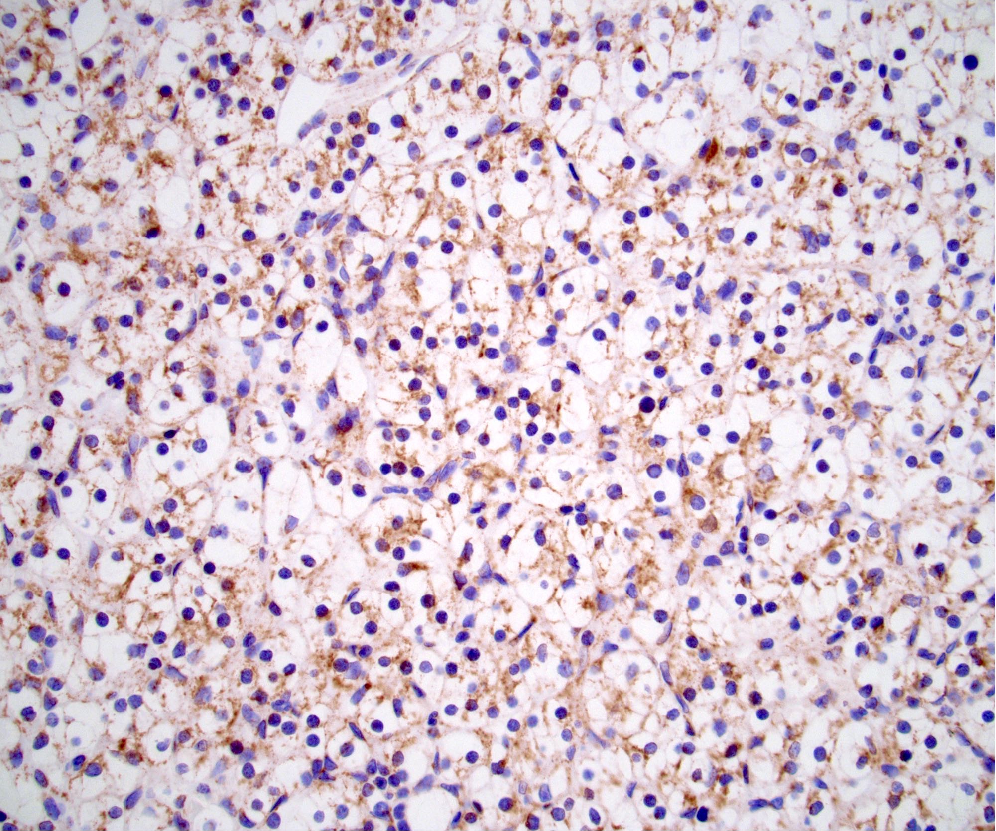
Interpretation
- Normal expression of SDHB is cytoplasmic and granular, indicative of mitochondrial staining; this can be appreciated in normal endothelial cells when evaluating tumors
- Loss of SDHB staining in neoplastic cells, in the presence of good positive internal control, is considered abnormal (Hum Pathol 2010;41:805)
- Sometimes a faint cytoplasmic blush is seen; in the context of a good positive internal control, this should be interpreted as loss of SDHB (Mod Pathol 2015;28:807)
Uses by pathologists
- Succinate dehydrogenase deficient tumors may show characteristic clinical and histologic features
- Pheochromocytomas / paragangliomas with succinate dehydrogenase deficiency
- With SDHB mutation, tend to form intra-abdominal, extra-adrenal tumors
- Those with SDHD mutations involve the head and neck
- SDHC related tumors are associated with the carotid body
- Cytologically, they show more rounded cells and distinct Zellballen nested pattern with prominent vasculature (Endocr Relat Cancer 2015;22:T91)
- Gastrointestinal stromal tumors with succinate dehydrogenase deficiency
- Almost always arise in the stomach
- Show positivity for KIT and DOG1
- Usually epithelioid and show a multilobular growth pattern (Am J Surg Pathol 2011;35:1712)
- KIT / PDGFRA wild type
- 3 molecular categories:
- ~30% germline SDHA mutations (Am J Surg Pathol 2013;37:234)
- ~20% mutations involving SDHB, SDHC and SDHD; inactivating mutations of SDHA, SDHB, SDHC or SDHD are characteristic of the Carney dyad (Carney-Stratakis syndrome) (Mol Cell Endocrinol 2018;469)
- ~50% show hypermethylation of SDHC, which is the molecular signature for the Carney triad, a syndromic, nonhereditary condition comprising succinate dehydrogenase deficient gastrointestinal stromal tumors, paraganglioma and pulmonary chondroma (Endocr Relat Cancer 2014;21:567)
- Renal cell carcinoma, succinate dehydrogenase deficient type
- Often show cystic change and entrapment of residual renal tubules; they also show eosinophilic cytoplasm, cytoplasmic vacuolization and bland nuclei (Mod Pathol 2015;28:80)
- Pituitary adenomas with succinate dehydrogenase deficiency
- Exceedingly rare but may be larger in size (Am J Surg Pathol 2014;38:560)
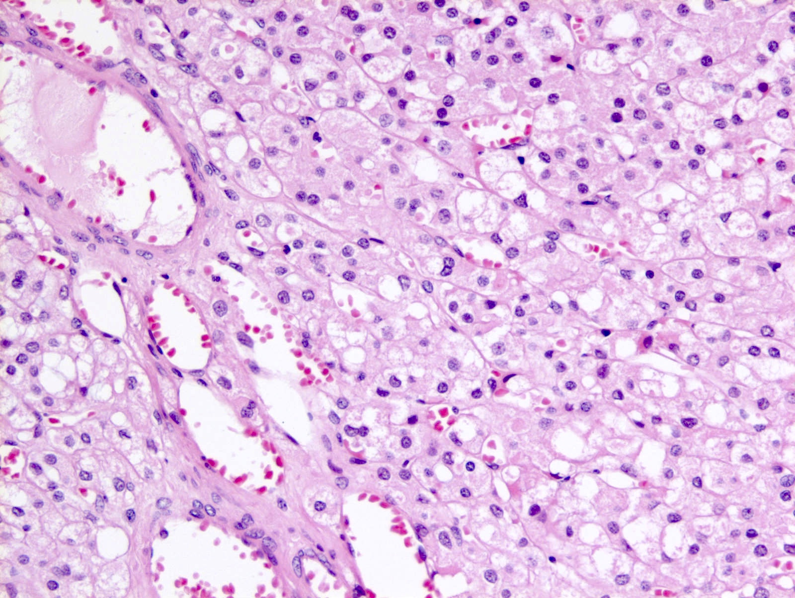
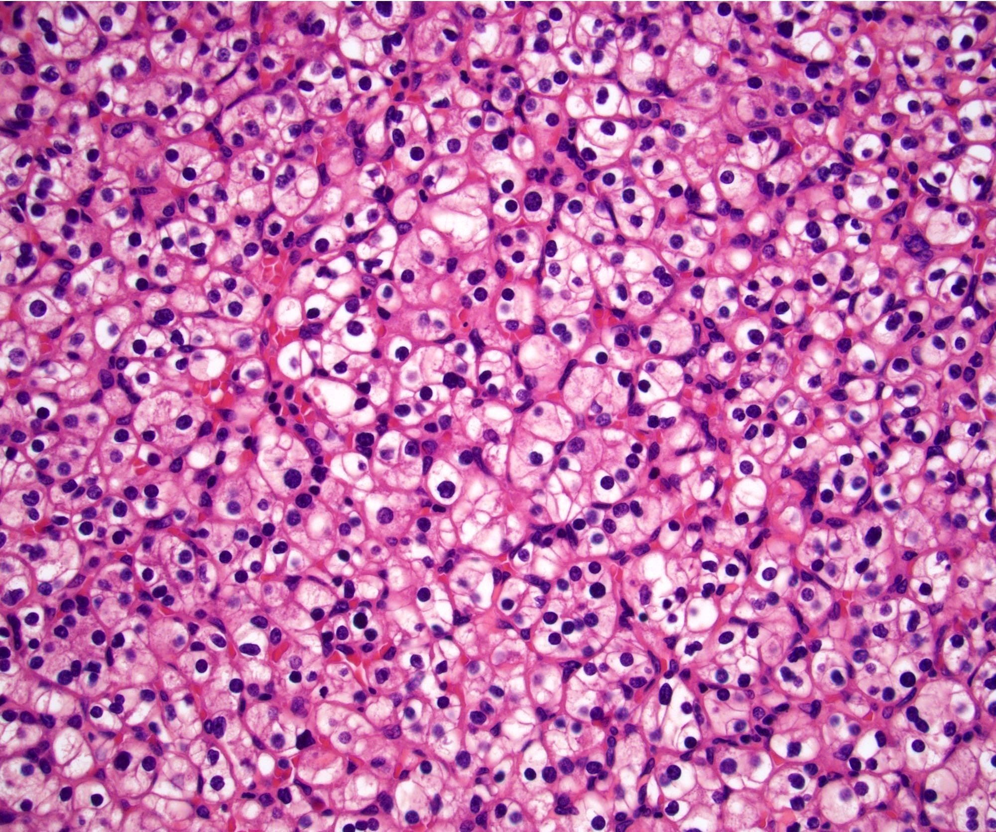
Microscopic (histologic) images
Contributed by Girish Venkataraman, M.D., Debra Zynger, M.D.
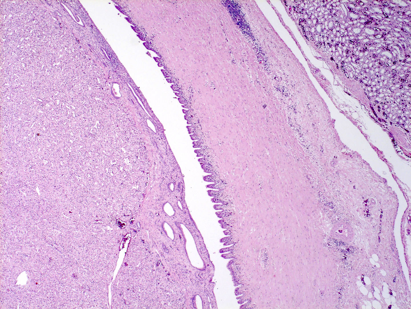
SDHB deficient renal cell carcinoma

SDHB retained in adrenal cortex
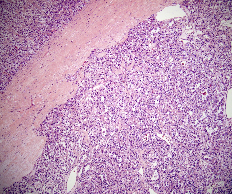

SDHB deficient paraganglioma
Clear cell renal cell carcinoma
Positive staining - normal
- Most tissues will be positive for SDHB (i.e. retain normal expression of SDHB), including endothelial cells, stromal cells and inflammatory cells and can be used as positive internal controls when assessing for loss of normal staining when suspecting succinate dehydrogenase deficient tumors
Positive staining - tumors
- Most pheochromocytomas / paragangliomas and gastrointestinal stromal tumors are nonsyndromic and will retain normal SDHB expression
- Epithelioid morphology in these tumors would suggest loss of SDHB
- PDGFRA associated gastrointestinal stromal tumors, which also may show epithelioid morphology, retain SDHB expression but are usually negative for KIT, in contrast to succinate dehydrogenase deficient gastrointestinal stroll tumors
- Renal cell carcinomas with SDHB loss may show clear or eosinophilic cytoplasm; clear cell renal cell carcinomas retain expression of SDHB
Negative staining - tumors
- 15% of pheochromocytomas / paragangliomas
- 3% of gastrointestinal stromal tumors
- < 1% of renal cell carcinomas
- < 1% of pituitary adenomas
- It is recommended that all pheochromocytomas / paragangliomas be assessed for loss of SDHB; gastrointestinal stromal tumors with epithelioid morphology and positive staining for KIT and DOG1 are suspicious for succinate dehydrogenase deficiency and should also be assessed
Molecular / cytogenetics description
- In SDH deficient neoplasms, next generation sequencing and PCR reveal inactivating mutations in SDHA, SDHB, SDHC or SDHD
- These are either nonsense mutations or missense mutations and likely lead to loss of protein function (Mod Pathol 2015;28:807, Hum Pathol 2010;41:805)
- SDHB immunohistochemistry is also useful to screen for these mutations
- 50% of succinate dehydrogenase deficient gastrointestinal stromal tumors show hypermethylation of SDHC, which causes loss of SDHB detectable by immunohistochemistry (Sci Transl Med 2014;6:268)
Board review style question #1
The normal immunohistochemical staining pattern for SDHB is
- Cytoplasmic
- Cytoplasmic and membranous
- Membranous
- Nuclear
Board review style answer #1
A. SDHB is a mitochondrial enzyme found in all normal cells and its usual immunostaining pattern is cytoplasmic and granular. Loss of SDHB suggests a genetic defect in one of the SDH related genes and is seen in SDH deficient neoplasms.
Comment Here
Reference: SDHB (succinate dehydrogenase)
Comment Here
Reference: SDHB (succinate dehydrogenase)
Board review style question #2
Which of the following tumors are associated with loss of SDHB by immunohistochemistry in a
subset of cases?
- Gastric adenocarcinoma
- Gastrointestinal stromal tumor
- Lung adenocarcinoma
- Neuroendocrine tumor
Board review style answer #2
B. Immunohistochemical loss of the SDHB protein is seen in a subset of the following tumors:
pheochromocytoma, paraganglioma, gastrointestinal stromal tumor, renal cell carcinoma and
rarely, pituitary adenoma. Loss of SDHB can be due to a variety of genetic mechanisms, including
mutations of any of the SDH subunits or due to hypermethylation of SDHC (particularly associated
with Carney triad).
Comment Here
Reference: SDHB (succinate dehydrogenase)
Comment Here
Reference: SDHB (succinate dehydrogenase)

