
Stains & CD markers
GATA3

Copyright: 2003-2024, PathologyOutlines.com, Inc.
PubMed Search: GATA3[TI] pathology free full text[sb]
GATA3
Author: Emily S. Reisenbichler, M.D.
Editorial Board Member: Maria Tretiakova, M.D., Ph.D.
Deputy Editor-in-Chief: Debra L. Zynger, M.D.
Last author update: 1 March 2018
Last staff update: 7 June 2022
Copyright: 2003-2024, PathologyOutlines.com, Inc.
PubMed Search: GATA3[TI] pathology free full text[sb]

Table of Contents
Definition / general | Essential features | Clinical features | Interpretation | Uses by pathologists | Prognostic factors | Microscopic (histologic) images | Positive staining - normal | Positive staining - tumors | Negative staining - normal | Negative staining - tumors | Board review style question #1 | Board review style answer #1Cite this page: Reisenbichler ES. GATA3. PathologyOutlines.com website. https://www.pathologyoutlines.com/topic/stainsgata3.html. Accessed April 24th, 2024.
Definition / general
- One of 6 members of the GATA family of transcription factors
- Involved in the luminal differentiation of breast epithelium, development of collecting system / urothelium and trophoblastic differentiation
- Also master regulator of type 2 helper T cells
Essential features
- Nuclear marker with expression in many epithelial neoplasms (including most breast, urothelial, paraganglioma / pheochromocytoma and skin carcinoma; smaller percentages of lung, liver, pancreatic, gastric, renal, thyroid, endometrial, ovarian and salivary gland carcinoma)
- Variable expression seen in selected germ cell tumors, mesotheliomas and rare sarcomas
- Due to the increasing number of tumors found to express GATA3, immunohistochemical staining with additional markers is necessary to determine the etiology of metastatic lesions of unknown primary
Clinical features
- GATA3 mutations cause HDR (hypoparathyroidism, sensorineural deafness and renal dysplasia) syndrome (Ann N Y Acad Sci 2011;1237:24, Endocr J 2011;58:117)
Interpretation
- Nuclear stain
Uses by pathologists
- Differentiate metastatic urothelial and breast carcinomas (GATA3+) from many other metastatic carcinomas (Am J Surg Pathol 2014;38:13)
- Differentiate urothelial carcinoma (> 80% GATA3+) from prostatic carcinoma (2% GATA3+)
- Differentiate metastatic lobular carcinoma of the breast (GATA3+) from gastric signet ring cell carcinoma (GATA3-)
- Differentiate squamous cell carcinoma of the skin (GATA3+) from squamous cell carcinoma of the lung (GATA3-)
- Differentiate mesothelioma (81% GATA3+) from pulmonary adenocarcinoma (12% GATA3+)
- Differentiate acute leukemias with T cell differentiation (GATA3+) from acute myeloid leukemia (< 10% GATA3+) and B lymphoblastic leukemias (GATA3-) (Hum Pathol 2017;65:166)
- Subtyping renal neoplasms: GATA3+ in subset of clear cell papillary renal carcinomas and chromophobe renal cell carcinomas (Appl Immunohistochem Mol Morphol 2018;26:316, Am J Surg Pathol 2014;38:13)
- Subtyping salivary gland neoplasms: diffuse GATA3 staining in 100% of mammary analogue secretory carcinoma and salivary duct carcinomas; GATA3 positive, but not diffuse staining, in other salivary tumors (Head Neck Pathol 2013;7:311)
Prognostic factors
- Low nuclear GATA3 immunoexpression may be a poor prognostic factor for invasive breast cancer (Hum Pathol 2010;41:1794)
- GATA3 expression is associated with worse prognosis in soft tissue sarcoma (PLoS One 2016;11:e0156524)
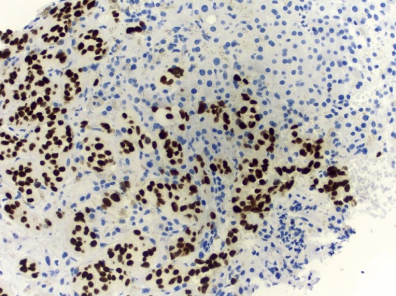
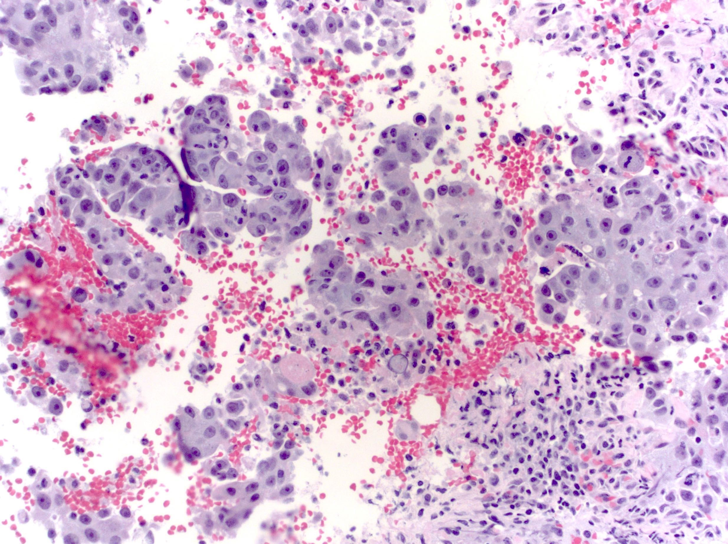
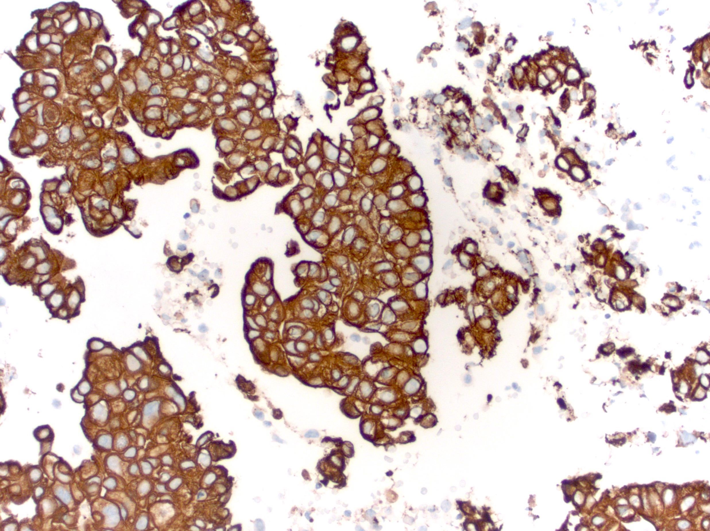
Microscopic (histologic) images
Contributed by Emily S. Reisenbichler, M.D., Andrey Bychkov, M.D., Ph.D., Maria Tretiakova, M.D., Ph.D. and Debra Zynger, M.D.
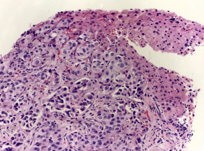
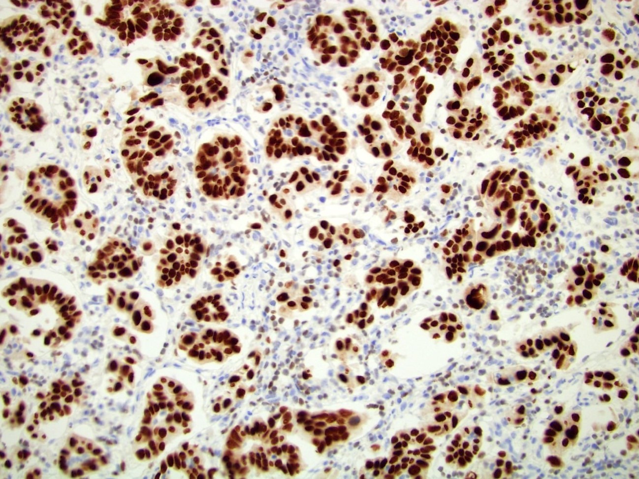
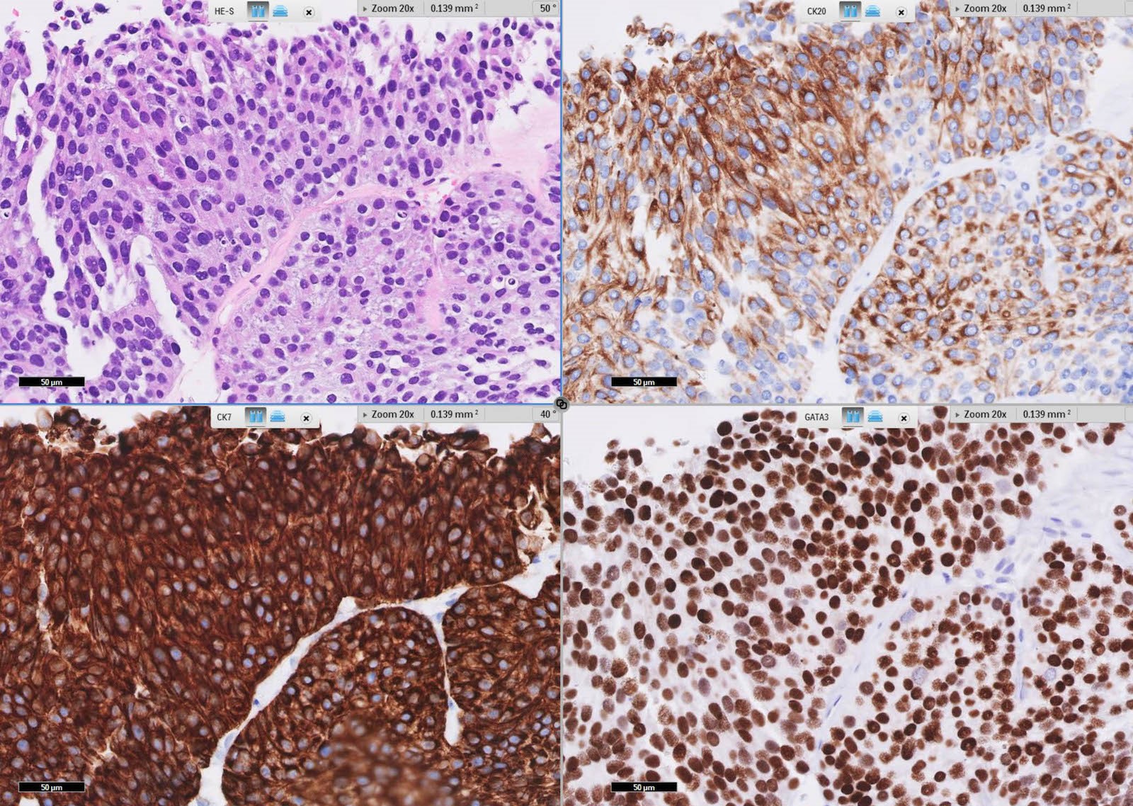
Metastatic breast carcinoma

Poorly differentiated breast carcinoma
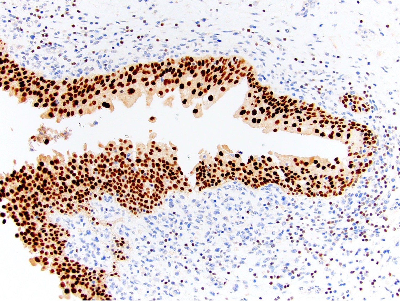
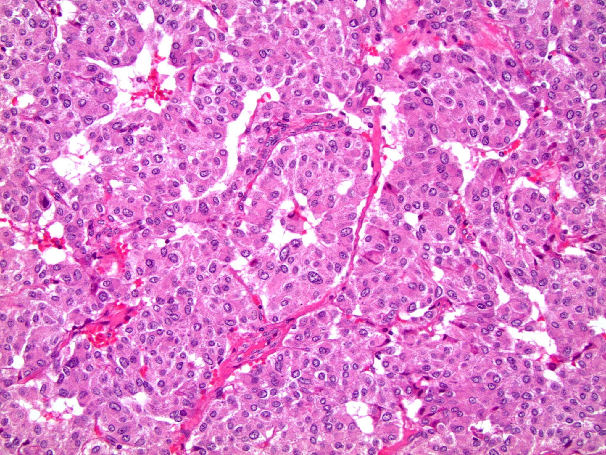
Invasive urothelial carcinoma
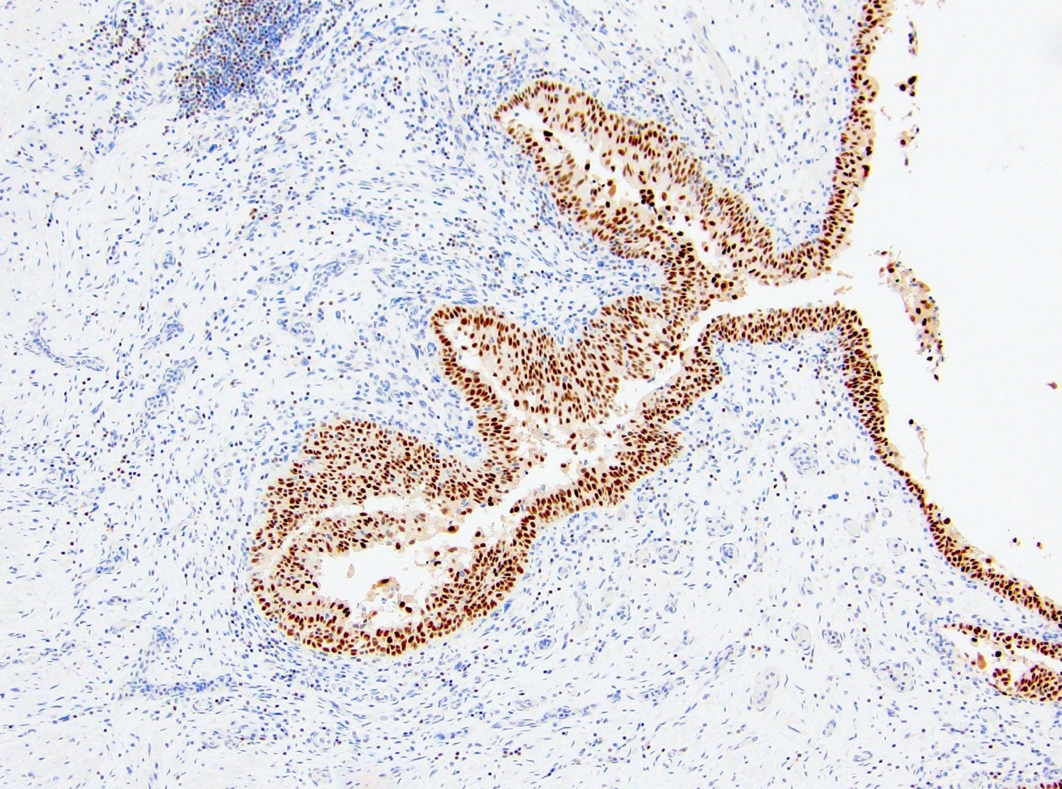
Nonneoplastic urothelium
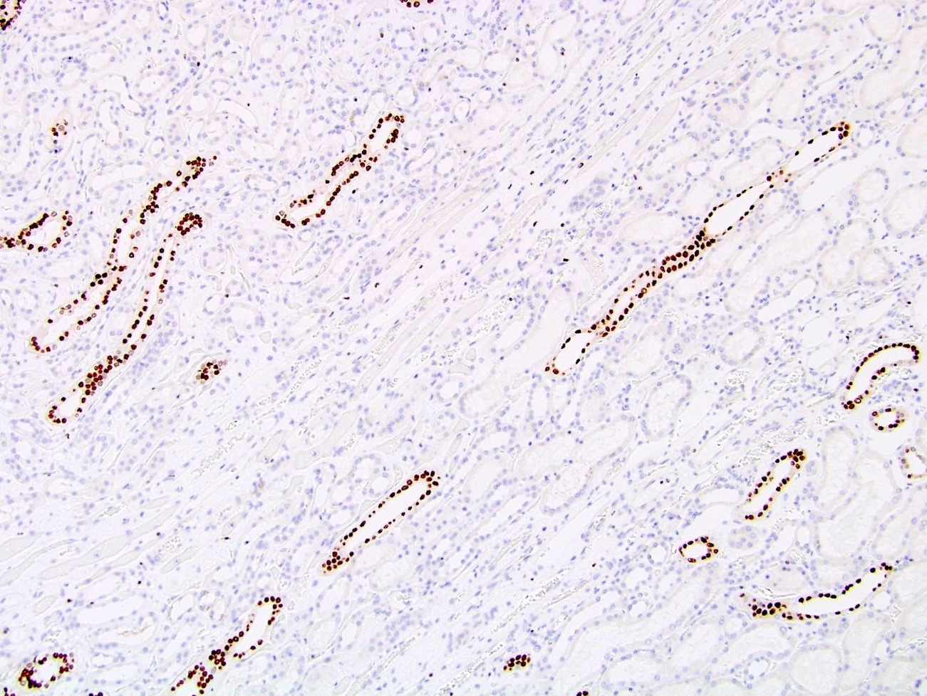
Nonneoplastic collecting ducts
Urothelial carcinoma
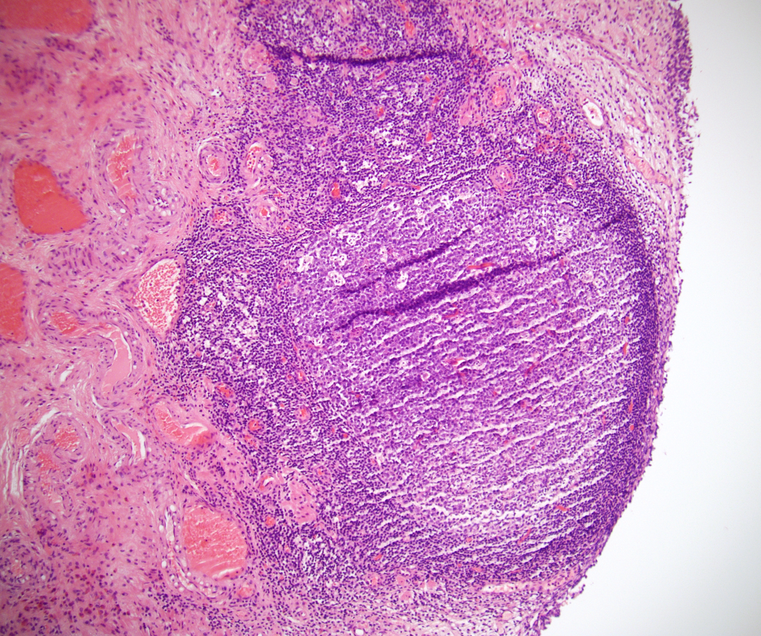
Pheochromocytoma
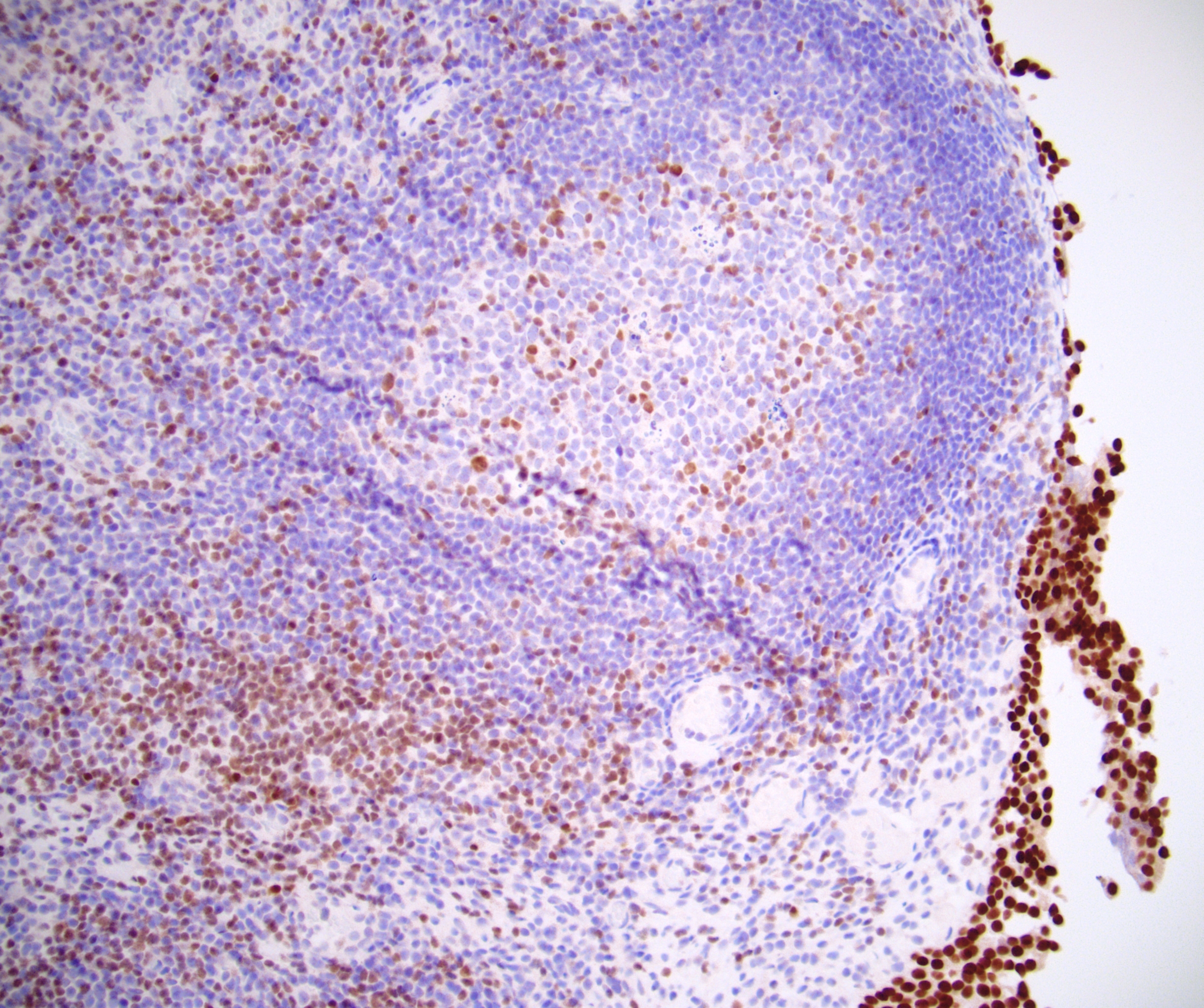
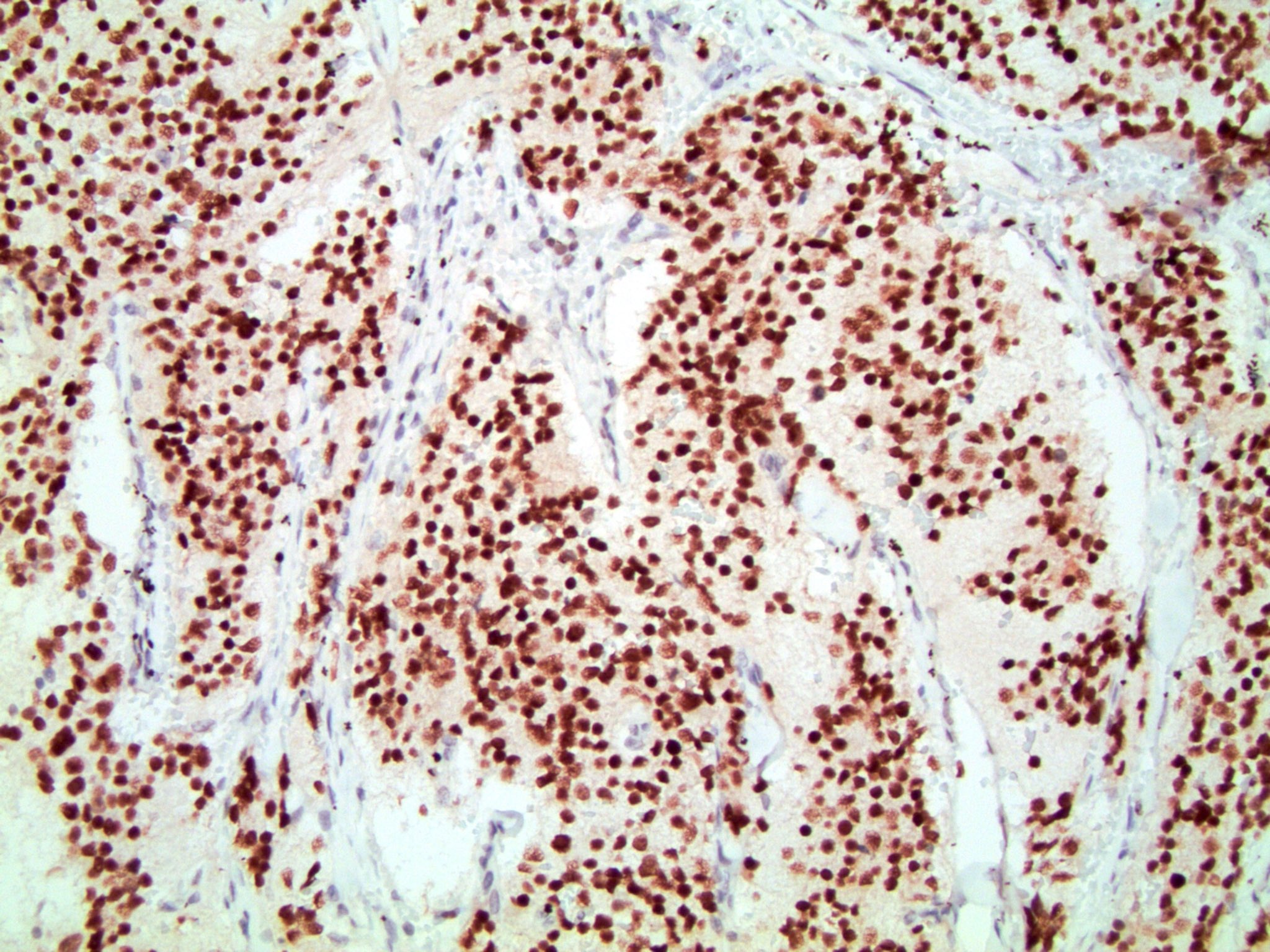
Nonneoplastic lymphocytes
Positive staining - normal
- Skin: basal and spinous layers of the epidermis, outer root sheath of the hair follicle, sebaceous sweat and apocrine glands (Am J Dermatopathol 2015;37:885)
- Distal convoluted tubules of kidney (Hum Pathol 2017;66:152)
- Parathyroid
- Radiated nonneoplastic prostate glands (basal and luminal cells) (Histopathology 2017;71:150, Am J Surg Pathol 2017;41:557)
Positive staining - tumors
- Breast cancer, invasive (72 - 94%) (Mod Pathol 2010;23:654, Am J Clin Pathol 2012;138:57), well differentiated > poorly differentiated; more sensitive than GCDFP-15 and mammoglobin in staining of metastatic breast carcinoma (Ann Diagn Pathol 2015;19:6)
- Primary and metastatic urothelial carcinoma (67 - 93%) (Am J Surg Pathol 2007;31:673, Am J Surg Pathol 2013;37:1876)
- Paraganglioma (78%) (Mod Pathol 2013;26:1365) and pheochromocytoma (71%) (Histopathology 2017;71:475)
- Choriocarcinomas and gestational trophoblastic tumors (100%) (Hum Pathol 2016;48:18, Histopathology 2015;67:636)
- Renal cell carcinoma, clear cell papillary type (76%) (Hum Pathol 2017;66:152), variable in chromophobe renal cell carcinoma (6 - 51%), less in oncocytoma (17 - 19%) (Hum Pathol 2014;45:244, Am J Surg Pathol 2014;38:13)
- Salivary gland neoplasms (51% overall), particularly salivary duct carcinoma and mammary analogue secretory carcinoma (100%) (Head Neck Pathol 2013;7:311)
- Malignant mesothelioma, including sarcomatoid and desmoplastic types (58 - 100%) (Am J Surg Pathol 2014;38:13, Am J Surg Pathol 2017;41:1221)
- Skin squamous cell, basal cell and sebaceous carcinoma (> 85%) (Am J Dermatopathol 2015;37:885)
- Neuroblastoma (100%) (Cancer Cytopathol 2017;125:940)
- Many adnexal tumors, including apocrine and follicular neoplasms (Am J Dermatopathol 2017;39:279)
- Peripheral T cell lymphoma, NOS (30%) (Am J Clin Pathol 2010;133:281)
- Selected sarcomas (Am J Surg Pathol 2014;38:13)
Negative staining - normal
- Breast myoepithelial cells
- Skin - epidermal granular cell layer, matrix cells of hair bulb and eccrine glands (Am J Dermatopathol 2015;37:885)
- Thyroid follicular cells
Negative staining - tumors
- Renal cell carcinoma, clear cell type (Histopathology 2017;71:475, Hum Pathol 2017;66:152)
- Renal cell carcinoma, papillary type (Hum Pathol 2017;66:152)
- Adrenal cortical carcinoma (Histopathology 2017;71:475)
- Seminoma
- Small cell carcinoma of lung
- Thymoma
- Neuroendocrine tumors of pancreas, small intestine and lung
- Rectal adenocarcinoma
- Melanoma
- Thyroid tumors (Histopathology 2014;65:288)
Board review style question #1
A biopsied liver lesion is radiographically suspected to be a metastasis of unknown primary. Which stain combination would be most consistent with metastasis from a breast primary?
- GATA3 negative, CK7 positive, TTF1 positive, CK20 negative
- GATA3 negative, S100 positive, SOX10 positive, AE1 / AE3 negative
- GATA3 positive, ER positive, CK7 positive, p63 negative
- GATA3 positive, p63 positive, ER negative, CK5 / 6 positive
Board review style answer #1
C. GATA3 positive, ER positive, CK7 positive, p63 negative.
While none of these markers alone is specific for breast carcinoma, most breast carcinomas are positive, particularly estrogen receptor (ER) positive tumors. The combined immunohistochemical findings in the other answer choices are most indicative of melanoma and carcinomas of urothelial and lung primary.
Comment Here
Reference: GATA3
Comment Here
Reference: GATA3

