
Stains & CD markers
MelanA / MART1

Copyright: 2002-2024, PathologyOutlines.com, Inc.
PubMed Search: MelanA MART1
MelanA / MART1
Author: Yao-Tseng Chen, M.D., Ph.D.
Editor-in-Chief: Debra L. Zynger, M.D.
Last author update: 1 May 2018
Last staff update: 14 June 2022
Copyright: 2002-2024, PathologyOutlines.com, Inc.
PubMed Search: MelanA MART1

Table of Contents
Definition / general | Essential features | Pathophysiology | Interpretation | Uses by pathologists | Microscopic (histologic) images | Positive staining - normal | Positive staining - disease | Negative staining - disease | Board review style question #1 | Board review style answer #1 | Board review style question #2 | Board review style answer #2Cite this page: Chen YT. MelanA / MART1. PathologyOutlines.com website. https://www.pathologyoutlines.com/topic/stainsmart1.html. Accessed April 26th, 2024.
Definition / general
- Melanocyte specific cytoplasmic protein involved in the formation of stage II melanosomes
- MelanA was discovered as an antigen recognized by tumor infiltrating cytotoxic T cells from a melanoma patient, hence the name Melanoma Antigen (J Exp Med 1994;180:35)
- Same gene was independently identified by a different research group who named it MART1 (Melanoma Antigen Recognized by T cells 1) (Proc Natl Acad Sci USA 1994;91:3515); MelanA and MART1 are synonyms
- Two antibodies are commercially available: A103 and M2-7C10
- A103: mouse monoclonal antibody against MelanA recombinant protein (Proc Natl Acad Sci USA 1996;93:5915, US patent 5,674,749)
- In addition to melanocytes, A103 also stains adrenal cortical cells and steroid producing cells in testis and ovary
- This is due to antibody cross reactivity to an unknown molecule in these cells; these cells do not produce MelanA / MART1 mRNA or protein (Am J Surg Pathol 1998;22:595)
- M2-7C10: mouse monoclonal antibody clone produced against MART1 protein
- This antibody stains only melanocytes among normal tissues; negative for adrenal cortical cells and steroid producing cells in testis or ovary (J Immunother 1997;20:60)
- A103: mouse monoclonal antibody against MelanA recombinant protein (Proc Natl Acad Sci USA 1996;93:5915, US patent 5,674,749)
Essential features
- Melanocyte lineage specific marker, more sensitive than HMB45 in the diagnosis of metastatic melanoma
- Cytoplasmic protein
- Positive in most primary melanomas but desmoplastic melanoma can be negative
- Also positive in nonmelanocytic tumors with melanosomes, including angiomyolipoma, PEComa, lymphangioleiomyomatosis
- Depending on antibody, positive in adrenal cortical tumors and sex cord stromal tumors
Pathophysiology
- Melanosome specific protein
- Plays a vital role in the expression, stability, trafficking and processing of Pmel17, which is critical to the formation of stage II melanosomes (J Biol Chem 2005;280:14006)
Interpretation
- Cytoplasmic staining
- Staining in any percentage of tumor cells is interpreted as positive
Uses by pathologists
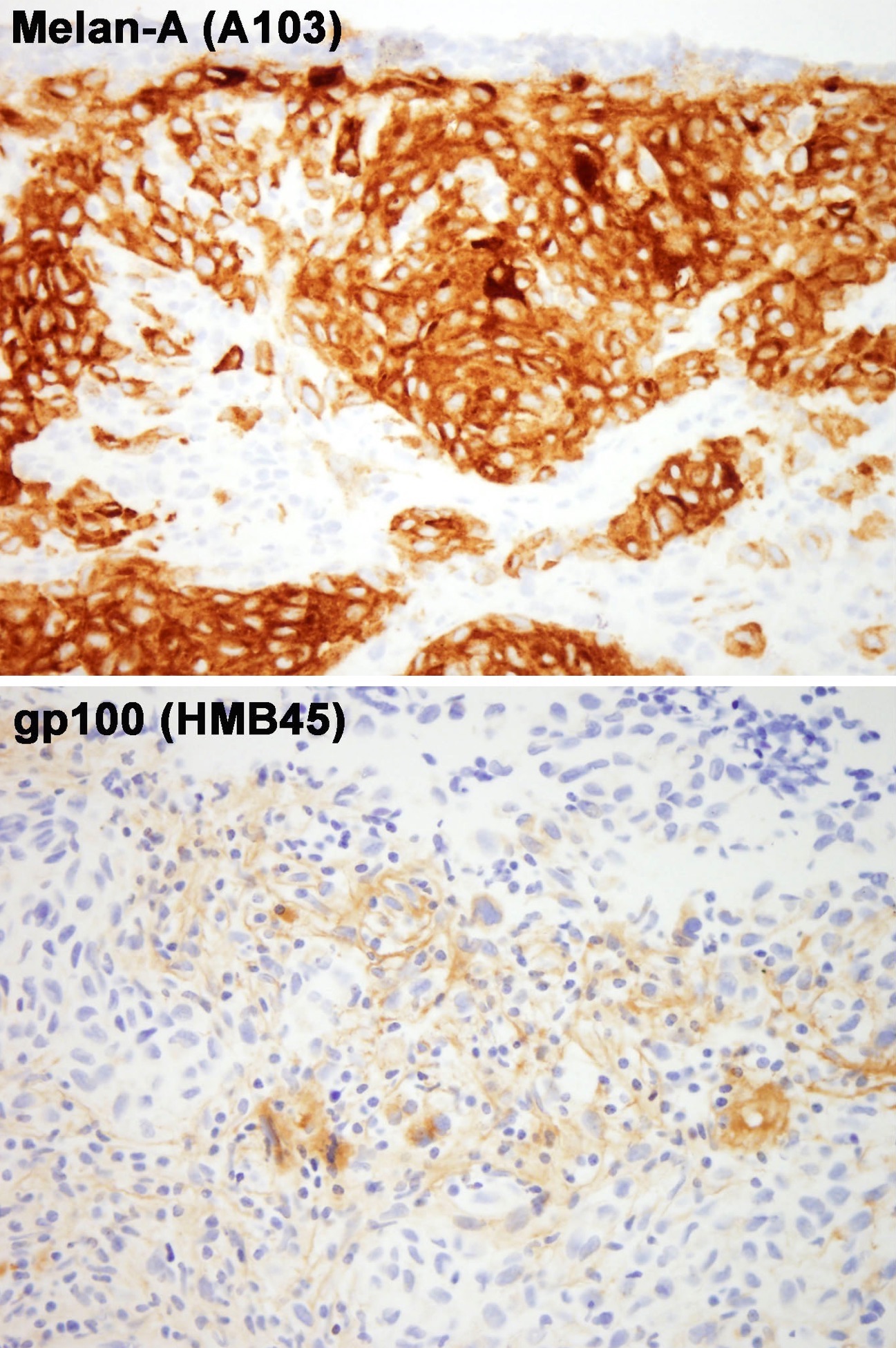
- To establish the diagnosis of metastatic melanoma, more sensitive than anti-gp100 antibody (HMB45) (Am J Surg Pathol 1998;22:976)
- Used in conjunction with S100 as a standard panel for evaluating sentinel lymph nodes for melanoma; HMB45 can be added to the panel but is not essential (Am J Surg Pathol 2001;25:1039)
- To confirm the diagnosis of angiomyolipoma, lymphangioleiomyomatosis (LAM) and other PEComas
- To distinguish adrenal cortical tumors from renal cell carcinoma (clone A103 only); inhibin A can also be used for this purpose
- To confirm the diagnosis of a sex cord stromal tumor (clone A103 only); inhibin A can also be used for this purpose
- To distinguish neurotized melanocytic nevi (MelanA+) from neurofibroma (MelanA-) (Arch Pathol Lab Med 2012;136:810)
- Caution: stains all cells derived from melanocytes, does not distinguish benign nevi from melanoma or premalignant melanocytic lesions
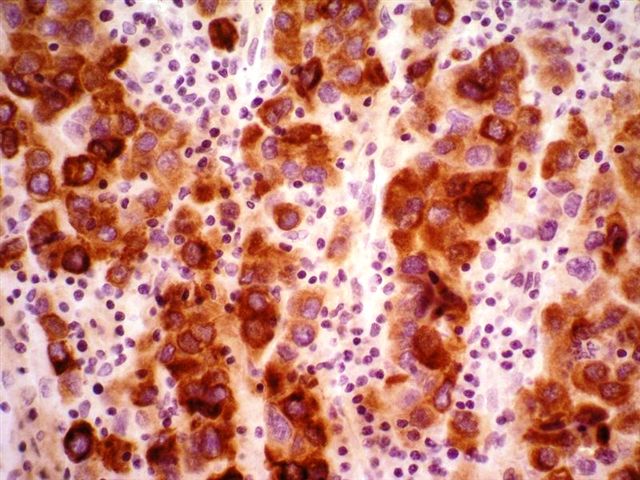
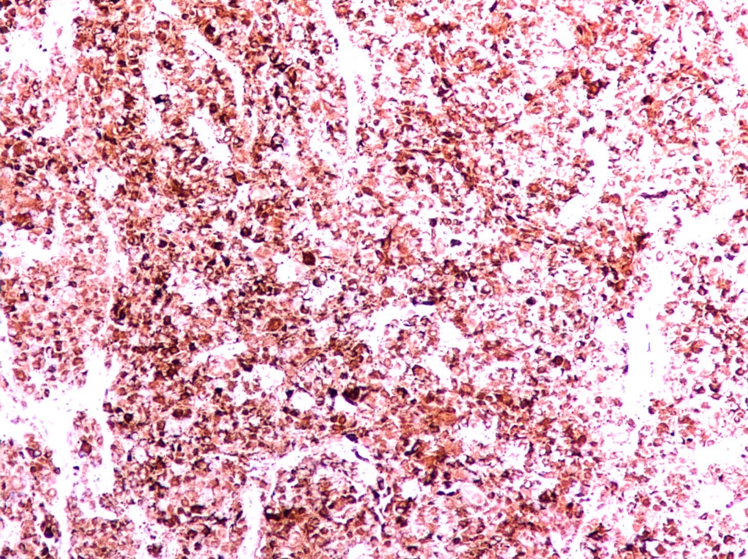
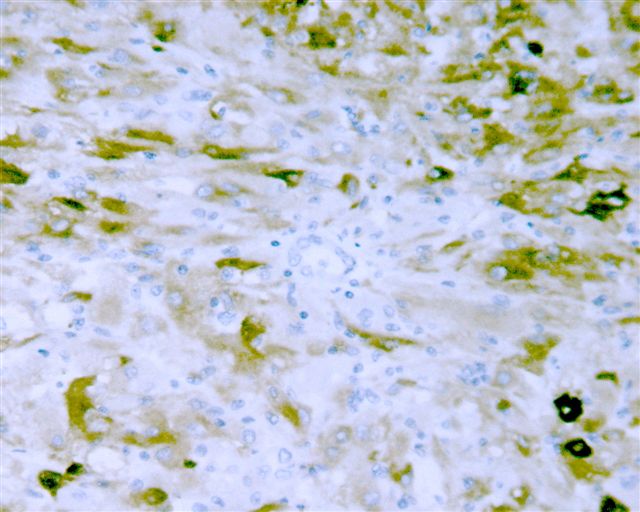
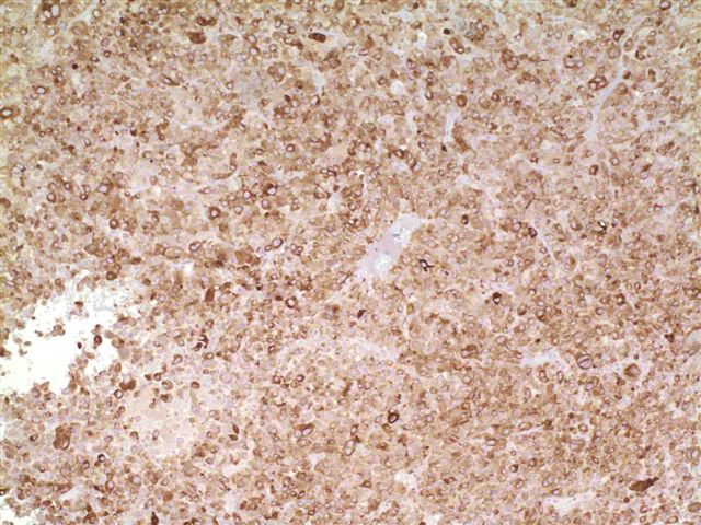
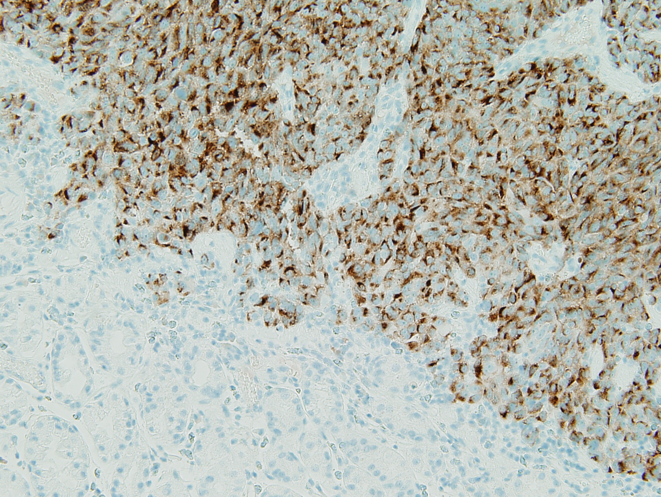
Microscopic (histologic) images
Contributed by Yao-Tseng Chen, M.D., Ph.D.
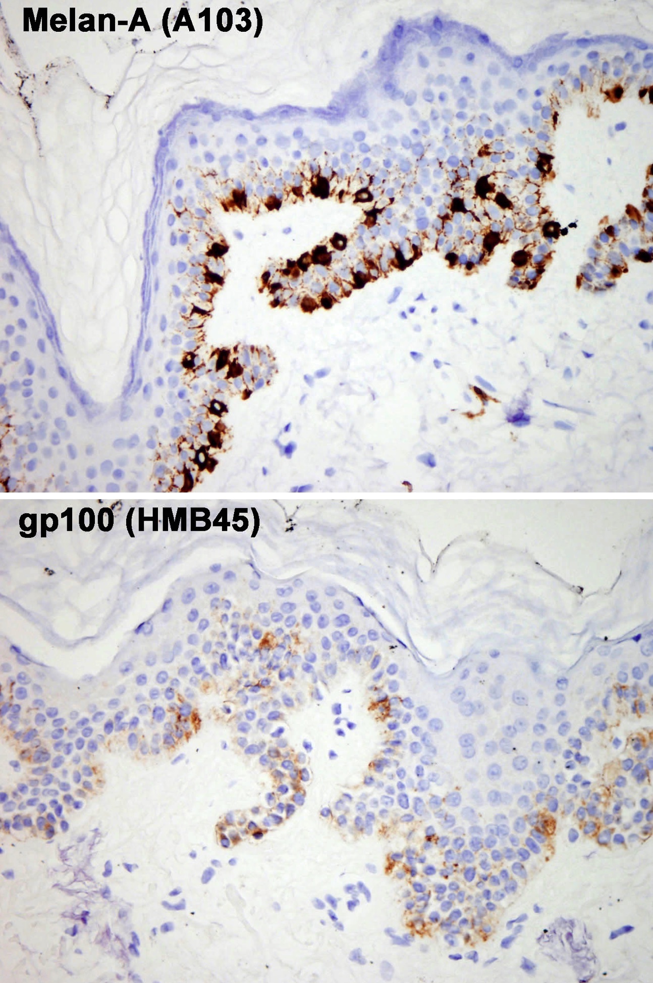
Lentiginous lesion, A103 versus HMB45

Primary melanoma, A103 versus HMB45
Metastatic melanoma, A103 versus HMB45

Angiomyolipoma of the liver, A103
Contributed by Semir Vranić, M.D., Ph.D., Kristine Cornejo, M.D., Case #32 and Case #109
Small bowel, metastatic melanoma
Testis, Leydig cell tumor
Kidney, atypical epithelioid angiomyolipoma
Uterus, melanoma
Images hosted on other servers:

Bladder PEComa, HMB45+, MelanA+

Skin, dermal nevus

Skin, melanoma, fibrohistiocytic proliferation


Skin, melanoma, sentinel lymph nodes with "stealth" metastases

Small bowel, metastatic melanoma
Stomach, metastatic melanoma, MelanA
Positive staining - normal
- Melanocytes in skin, retina - both A103 and M2-7C10 clones
- Adrenal cortex, testis (Leydig cells and occasional Sertoli cells), ovary (granulosa / theca cells, hilus and stromal cells) - clone A103 only
Positive staining - disease
- Nevi
- Most primary and metastatic melanomas but only some (4 of 13) desmoplastic melanomas (Am J Surg Pathol 1998;22:976)
- Angiomyolipoma (Virchows Arch 1999;434:429)
- Lymphangioleiomyomatosis (LAM) (Adv Anat Pathol 1999;6:12)
- Other perivascular epithelioid cell tumors (PEComa)
- Clear cell sarcoma of the soft tissue (23 of 32) (Am J Surg Pathol 2008;32:452)
- Adrenal cortical adenoma and carcinoma (clone A103 only) (Am J Surg Pathol 1998;22:57)
- Sex cord stromal tumors (clone A103 only) (Am J Surg Pathol 1998;22:57)
- t(6;11) / TFEB renal cell carcinoma (Am J Surg Pathol 2016;40:1484, Am J Surg Pathol 2014;38:604)
- Melanotic schwannoma (Am J Surg Pathol 2014;38:94)
Negative staining - disease
- All tumors not listed above, including all carcinomas, lymphomas, etc.
Board review style question #1
Which of the following lesions will stain positively for MelanA antibody (clone A103) but does not express MelanA mRNA or protein?
- Adrenal cortical adenoma
- Angiomyolipoma
- Benign melanocytic nevus
- Lymphangiomyomatosis
- PEComa
Board review style answer #1
A. Adrenal cortical adenoma. A103 stains adrenal cortical cells and steroid producing cells in testis and ovary due to antibody cross reactivity to an unknown molecule in these cells; these cells do not produce MelanA / MART1 mRNA or protein.
Comment Here
Reference: MelanA / MART1
Comment Here
Reference: MelanA / MART1
Board review style question #2
Which of the following statements is false about gp100 (HMB45 antibody) and MelanA (A103 antibody)?
- Both are likely to be positive in angiomyolipoma
- Both are markers for melanocyte differentiation
- HMB45 is more sensitive than MelanA (A103) in detecting metastatic melanoma in sentinel lymph nodes
- Neither can be used to distinguish melanomas from benign nevi
- Only MelanA (A103 antibody) is positive in adrenal cortical adenoma
Board review style answer #2
C. HMB45 is more sensitive than MelanA (A103) in detecting metastatic melanoma in sentinel lymph nodes is false. MelanA (A103) is more sensitive than HMB45 (gp100) in detecting metastatic melanoma in sentinel lymph nodes.
Comment Here
Reference: MelanA / MART1
Comment Here
Reference: MelanA / MART1

