
Stains & CD markers
Nonspecific esterase

Copyright: 2002-2024, PathologyOutlines.com, Inc.
PubMed Search: Nonspecific esterase stain
Nonspecific esterase
Author: Chunyu Cai, M.D., Ph.D.
Editor-in-Chief: Debra L. Zynger, M.D.
Last author update: 3 March 2022
Last staff update: 11 September 2023
Copyright: 2002-2024, PathologyOutlines.com, Inc.
PubMed Search: Nonspecific esterase stain

Table of Contents
Definition / general | Essential features | Terminology | Interpretation | Uses by pathologists | Microscopic (histologic) description | Microscopic (histologic) images | Positive staining - normal | Positive staining - disease | Negative staining - disease | Board review style question #1 | Board review style answer #1Cite this page: Cai C. Nonspecific esterase. PathologyOutlines.com website. https://www.pathologyoutlines.com/topic/stainsnonspecificesterase.html. Accessed April 23rd, 2024.
Definition / general
- Enzyme histochemical stain that relies on endogenous esterase activity to hydrolyze exogenous alpha naphthyl acetate substrate, which yields naphthol, a reddish brown product visible under light microscopy
Essential features
- Routinely used in skeletal muscle biopsies to detect macrophages, myophagocytosis and denervated myofibers
- Differentiates macrophages (positive) from lymphocytic inflammation (negative) in muscle biopsies
- Also highlights neuromuscular junctions and myotendinous junctions in normal skeletal muscle
- Used in hematopathology to differentiate the monocytic lineage (strongly positive) from the lymphoid and other myeloid lineage cell types (weak punctate or negative)
Terminology
- Other names: alpha naphthyl acetate, alpha naphthyl butyrate esterase
- Note: the abbreviation NSE also refers to neuron specific enolase
Interpretation
- Cytoplasmic
Uses by pathologists
- In muscle biopsies (Dubowitz: Muscle Biopsy - A Practical Approach - Expert Consult, 4th Edition, 2013):
- Identifies myophagocytosis as evidence of acute myopathic change
- Identifies macrophagic inflammation
- Identifies neuromuscular junctions
- Differentiates normal structural alteration at myotendinous junctions from chronic myopathic changes
- In hematopathology (Swerdlow: WHO Classification of Tumours of Haematopoietic and Lymphoid Tissues (Medicine), Revised Edition, 2017):
- Differentiates the monocytic lineage from the lymphoid and other myeloid lineage cell types on marrow aspirate smears and peripheral blood smears
- Recommended by expert opinion in the guidelines for the initial work up of acute leukemias by the College of American Pathologists and the American Society of Hematology to assist in the diagnosis and classification of acute myeloid leukemia (Arch Pathol Lab Med 2017;141:1342)
Microscopic (histologic) description
- Staining pattern varies depending on structure highlighted
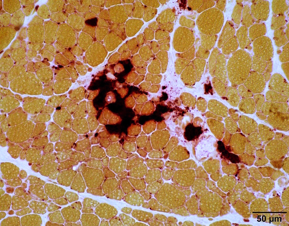
- Macrophages: strong cytoplasmic (lysosomes)
- Macrophage associated with myophagocytosis are located within the sarcoplasm of myofibers
- Macrophages associated with inflammatory myositis are typically located outside myofibers in the perimysial or endomysial connective tissue (Curr Protoc Immunol 2001;Appendix 3)
- Neuromuscular junction: strong, discrete, subsarcolemmal, clustered around intramuscular nerve (Histochem J 1998;30:7)
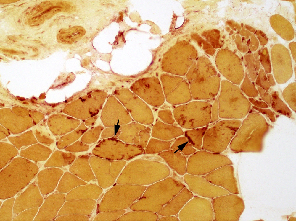
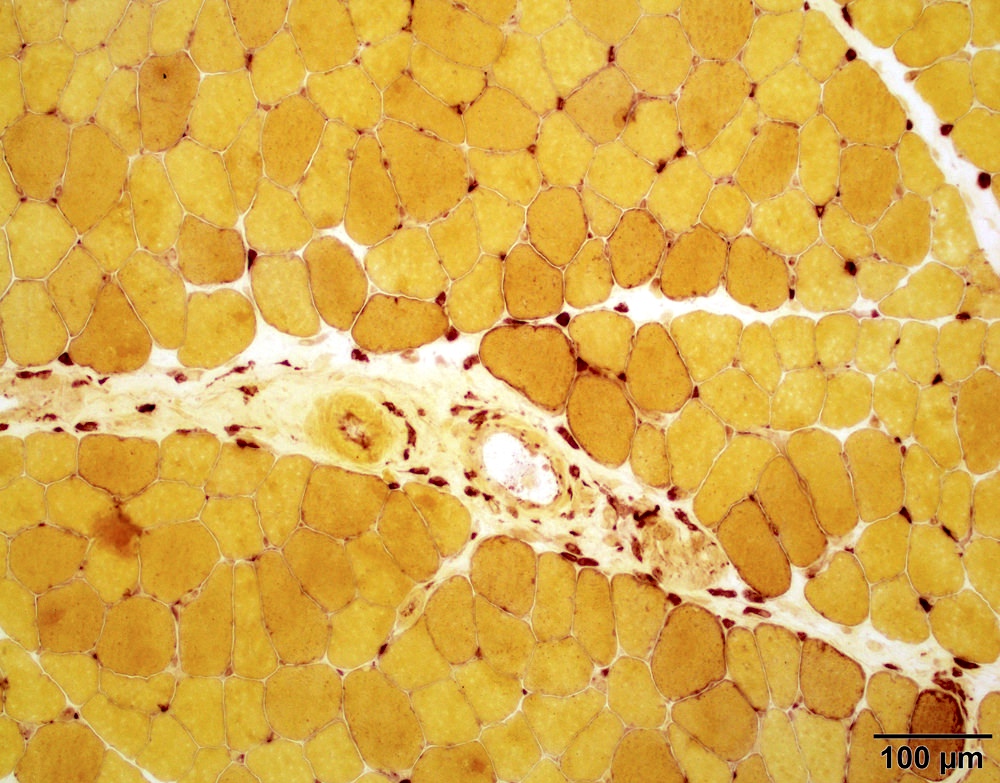
- Denervated myofibers: weak diffuse sarcoplasmic (J Neurol Sci 1975;26:133)
- Myotendinous junction: irregular linear (Neuromuscul Disord 1996;6:211)
- Slightly darker in type I than in type II muscle fibers (J Anat 1988;157:79)
- Macrophages: strong cytoplasmic (lysosomes)
Microscopic (histologic) images
Contributed by Chunyu "Hunter" Cai, M.D., Ph.D. and Genevieve M. Crane, M.D., Ph.D.
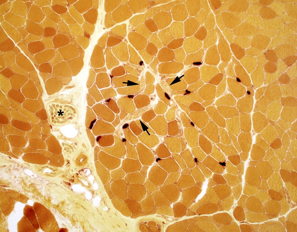
Neuromuscular junction
Myotendinous junction
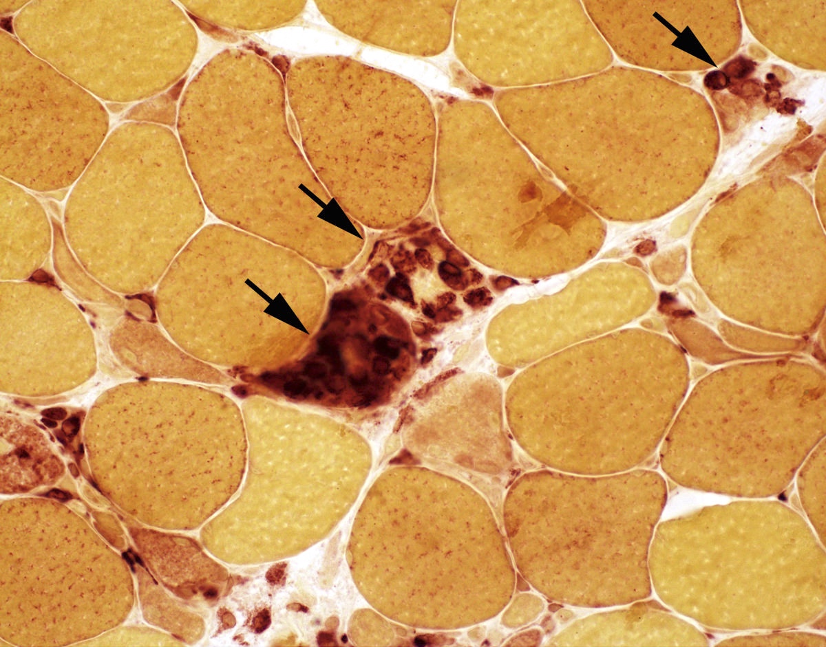
Myophagocytosis

Rhabdomyolysis
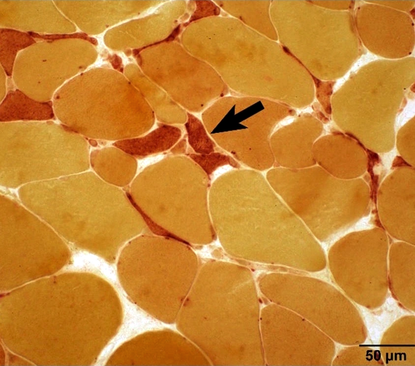
Denervated myofibers
Inflammatory myositis
Macrophagic myofasciitis

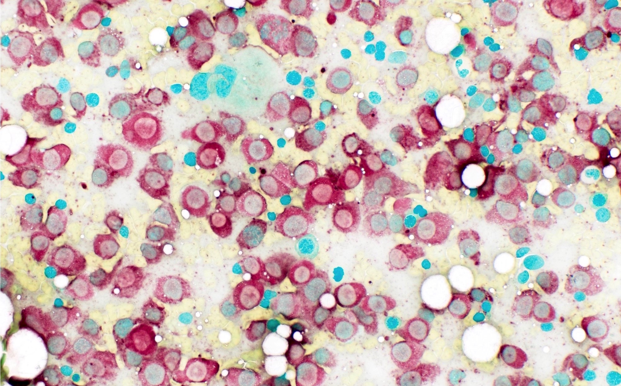
NSE highlights blasts of monocytic origin
Positive staining - normal
- Positive in neuromuscular junction (Dubowitz: Muscle Biopsy - A Practical Approach: Expert Consult, 4th Edition, 2013)
- Positive in myotendinous junction (Neuromuscul Disord 1996;6:211)
- Type 1 fibers generally stain a bit more strongly than type 2 fibers
Positive staining - disease
- In muscle biopsy:
- Denervated atrophic myofibers (Dubowitz: Muscle Biopsy - A Practical Approach: Expert Consult, 4th Edition, 2013)
- Macrophages in inflammatory myositis (Curr Rheumatol Rep 2009;11:295)
- Macrophages in dermatomyositis (Acta Neuropathol 2009;118:793)
- Macrophagic myofasciitis (J Neuropathol Exp Neurol 2017;76:323)
- In hematopathology:
- Promonocytes, monoblasts, monocytes, are strongly positive for nonspecific esterase, inhibited by sodium fluoride (NaF) (Swerdlow: WHO Classification of Tumours of Haematopoietic and Lymphoid Tissues, 4th Edition, 2008)
Negative staining - disease
- In muscle biopsy
- Negative in lymphocytes and viable myofibers
- In hematopathology:
- Weak punctate or negative in lymphoid and myeloid lineage tumors AML M0, M1, M2, M3, lymphoblastic lymphoma / leukemia (Swerdlow: WHO Classification of Tumours of Haematopoietic and Lymphoid Tissues, Revised Edition, 2017)
Board review style question #1

Which of the following correctly describes the finding in the pictured nonspecific esterase stained muscle?
- Denervation
- Myophagocytosis
- Myotendinous junction
- Neuromuscular junction
- Type II atrophy
Board review style answer #1

