
Stains & CD markers
Oil red O

Copyright: 2003-2024, PathologyOutlines.com, Inc.
PubMed Search: Oil red O [title]
Oil red O
Author: Chunyu Cai, M.D., Ph.D.
Editorial Board Member: Raul S. Gonzalez, M.D.
Editor-in-Chief: Debra L. Zynger, M.D.
Last author update: 24 August 2020
Last staff update: 17 June 2021
Copyright: 2003-2024, PathologyOutlines.com, Inc.
PubMed Search: Oil red O [title]

Table of Contents
Definition / general | Essential features | Terminology | Interpretation | Uses by pathologists | Frozen section images | Microscopic (histologic) images | Positive staining - normal | Positive staining - disease | Sample pathology report | Board review style question #1 | Board review style answer #1Cite this page: Cai C. Oil red O. PathologyOutlines.com website. https://www.pathologyoutlines.com/topic/stainsoilredo.html. Accessed April 24th, 2024.
Definition / general
- Oil red O is a fat soluble, hydrophobic diazo dye that stains neutral fat, fatty acids and triglycerides (Histochemistry 1992;97:493)
- Maximum absorption at 518 nm; appears red under light microscopy
- Unlike Sudan black, oil red O stains poorly for complex phospholipids and glycolipids that have polar groups, meaning it does not stain myelin / peripheral nerve or biological membranes
- In order to highlight fat droplets, oil red O staining needs to be performed on fresh or frozen tissue; paraffin embedding or alcohol based fixation removes most neutral lipids
Essential features
- In muscle biopsies, oil red O is mainly used to assess the amount of sarcoplasmic lipid droplets and assess for lipid storage diseases (Dubowitz: Muscle Biopsy: A Practical Approach, 4th Edition, 2013)
- Staining must be performed on fresh or frozen tissue
- In general or forensic / autopsy pathology, oil red O is used to
- Highlight fat emboli (Hum Pathol 1981;12:753, J Forensic Sci 1987;32:1426)
- Highlight lipid laden macrophages (Cytopathology 2010;21:245)
- Assess any other pathological lipid accumulation (Nat Protoc 2013;8:1149)
Terminology
- IUPAC name: 1-(2,5-dimethyl-4-(2,5-dimethylphenyl) phenyldiazenyl) azonapthalen-2-ol
- Chemical formula: C26H24N4O
Interpretation
- Red cytoplasmic / sarcoplasmic stain in muscle biopsy
- In fat emboli, red staining fat globules in the lumen of capillaries
Uses by pathologists
- In muscle biopsies, it is mainly used to assess the amount of sarcoplasmic lipid droplets and assess for lipid storage diseases (Dubowitz: Muscle Biopsy: A Practical Approach, 4th Edition, 2013)
- In general or forensic / autopsy pathology, oil red O is used to
- Highlight fat emboli (Hum Pathol 1981;12:753, J Forensic Sci 1987;32:1426)
- Highlight lipid laden macrophages (Cytopathology 2010;21:245)
- Assess any other pathological lipid accumulation (Nat Protoc 2013;8:1149)
Frozen section images
Contributed by Robert E. Schmidt, M.D., Ph.D.
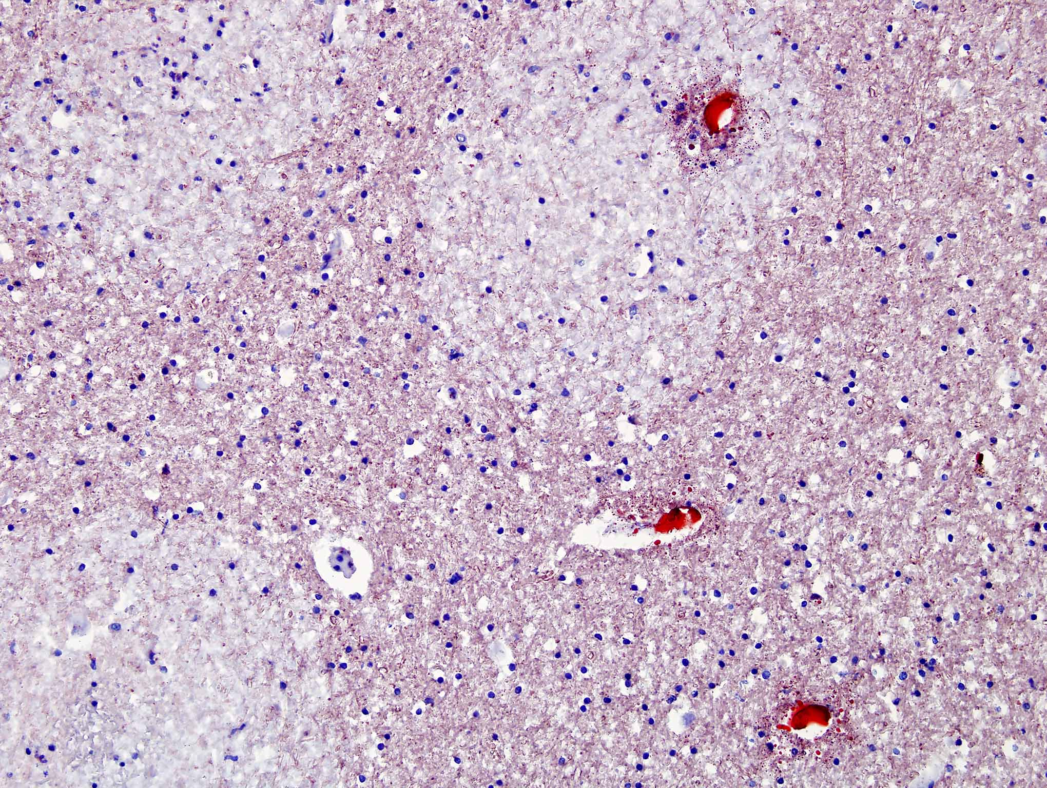
Fat embolism brain
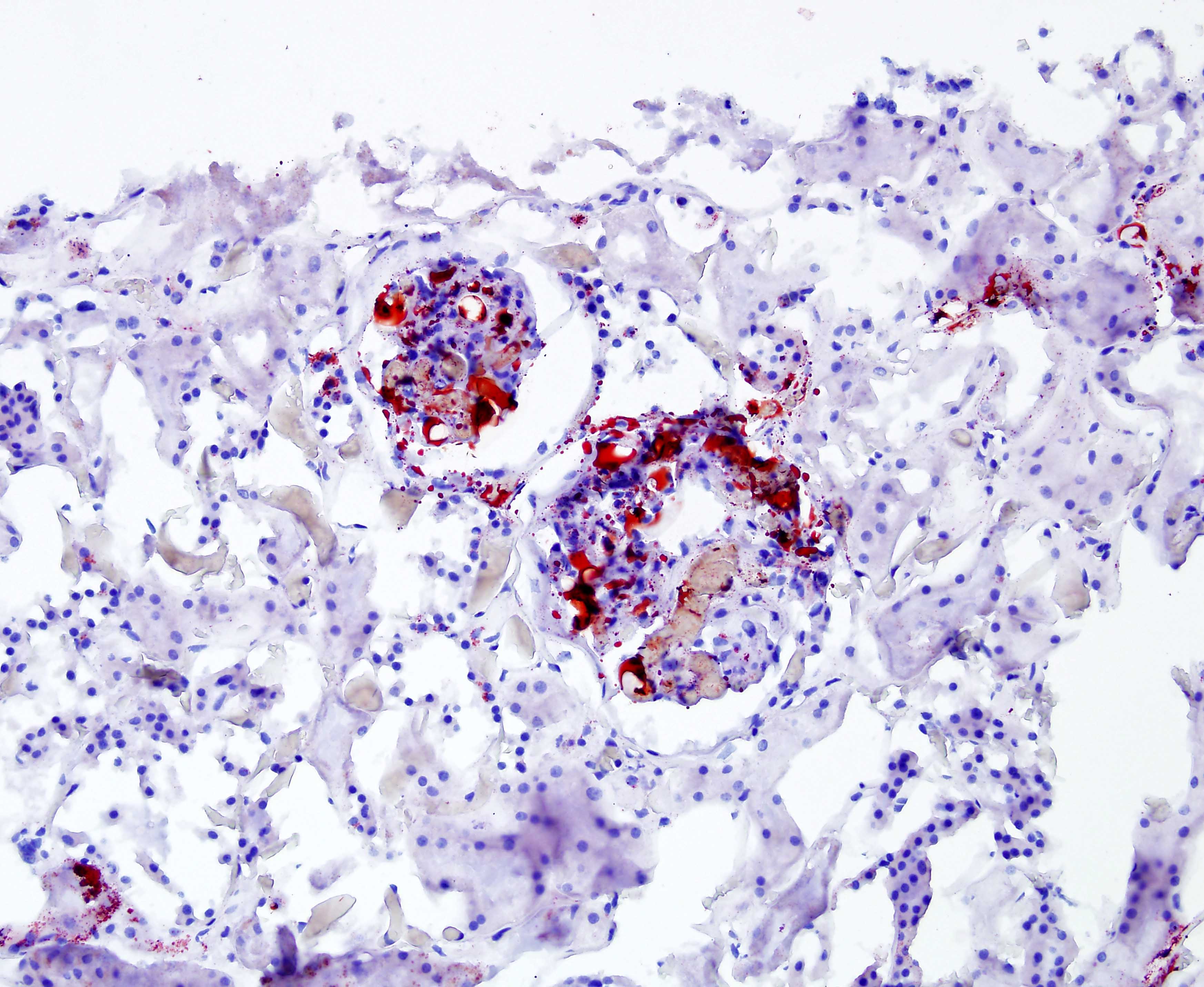
Fat embolism kidney
Microscopic (histologic) images
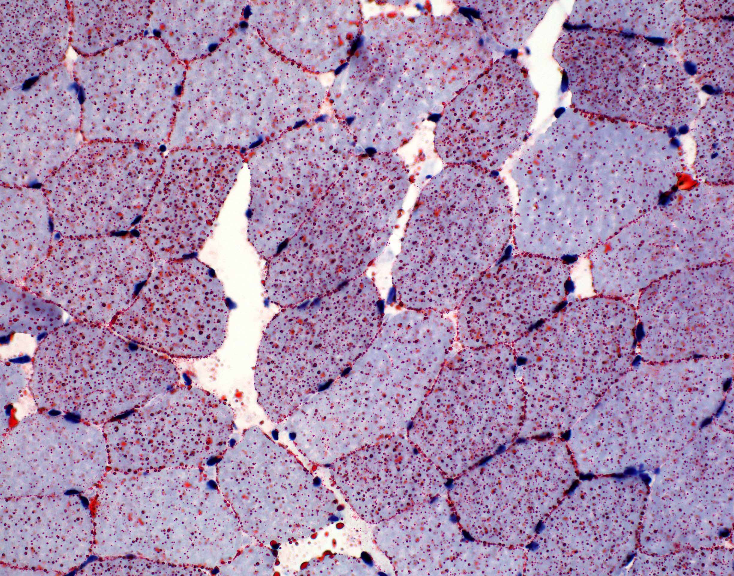
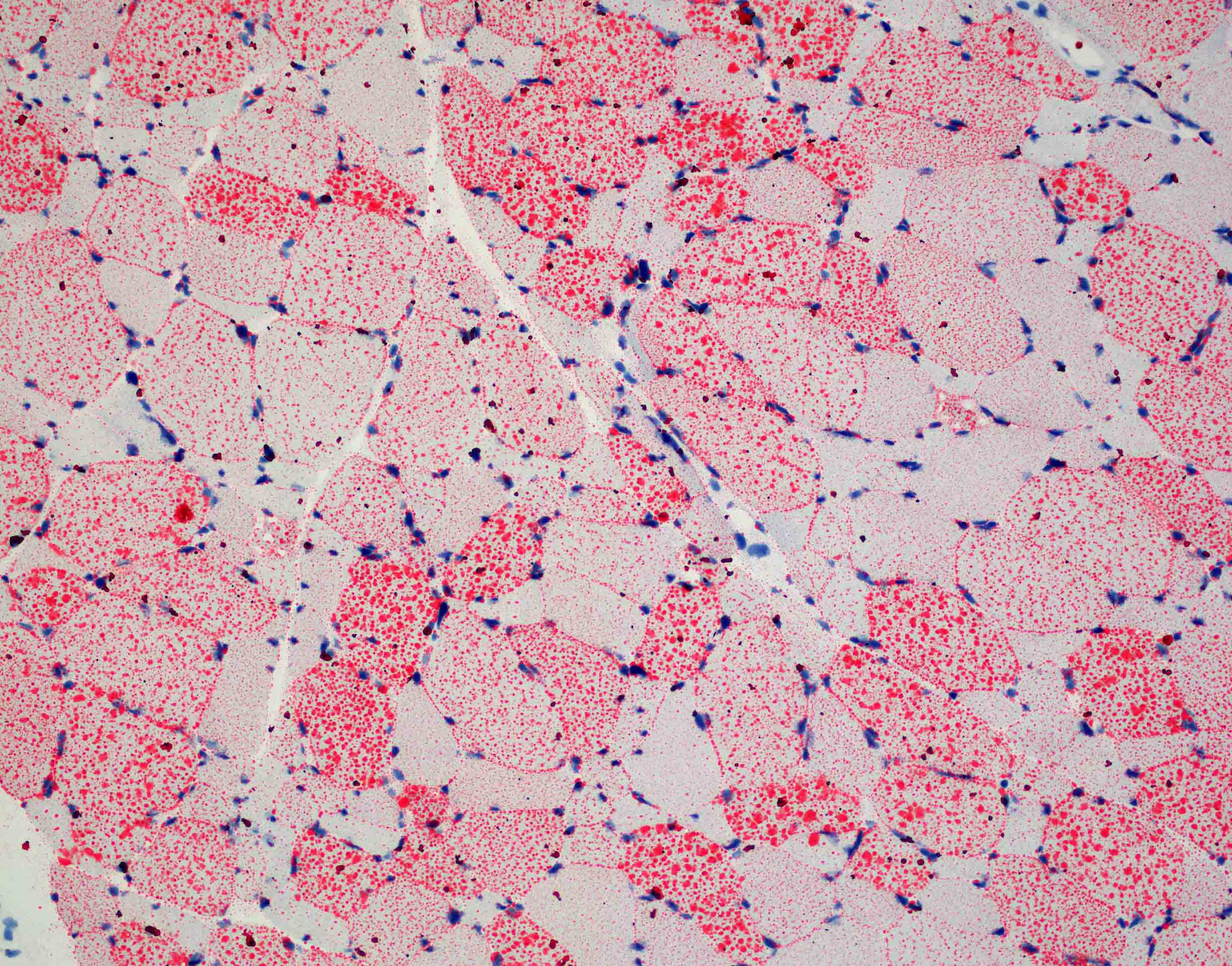
Positive staining - normal
- Skeletal muscle: see normal muscle image above
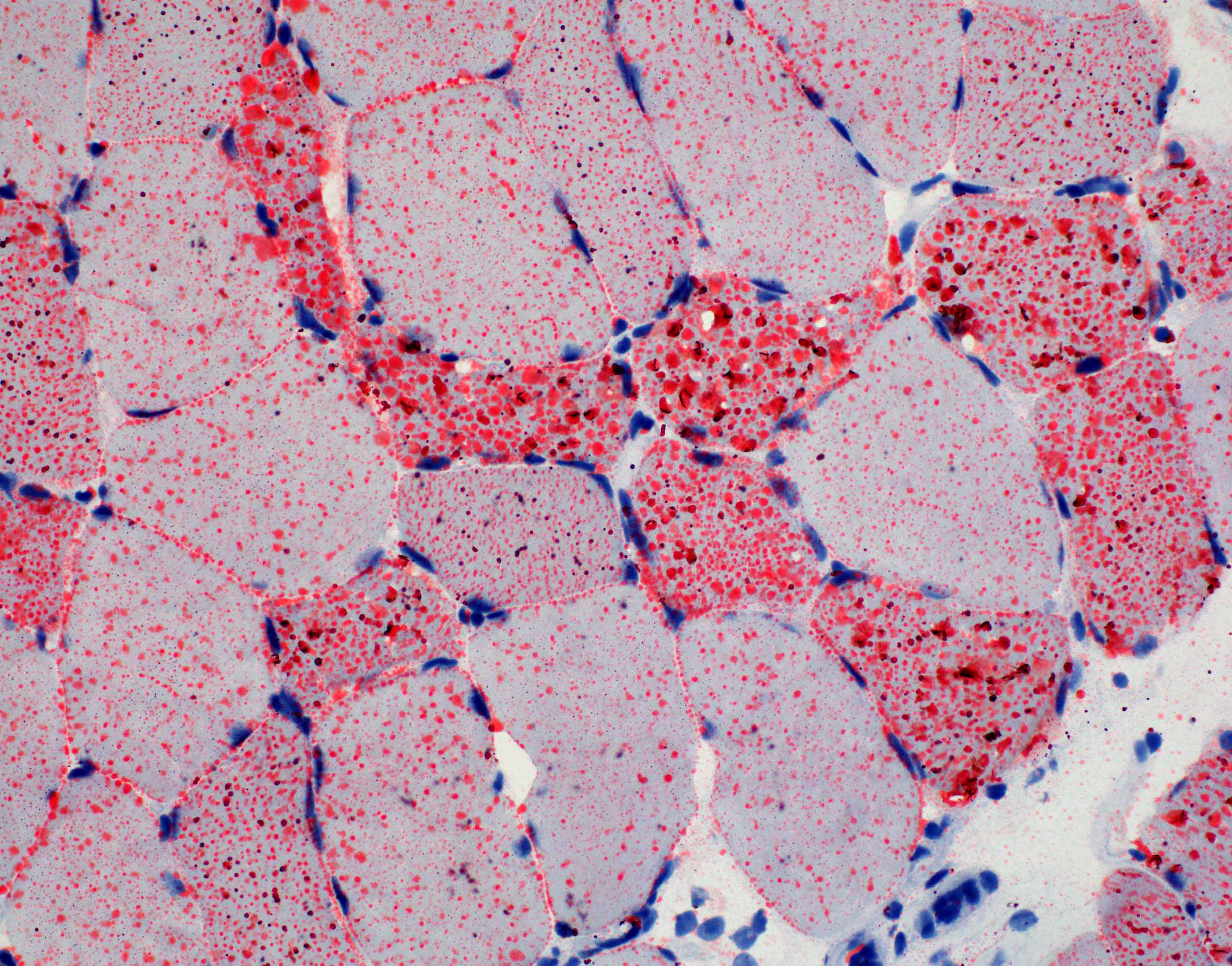
Positive staining - disease
- Lipid storage diseases in muscle biopsy
- Primary carnitine deficiency (Pathology 1985;17:161)
- Carnitine palmityl transferase deficiency (N Engl J Med 1975;292:443)
- Multiple acyl-CoA dehydrogenase deficiency (MADD) (Brain 1988;111:815)
- Neutral lipid storage disease with myopathy (Nat Genet 2007;39:28)
- Neutral lipid storage disease with ichthyosis (Biochem Biophys Res Commun 2008;369:1125)
- Lipin 1 deficiency (J Inherit Metab Dis 2012;35:1119)
- Use in general pathology
- Detection of fat embolism (Hum Pathol 1981;12:753, J Forensic Sci 1987;32:1426)
- Assessing steatosis in liver transplant biopsy (Transplant Proc 2018;50:3539, Virchows Arch 2014;464:165)
- Highlighting lipid laden macrophages in cytology or lung transplant biopsies (Cytopathology 2010;21:245, J Heart Lung Transplant 2010;29:859)
Sample pathology report
- Right quadriceps, biopsy:
- Lipid storage myopathy (see comment)
- Comment: The morphologic features of this biopsy are those of a lipid storage myopathy and include conspicuous vacuolar change, staining of the vacuoles in the oil red O stain and excess sarcoplasmic lipid vacuoles by ultrastructural examination. The cause of the lipid accumulation is not apparent in this biopsy. The differential diagnosis of adult onset lipid storage myopathy include primary and secondary carnitine deficiency, as well as a number of rarer entities including multiple acyl-CoA dehydrogenase deficiency, lipin 1 deficiency, neutral lipid storage disease with ichthyosis and neutral lipid storage disease with myopathy. Serum carnitine and acylcarnitine analyses, as well as urine organic acid analysis may be of additional diagnostic value. Cardiomyopathy and fatty liver disease should be ruled out by appropriate clinical means.
Board review style question #1
Which of the following special stains can be used to highlight both lipid droplets and myelin in muscle biopsies?
- Gomori trichrome
- Oil red O
- Sudan black
- Verhoeff van Gieson (VVG) stain
Board review style answer #1
C. Sudan black. Gomori trichrome can stain myelin red but does not stain lipid. Oil red O stains lipid red but does not stain myelin. VVG stains elastin and myelin but not lipid.
Comment Here
Reference: Oil red O
Comment Here
Reference: Oil red O

