
Stains & CD markers
Treponema pallidum IHC


Copyright: 2020-2024, PathologyOutlines.com, Inc.
PubMed Search: Treponema pallidum IHC
Treponema pallidum IHC
Editorial Board Member: Raul S. Gonzalez, M.D.
Editor-in-Chief: Debra L. Zynger, M.D.
Last author update: 22 May 2023
Last staff update: 22 June 2023
Copyright: 2020-2024, PathologyOutlines.com, Inc.
PubMed Search: Treponema pallidum IHC

Table of Contents
Definition / general | Essential features | Terminology | Pathophysiology | Clinical features | Interpretation | Uses by pathologists | Prognostic factors | Microscopic (histologic) images | Virtual slides | Positive staining - normal | Positive staining - disease | Molecular / cytogenetics description | Board review style question #1 | Board review style answer #1Cite this page: Aghighi M, Gottesman SP. Treponema pallidum IHC. PathologyOutlines.com website. https://www.pathologyoutlines.com/topic/stainstreponemaihc.html. Accessed April 25th, 2024.
Definition / general
- Syphilis is a sexually transmitted disease caused by Treponema pallidum, a bacterium discovered in 1905 by Schaudinn and Hoffman who initially named it Spirochaeta pallida (J Med Life 2014;7:4)
- T. pallidum can be localized on formalin fixed paraffin embedded tissue; the antibody has a rabbit purified IgG fraction (J Cutan Pathol 2004;31:595)
Essential features
- Immunohistochemical stain for T. pallidum is more sensitive (71% sensitive) than silver stains - Warthin-Starry or Steiner (41% sensitive) (J Cutan Pathol 2004;31:595)
- Diaminobenzidine chromogen can be problematic (melanin pigment on dendritic melanocytes can be confused for spirochetes)
- Thus, some labs use fast red chromogen, in which the spirochetes stain red
Terminology
- Anti-Treponema pallidum immunohistochemical stain
Pathophysiology
- In patients with syphilis, T. pallidum spirochetes show an epitheliotropic and vasculotropic pattern (Hum Pathol 2009;40:624)
Clinical features
- Primary syphilis: painless chancre with nontender lymphadenopathy 1 - 3 weeks after exposure
- Secondary syphilis
- Papulosquamous thin papules on the trunk and extremities, palms and soles, fever and adenopathy
- Rash may resemble a drug eruption, pityriasis rosea and psoriasis
- May present as moth eaten alopecia on the scalp, mucous patches on tongue
- Tertiary syphilis: may present with gummatous lesions, neurologic or cardiovascular symptoms (Infect Dis Clin North Am 2013;27:705)
Interpretation
Uses by pathologists
- Although serologic studies remain the gold standard, for histologic evaluation, T. pallidum IHC is preferred in evaluating mucocutaneous biopsies suspicious for syphilis (J Cutan Pathol 2004;31:595)
Prognostic factors
- Missed and delayed syphilis diagnosis is a public health hazard as well as a personal health risk, as neurosyphilis, vision loss, aortitis, aortic aneurysm, aortic insufficiency, coronary stenosis and myocarditis are serious complications (CDC: Syphilis - CDC Fact Sheet [Accessed 23 May 2023], Arch Cardiol Mex 2006;76:S189)
Microscopic (histologic) images
Contributed by Silvija P. Gottesman, M.D.
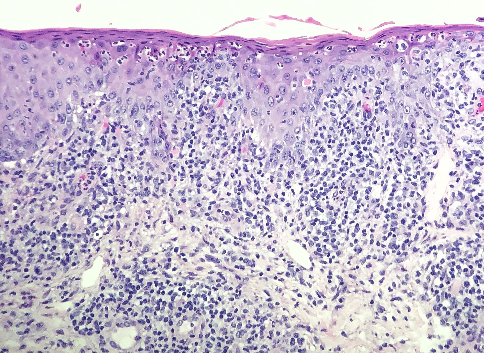
Lymphoplasmacytic infiltrate
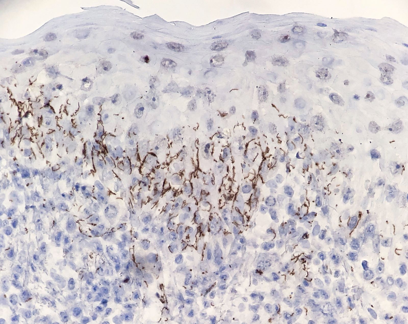
Spirochetes in epidermis
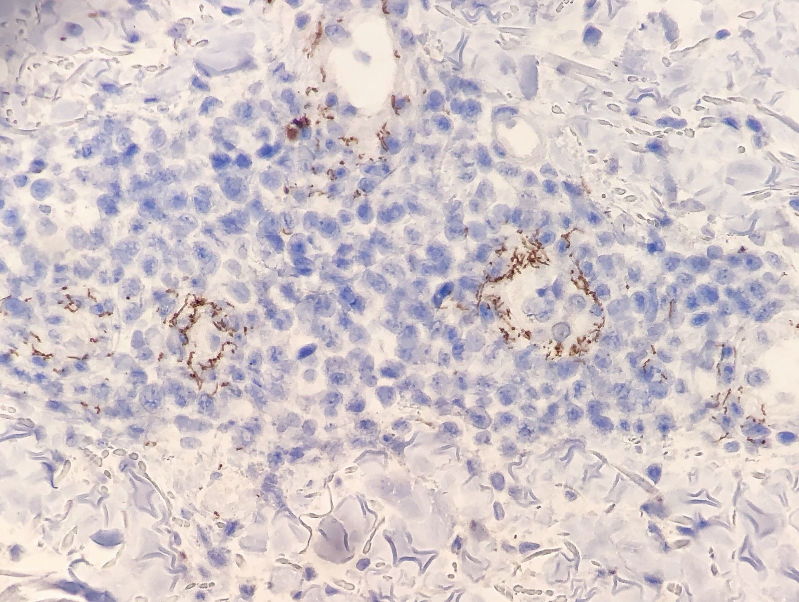
Spirochetes in dermal vessel walls
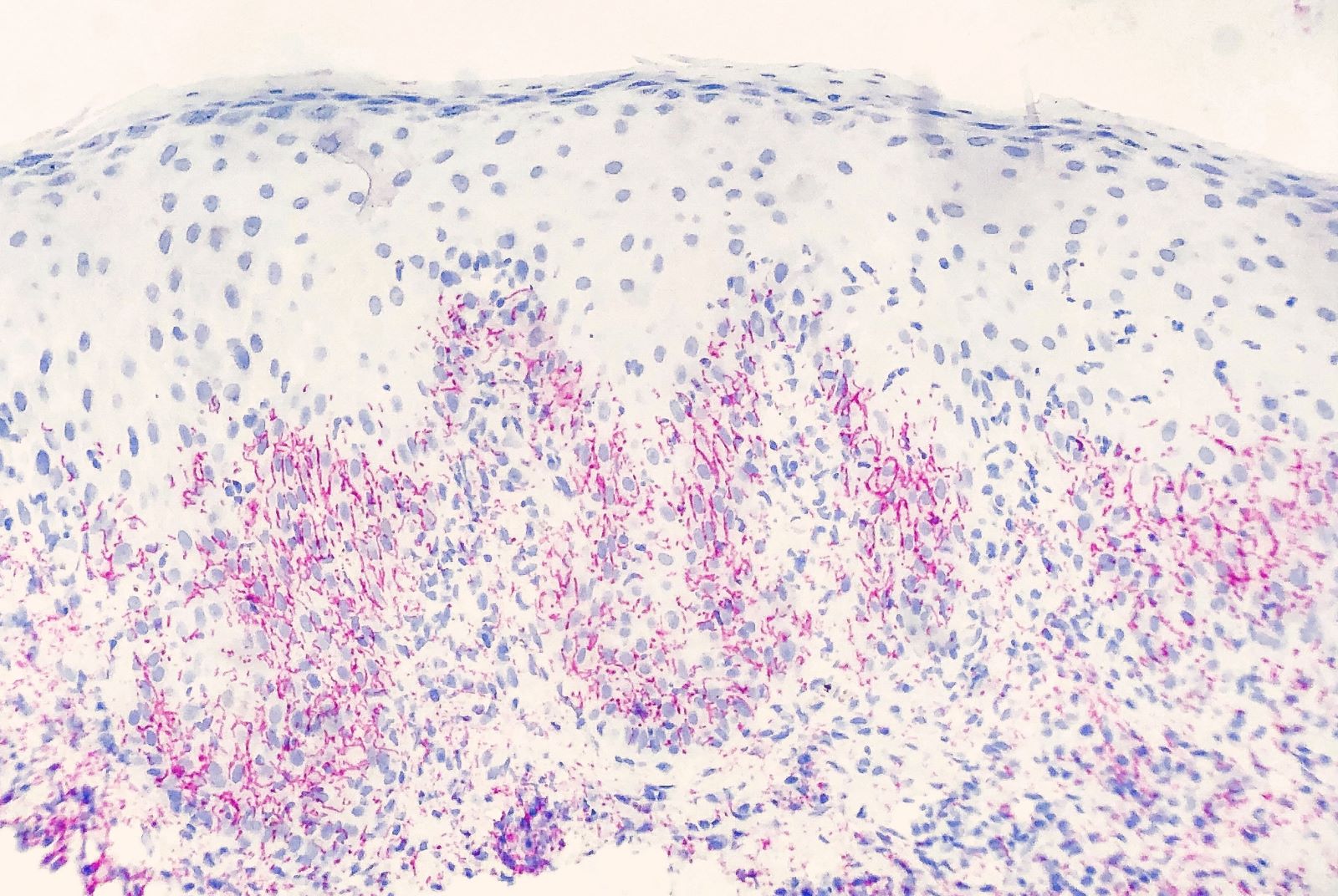
Spirochetes in epidermis
Virtual slides
Images hosted on other servers:

Spirochetes highlighted with IHC (brown)

Subtle spirochetes with IHC (red)

Spirochetes highlighted with IHC (brown)
Positive staining - normal
- None
Positive staining - disease
- Highlights spirochetes in lower half of epidermis, mucosa and dermal vessels (within vessel walls and in the perivascular space) (Hum Pathol 2009;40:624)
- Human intestinal spirochetosis (Diagn Pathol 2018;13:7)
- Other spirochetes such as Borrelia burgdorferi and Brachyspira intestinal spirochetes (Am J Dermatopathol 2019;41:924, Arch Pathol Lab Med 2016;140:1021)
- Cross reactivity has been seen in Mycobacteria marinum and Mycobacterial leprae skin specimens (Am J Dermatopathol 2012;34:559)
Molecular / cytogenetics description
- Polymerase chain reaction (PCR) may be used to detect T. pallidum in mucocutaneous lesions and is more sensitive and specific than T. pallidum immunohistochemistry (Br J Dermatol 2011;165:50)
Board review style question #1
A 35 year old pregnant woman presented to clinic with fever, fatigue and a papulosquamous eruption on the trunk and extremities that includes the palms and soles. Which one of the following stains is the most sensitive for diagnosis of syphilis in biopsy specimens?
- Giemsa stain
- Gram stain
- Steiner stain
- T. pallidum IHC stain
- Warthin-Starry stain
Board review style answer #1
D. T. pallidum IHC stain. Answer A is incorrect because, while Giemsa stain can highlight spirochetes, it has not been routinely used in skin tissue preparations. Its spirochete sensitivity in blood smear preparations has been estimated at 25% and higher in thick smear preparations (J Clin Microbiol 1999;37:2027). Answer B is incorrect because Gram stain does not highlight Treponema pallidum spirochetes. Answers C and E are incorrect because immunohistochemical stain for T. pallidum is more sensitive (71% sensitive) than silver stains Warthin-Starry or Steiner (41% sensitive).
Comment Here
Reference: Treponema pallidum IHC
Comment Here
Reference: Treponema pallidum IHC

