
Stains & CD markers
Villin

Copyright: 2003-2024, PathologyOutlines.com, Inc.
PubMed Search: Villin[TI] pathology
Villin
Author: Wafaey Gomaa, M.D., Ph.D.
Deputy Editor-in-Chief: Raul S. Gonzalez, M.D.
Last author update: 30 June 2020
Last staff update: 12 August 2021
Copyright: 2003-2024, PathologyOutlines.com, Inc.
PubMed Search: Villin[TI] pathology

Table of Contents
Definition / general | Essential features | Interpretation | Uses by pathologists | Prognostic factors | Microscopic (histologic) description | Microscopic (histologic) images | Positive staining - normal | Positive staining - disease | Negative staining | Board review style question #1 | Board review style answer #1Cite this page: Gomaa W. Villin. PathologyOutlines.com website. https://www.pathologyoutlines.com/topic/stainsvillin.html. Accessed April 20th, 2024.
Definition / general
- One of the gelsolin family of calcium regulated actin binding proteins
- First isolated and characterized in the microvilli of intestinal epithelium and later found in the brush of many absorptive epithelia
- Intestinal microvilli in the apical membrane (brush border) are maintained by bundles of parallel actin filaments that are organized by multiple actin binding proteins including villin (Am J Pathol 2012;180:1509)
- Expressed in tumors with enteric differentiation
Essential features
- Normally expressed in the brush border of epithelial cells lining the gastrointestinal tract, hepatobiliary tract and renal proximal convoluted tubules
- In colorectal carcinoma, villin is highly expressed
- In practice, villin can be included in the panel used for metastatic carcinoma to detect colorectal origin
Interpretation
- Apical membranous (brush border) immunostaining in normal epithelium and tumors
- Cytoplasmic immunostaining in tumors
Uses by pathologists
- Main use is confirming the site of origin of metastatic carcinoma (Dabbs: Diagnostic Immunohistochemistry: Theranostic and Genomic Applications, 5th Edition, 2018)
- The following tumors are villin positive in varying percentage:
- Colorectal, gastric, duodenal and esophageal carcinoma (sensitive and relatively specific pan-GI tract marker)
- Gastrointestinal neuroendocrine marker
- Endometrial carcinoma
- Hepatocellular carcinoma
- Pulmonary adenocarcinoma, enteric type
Prognostic factors
- Loss of villin immunostaining in colorectal carcinoma is associated with poor differentiation and survival (ISRN Gastroenterol 2013;2013:679724)
Microscopic (histologic) description
- In normal colonic crypts, shows a well organized brush border pattern together with small cytoplasmic dots in all crypts (ISRN Gastroenterol 2013;2013:679724)
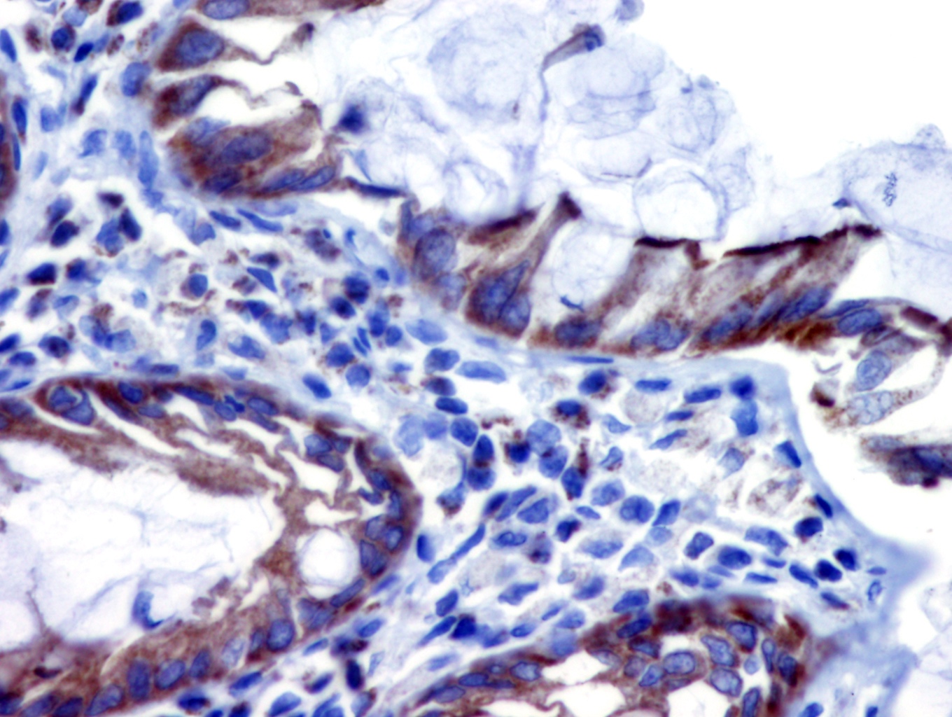
- Can highlight brush border or cytoplasm in positive tumors
Microscopic (histologic) images
Contributed by Wafaey Gomaa, M.D., Ph.D.
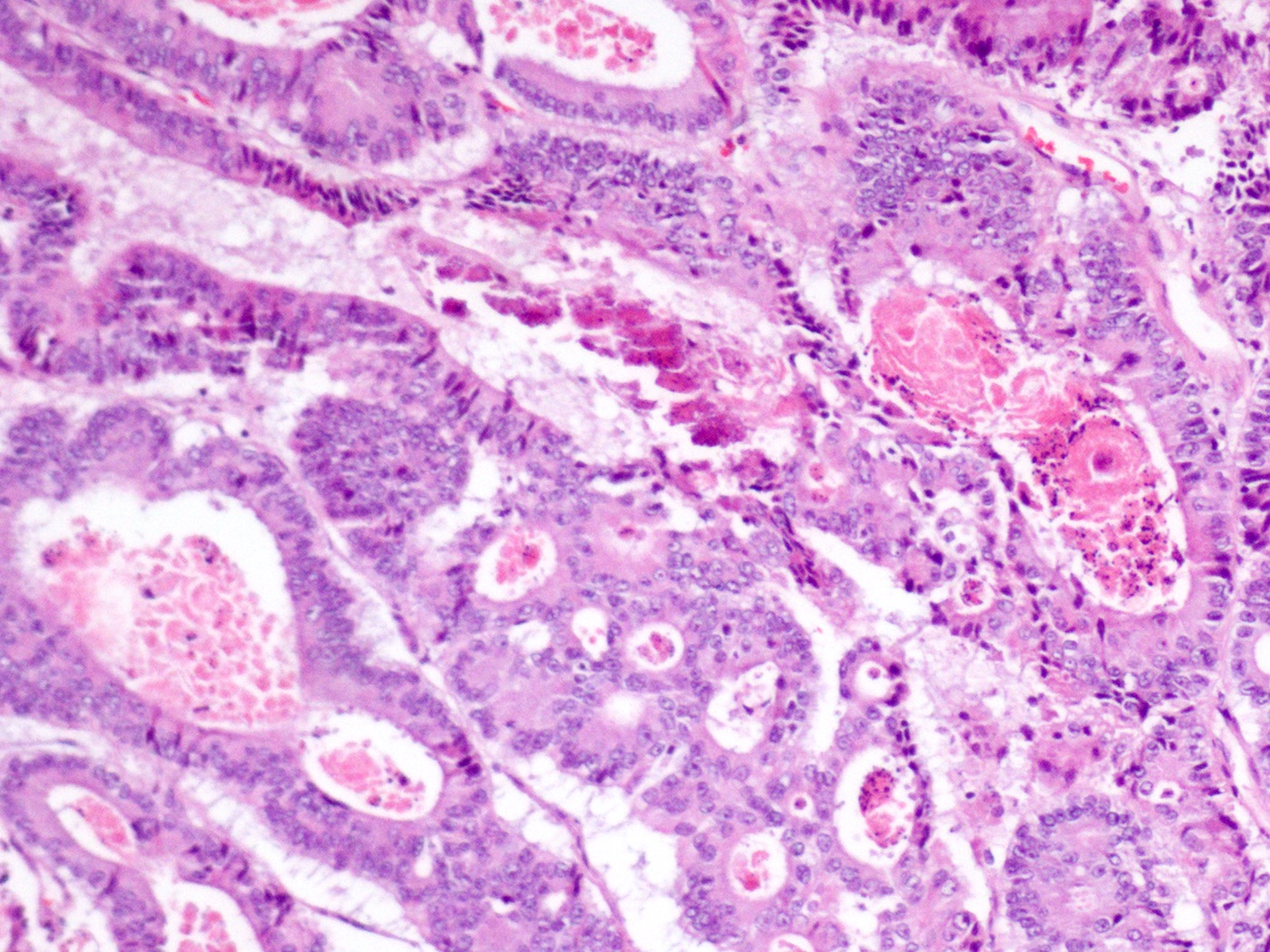
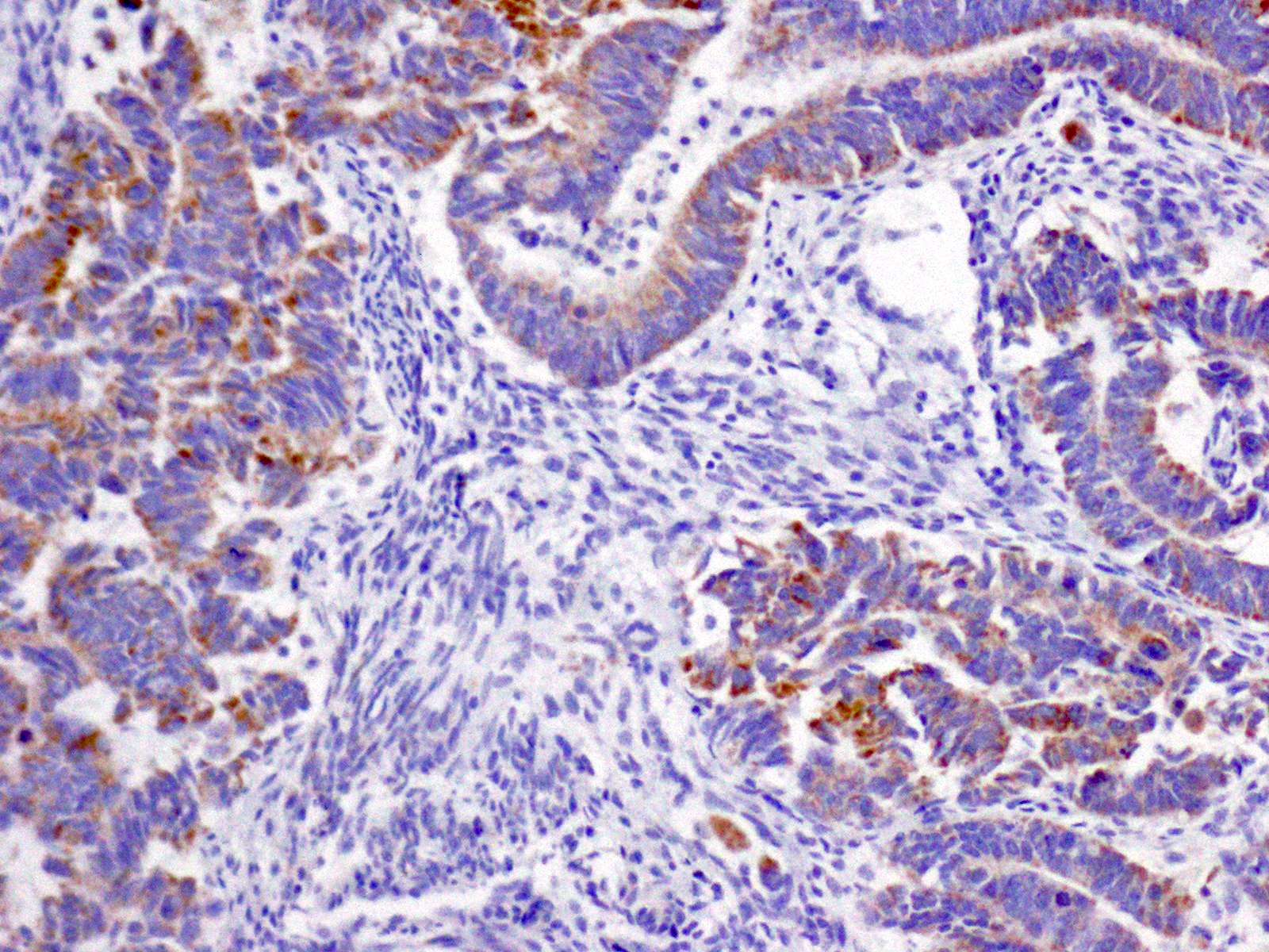
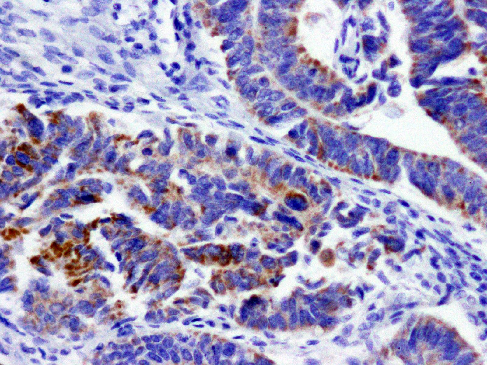
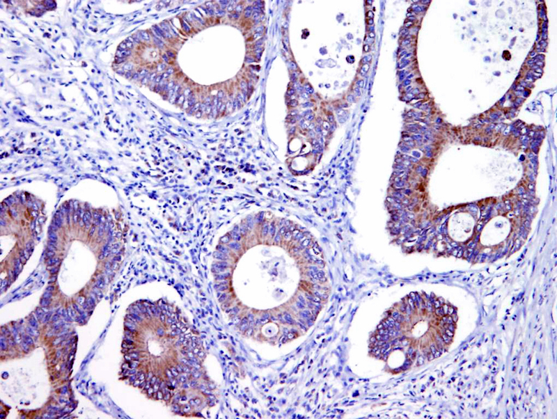
Moderately differentiated colorectal carcinoma
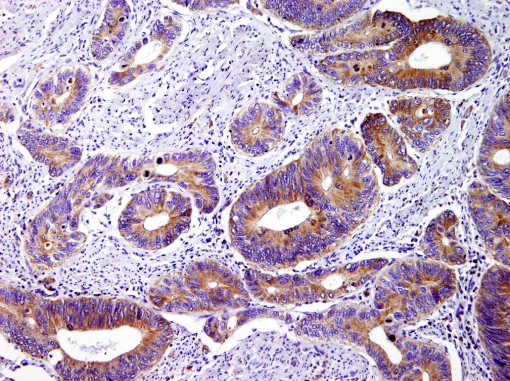
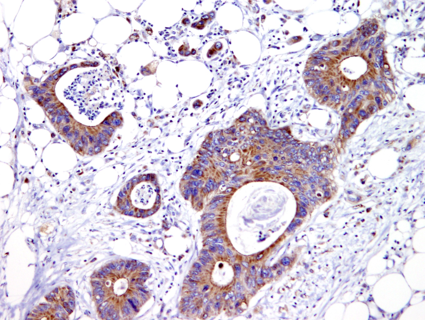
Colorectal carcinoma, villin
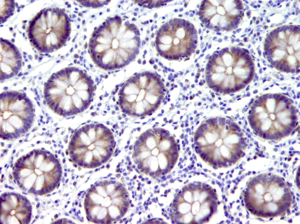

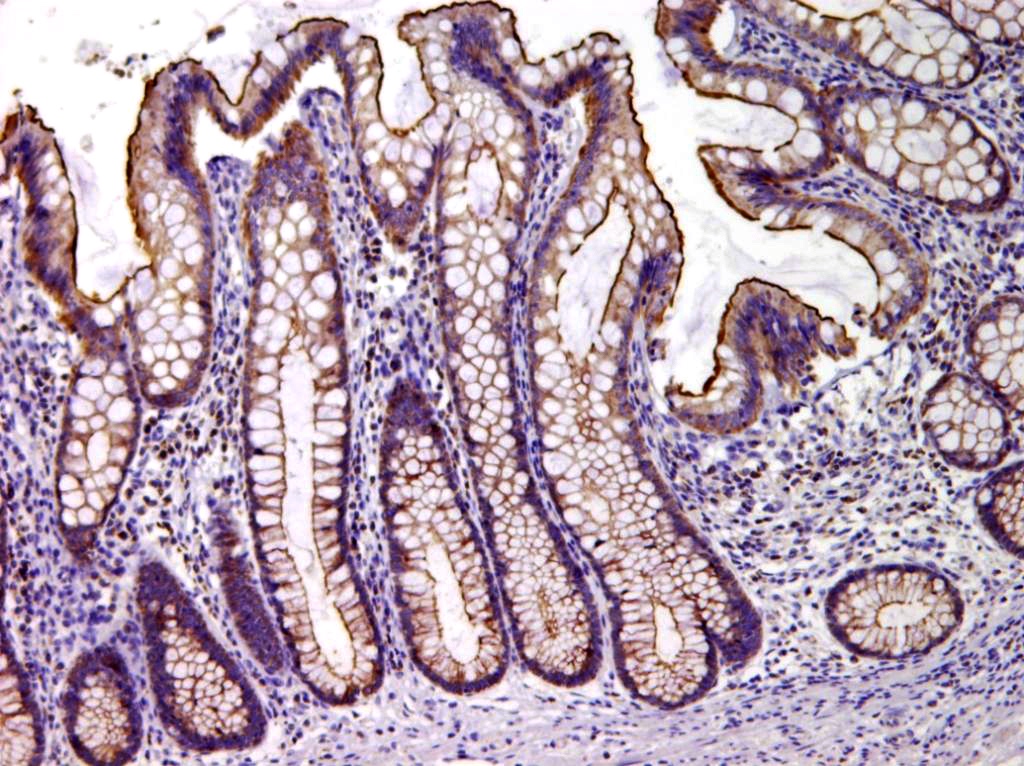
Normal colonic mucosa, villin
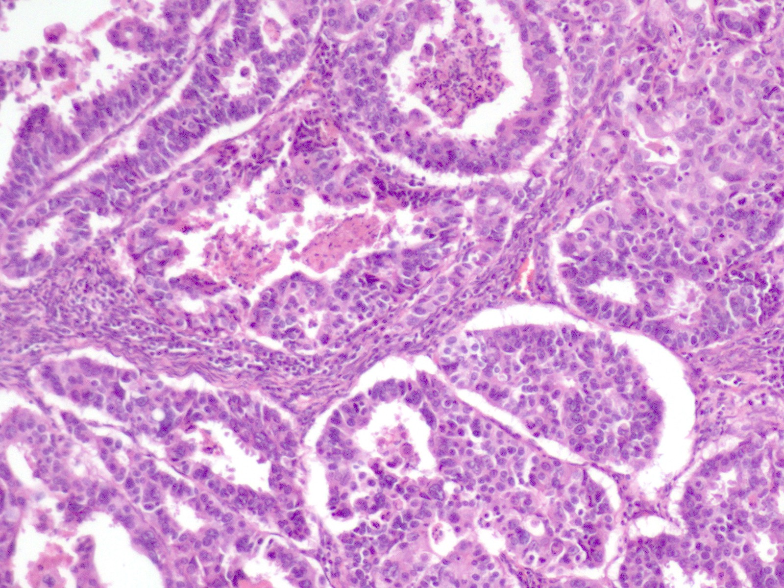
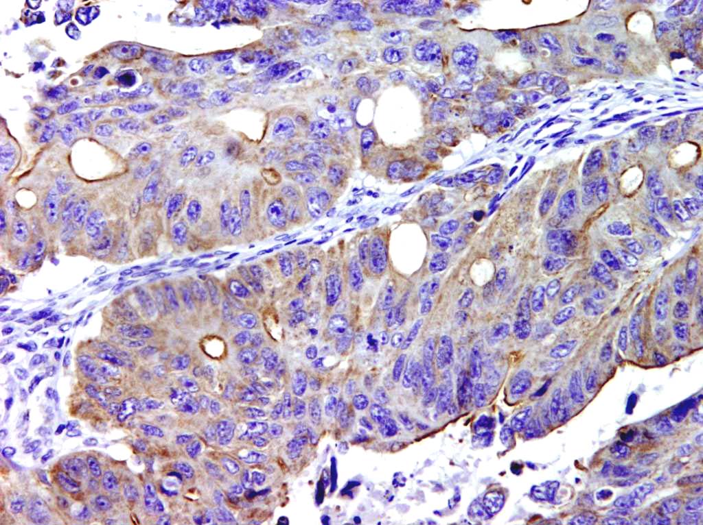
Endometrial carcinoma
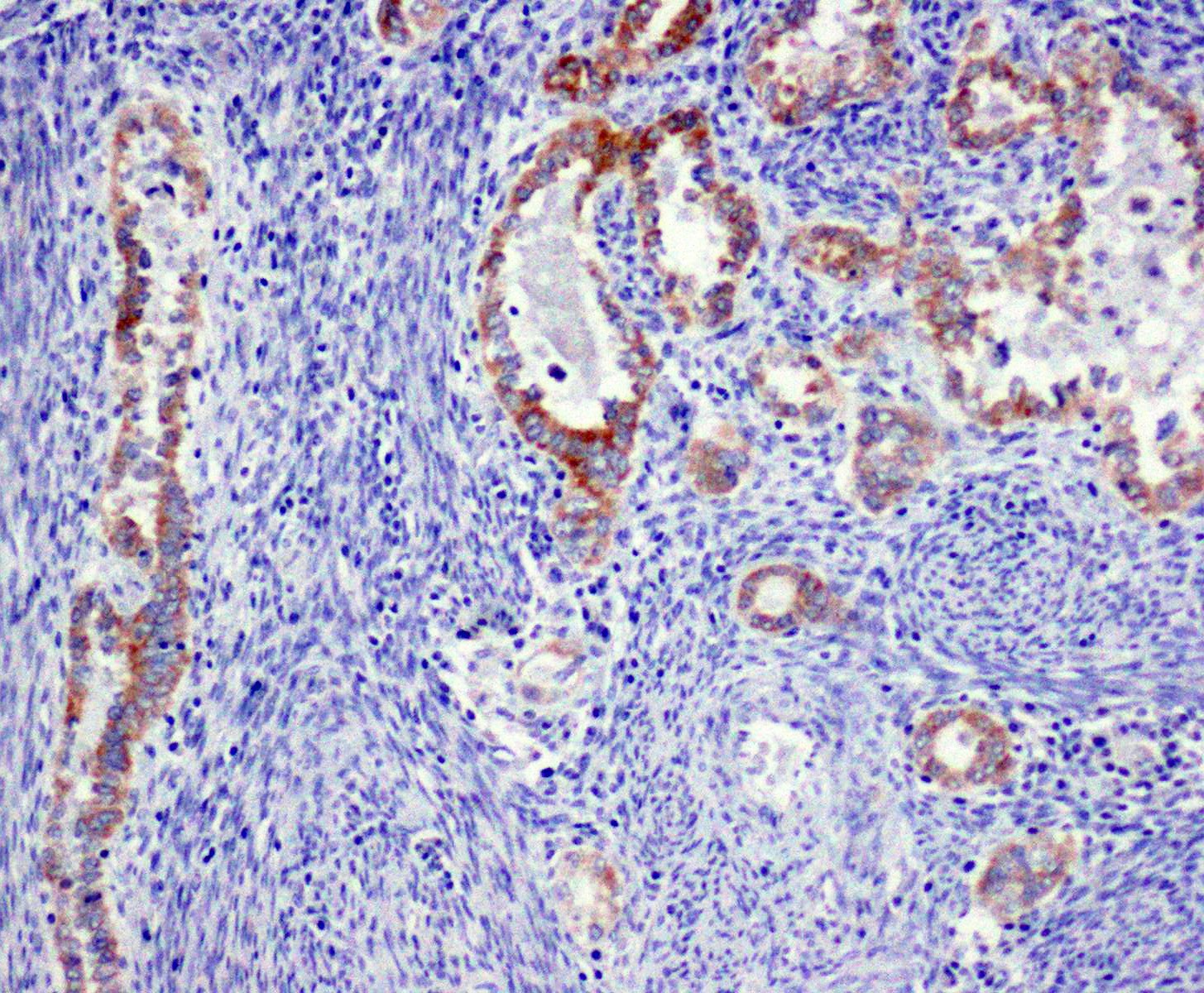
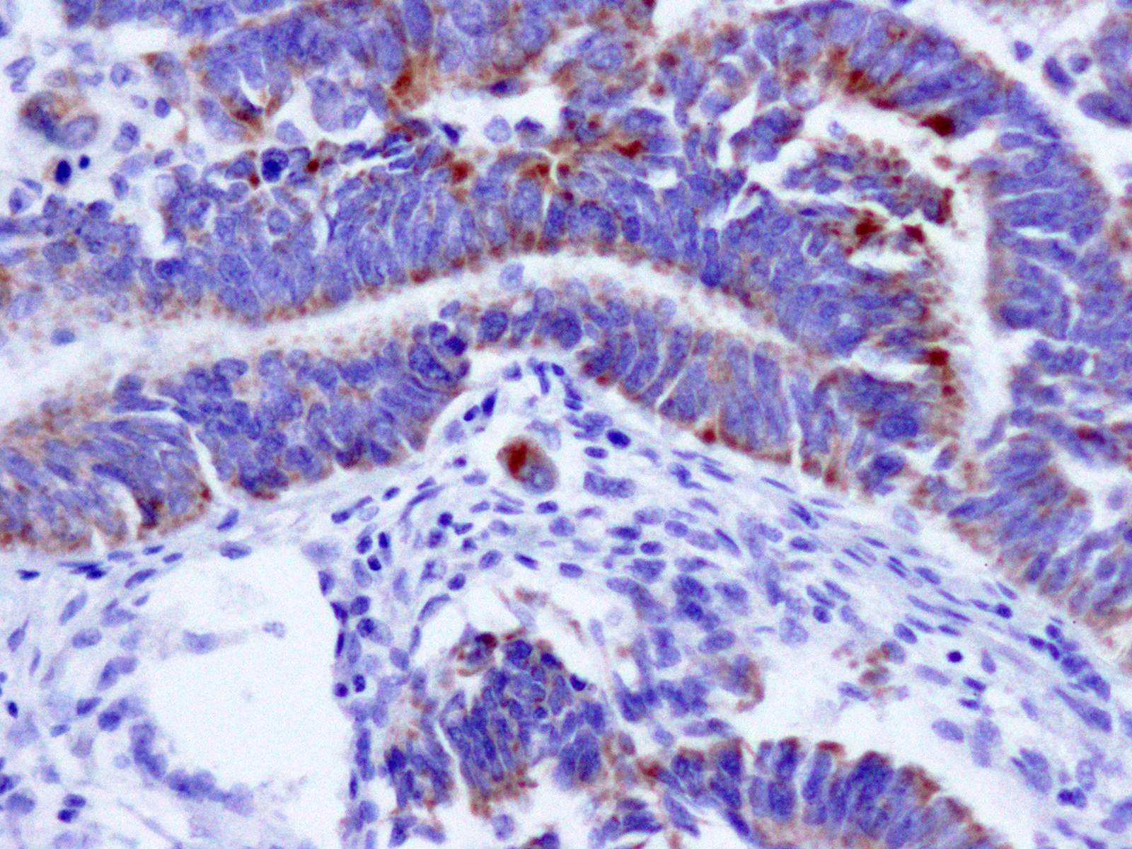
Endometrial carcinoma, villin
Positive staining - normal
- Normal colonic mucosa (ISRN Gastroenterol 2013;2013:679724)
- Normal small bowel (Ann Hepatol 2011;10:508, Lin: Handbook of Practical Immunohistochemistry, 2011)
- Brush border of bronchial glandular cells (Lin: Handbook of Practical Immunohistochemistry, 2011)
- Gastric pylorus (Dev Dyn 2002;224:90)
- Proximal renal tubules (Lin: Handbook of Practical Immunohistochemistry, 2011)
Positive staining - disease
- Colorectal carcinoma (82 - 84.9%) (ISRN Gastroenterol 2013;2013:679724, Lin: Handbook of Practical Immunohistochemistry, 2011)
- Gastrointestinal neuroendocrine tumors (72%) (Arch Pathol Lab Med 1999;123:812)
- Endocervical adenocarcinoma (93.3%) (Turk Patoloji Derg 2017;1:29)
Negative staining
- Bronchial and bronchiolar basal (reserve cells), bronchial epithelium in conducting airway, bronchiolar epithelium, Clara cells and type I and II pneumocytes (Hum Pathol 1998;29:390, Chu: Modern Immunohistochemistry, 1st Edition, 2009)
- Normal endometrium (EJGO 2017;38:560)
- Distal renal tubules (Lin: Handbook of Practical Immunohistochemistry, 2011)
- Renal glomeruli (Lin: Handbook of Practical Immunohistochemistry, 2011)
- Gastric mucosa (body) (Lin: Handbook of Practical Immunohistochemistry, 2011)
- Urothelial carcinoma (Lin: Handbook of Practical Immunohistochemistry, 2011, Arch Pathol Lab Med 2002;126:1057)
- Prostatic adenocarcinoma (Am J Surg Pathol 2003;27:303, Medicine (Baltimore) 2018;97:e13697)
- Breast carcinoma (Am J Surg Pathol 2003;27:303)
- Hepatocellular carcinoma (canalicular) (9% and 31%) (Ann Hepatol 2011;10:508, Hum Pathol 2002;33:1175)
- Lung adenocarcinoma (6% and 31.6%) (Chu: Modern Immunohistochemistry, 1st Edition, 2009, Hum Pathol 1998;29:390, Mol Carcinog 1998;23:234)
- Cholangiocarcinoma (20% and 22%) (Ann Hepatol 2011;10:508, Hum Pathol 2002;33:1175)
- Esophageal adenocarcinoma (17%) (Lin: Handbook of Practical Immunohistochemistry, 2011)
- Endometrial carcinoma (12.3% and 20%) (EJGO 2017;38:560, Turk Patoloji Derg 2017;1:29)
Board review style question #1
- A 61 year old woman presented with an abdominal nodule. The patient had multiple liver nodules that were previously biopsied and tumor cells were positive for CDX2, CK20 and the stain depicted in the image. The depicted stain is most likely which of the following?
- CK5/6
- CK7
- DOG1
- MelanA
- Villin
Board review style answer #1
E. Villin. The patient has metastasis to the liver. Positivity to CDX2 and CK20 suggests colorectal origin. Villin is the most relevant of the provided markers to confirm colorectal origin.
Comment Here
Reference: Villin
Comment Here
Reference: Villin

