

Thyroid & parathyroid
Other thyroid nonneoplastic
Calcification
Author: Andrey Bychkov, M.D., Ph.D.

Last author update: 1 February 2015
Last staff update: 5 September 2023
Copyright: 2003-2024, PathologyOutlines.com, Inc.
PubMed search: calcification thyroid

Table of Contents
Definition / general | Epidemiology | Sites | Pathophysiology | Diagnosis | Laboratory | Radiology description | Prognostic factors | Case reports | Clinical images | Gross images | Microscopic (histologic) images | Positive stains | Differential diagnosisCite this page: Bychkov A. Calcification. PathologyOutlines.com website. https://www.pathologyoutlines.com/topic/thyroidcalcification.html. Accessed April 19th, 2024.
Definition / general
- Deposition of calcium salts in thyroid gland
- More important to radiologists than pathologists
Epidemiology
- Prevalence in thyroidectomy specimens is 15% (Head Neck 2002;24:651)
- Found by ultrasound in 8% of benign (multinodular goiter) and 26% of malignant nodules (Head Neck 2002;24:651)
- Increases with advancing age
Sites
- Retrosternal thyroid / goiter tends to be calcified more heavily
Pathophysiology
- Dystrophic calcification of thyroid results from degenerative changes (calcified colloid and degenerated epithelium, psammoma bodies, old hemorrhage, vessel wall, etc.)
- Metastatic calcification is caused by elevated blood calcium / phosphate
- Stromal calcification may progress to bone formation (Mod Pathol 2009;22:887)
Diagnosis
- Core needle biopsy is superior to FNA for thyroid nodules with macrocalcification (Thyroid 2015;25:657)
Laboratory
- Hypercalcemia due to hyperparathyroidism, in rare cases of metastatic calcification
Radiology description
- Microcalcifications (< 2 mm and without acoustic shadow by ultrasound) in thyroid nodules are usually psammoma bodies
- Macrocalcifications (≥ 2 mm and with acoustic shadow) are secondary to tumor necrosis and can be seen in both benign and malignant nodules
- Peripheral (eggshell) calcifications surrounding the nodule are secondary to chronic degenerative changes
- Various patterns of calcification may be observed on Xray: nodular, flat, curvilinear, cloudy and a mixed type (Clin Radiol 1981;32:571)
Prognostic factors
- Microcalcification due to psammoma bodies is a strong predictor of thyroid carcinoma (Head Neck 2002;24:651, J Int Med Res 2012;40:350), but the association of macrocalcifications with malignancy is controversial (Thyroid 2013;23:1106)
Case reports
- 41, 49 and 72 year old women with osseous metaplasia and mature bone formation (Oncol Lett 2013;6:977)
- 49 year old woman with long standing goiter (Minerva Endocrinol 2000;25:81)
- 67 year old man with eggshell calcification (Cases J 2008;1:11)
- 74 year old man with metastatic calcification (J Nucl Med 1986;27:373)
- Unusual calcification in mixed papillary and follicular carcinoma (Radiology 1976;119:554)
- Association with hypothyroidism (Clin Radiol 1992;45:209)
Clinical images
Images hosted on other servers:

Eggshell calcified retrosternal thyroid
Gross images
Images hosted on other servers:

Muddy appearance
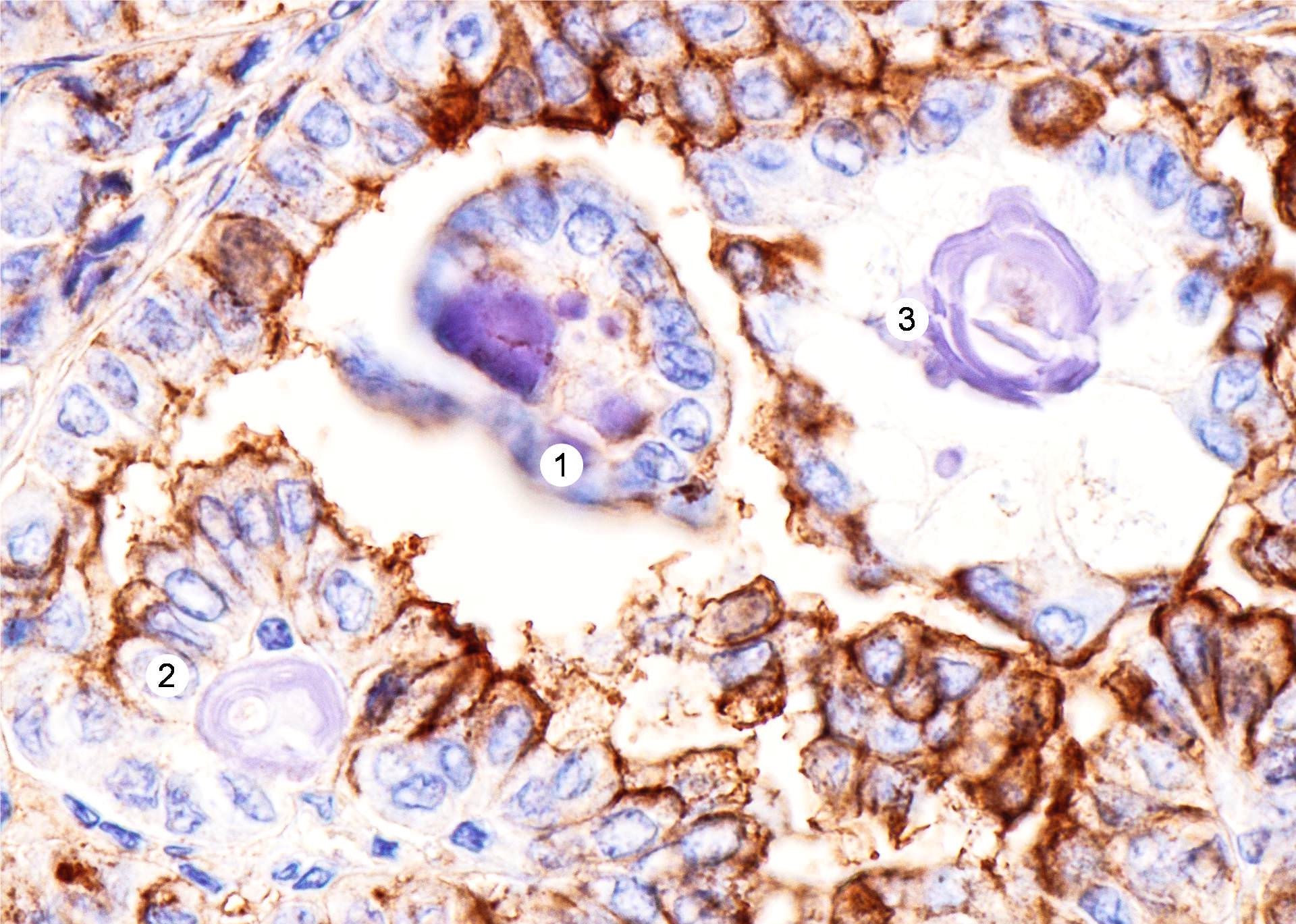
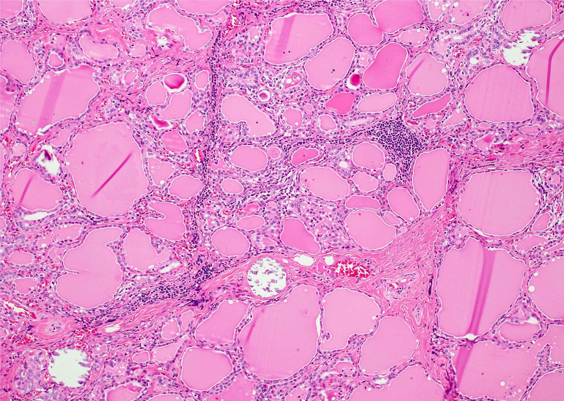
Microscopic (histologic) images
Contributed by Andrey Bychkov, M.D., Ph.D.
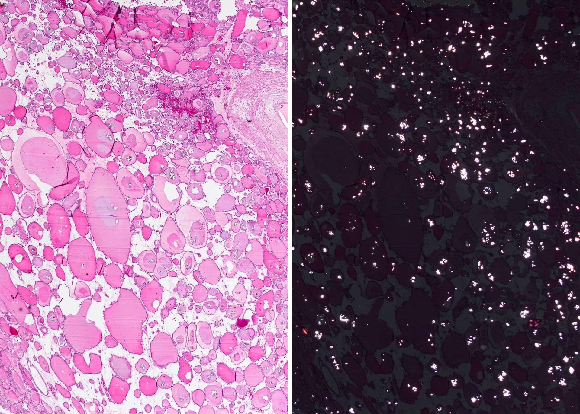
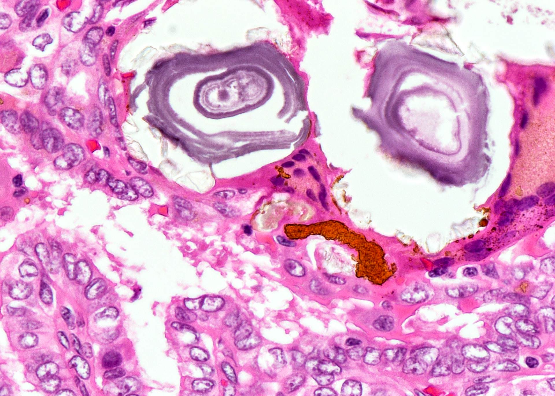
Calcium oxalate crystals (abundant)
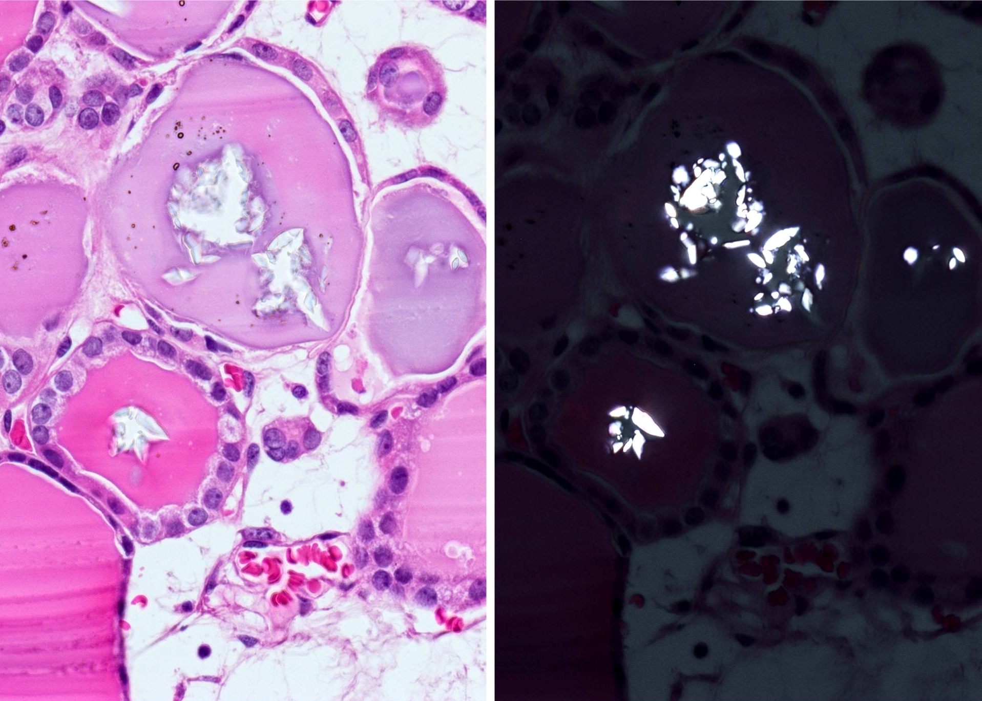
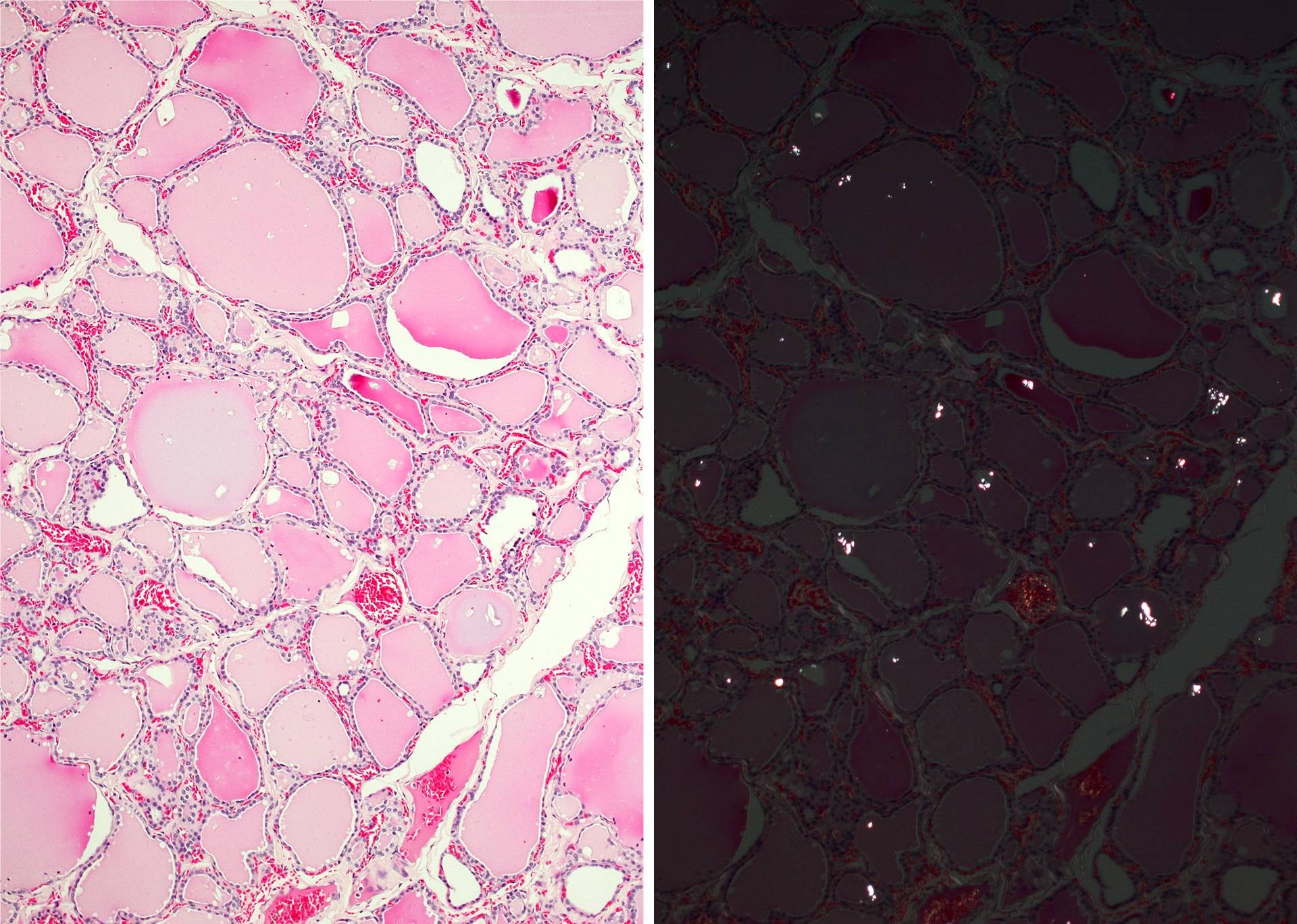
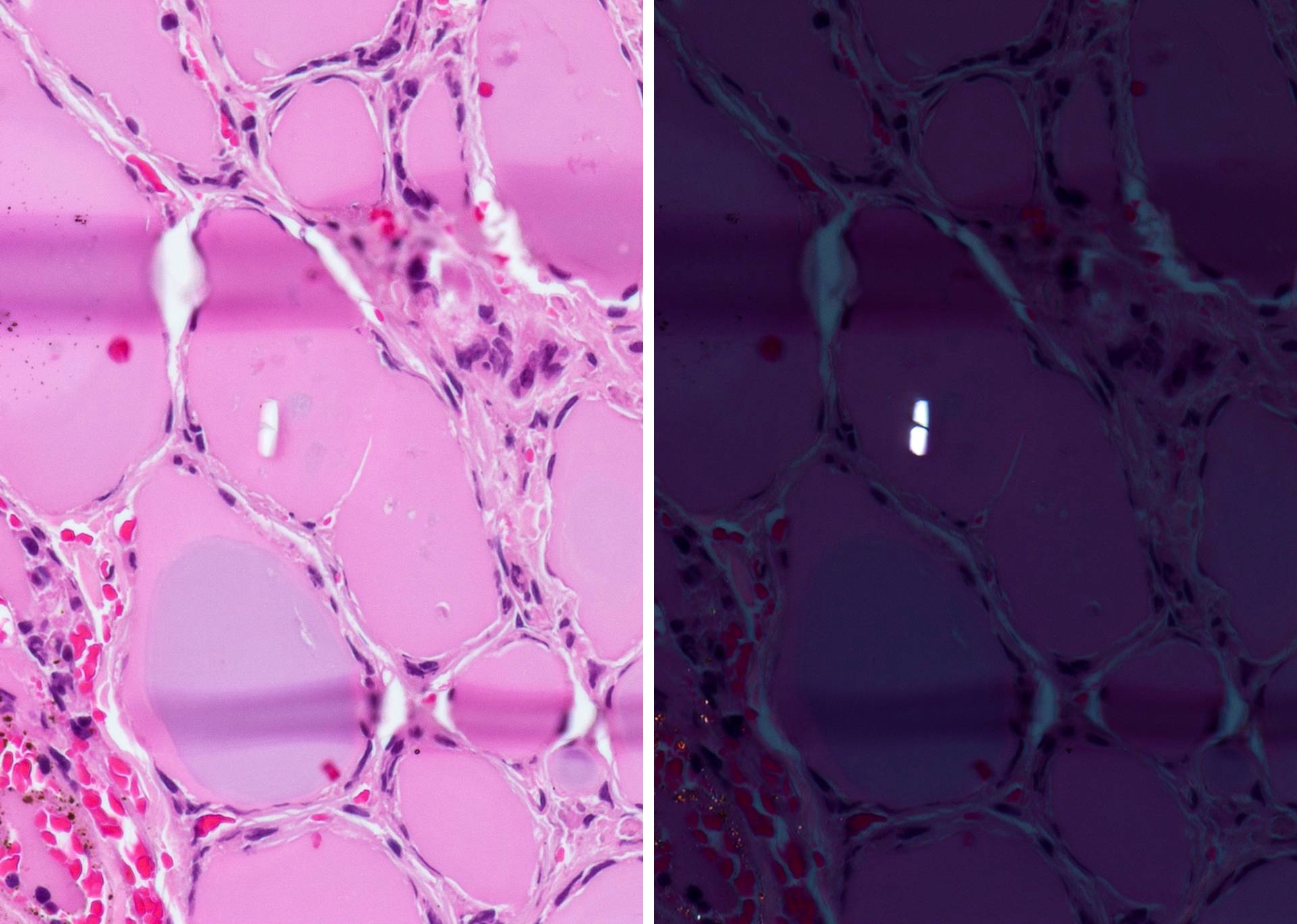
Calcium oxalate crystals
Papillary thyroid carcinoma
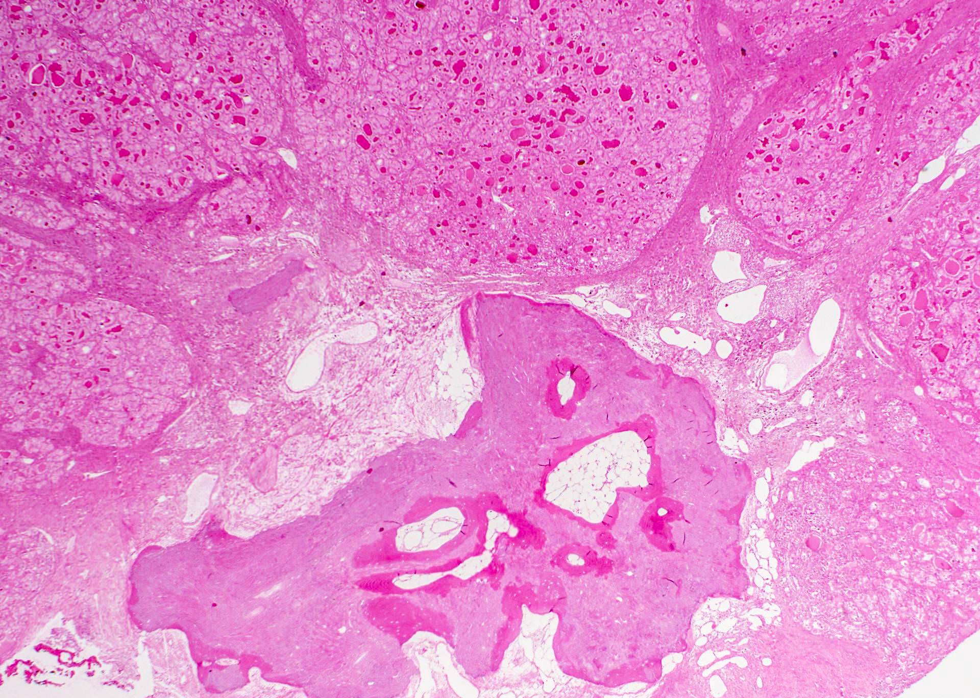
PTC follicular variant with ossification
Concentric layers of calcium deposition in tumor stroma
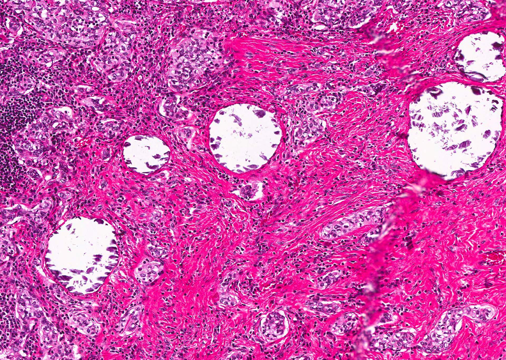
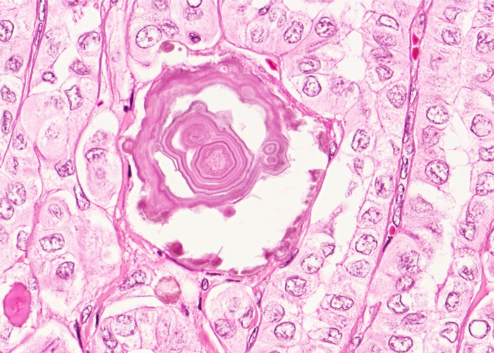
Psammoma bodies, laminated calcifications
Evolution in psammoma body in PTC
Psammoma bodies in benign appearing thyroid
Images hosted on other servers:

Extensive dystrophic calcification


Osseous metaplasia with fatty marrow

Calcification induced artifact
Positive stains
Differential diagnosis
- Psammoma bodies are highly specific for papillary thyroid carcinoma, but should be differentiated from dystrophic stromal calcifications and inspissated colloid, both of which lack concentric laminations
- Thyroid calcium oxalate crystals

