

Vulva & vagina
General
Anatomy & histology - vulva, vagina & female urethra
Editorial Board Member: Kyle Devins, M.D.
Deputy Editor-in-Chief: Gulisa Turashvili, M.D., Ph.D.


Last author update: 8 December 2023
Last staff update: 8 December 2023
Copyright: 2002-2024, PathologyOutlines.com, Inc.
PubMed Search: Vulva / vagina anatomy & histology


Table of Contents
Definition / general | Essential features | Terminology | Physiology / embryology | Diagrams / tables | Clinical images | Gross description | Microscopic (histologic) description | Microscopic (histologic) images | Virtual slides | Videos | Board review style question #1 | Board review style answer #1 | Board review style question #2 | Board review style answer #2Cite this page: Srivastava P, Lanjewar S. Anatomy & histology - vulva, vagina & female urethra. PathologyOutlines.com website. https://www.pathologyoutlines.com/topic/vulvaanatomy.html. Accessed April 19th, 2024.
Definition / general
- Vulva constitutes the portion of female genitalia that is external to the hymen
- Vagina is a fibromuscular tube that extends from the vestibule of vulva to uterine cervix
- Female urethra extends from the bladder to midurethra and exits the body between clitoris and vagina
Essential features
- Vulva is composed of mons pubis, clitoris, labia minora, labia majora, vulvar vestibule, vestibulovaginal bulbs, urethral meatus, hymen, Bartholin and Skene glands and ducts, vaginal introitus
- Vagina extends from vulva to uterine cervix and is derived from paired Müllerian ducts
Terminology
- Vulva
- Lies external to hymen and is limited by mons pubis anteriorly, anus posteriorly and inguinal gluteal folds laterally
- Vagina
- Fibromuscular canal that extends from the vestibule of vulva, between labia minora, to the uterine cervix
- Wolffian (mesonephric) duct, also known as Gartner duct, runs deeply along lateral vaginal walls
- Lymphatic drainage: external iliac nodes (upper third of the vagina), the common and internal iliac nodes (middle third) and the superficial inguinal and perirectal nodes (lower third)
- Female urethra
- Fibromuscular tube that takes urine from the urinary bladder to the exterior through the external urethral meatus
Physiology / embryology
- Germ cells from yolk sac migrate to the urogenital ridge, forming the epithelium and stroma of the gonads; genital tubercle becomes the clitoris and the parallel ridges become the labia minora
- Urorectal septum divides the cloaca into the urogenital sinus and anal canal; degeneration of the central portion of the urogenital membrane forms the hymen opening
- Lateral Müllerian ducts (paramesonephric ducts) give rise to upper vagina while the lower vagina is formed by the urogenital sinus
- Vaginal vestibule develops by the joining of the distal vagina and urogenital sinus
- Originates from endoderm, except near the urethra (ectoderm)
- Vestibular line of Hart marks the boundary between these tissues
- Epithelium of female urethra is derived from endoderm of the urogenital sinus while the surrounding connective tissue and smooth muscle tissue is derived from splanchnic mesenchyme (Sadler: Langman's Medical Embryology, 15th Edition, 2023, Am J Obstet Gynecol 1976;126:769)
Diagrams / tables
Images hosted on other servers:
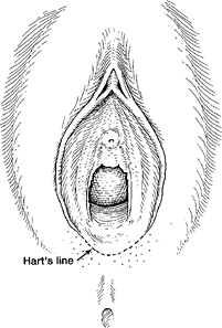
Hart line


Female urethra: embryology and anatomy
Clinical images
Images hosted on other servers:

Labia majora

Vestibule
Gross description
- Vulva
- Mons pubis
- Anteriormost region of vulva and is anatomically located over the prominence of pubic symphysis
- Hymen
- Corresponds to the distalmost extent of vagina and posterior aspect of vulvar vestibule
- Clitoris
- Erectile tissue similar to corpora cavernosa of penis and is located anterior to frenulum at the junction of labia minora
- Labia majora
- Form the lateral boundaries of the vulva
- Fuse anteriorly into mons pubis
- Posteriorly terminate 3 - 4 cm anterior to the anus where they are united by posterior commissure or fourchette
- Labia minora
- Are medial to labia majora and lateral to vulvar vestibule
- Anteriorly, labia minora divide into 2 parts; one part passes over clitoris to form prepuce and the other joins beneath clitoris and forms frenulum
- Posteriorly, they blend with medial surfaces of labia majora
- Hart line
- Lies at the inferior junction between vulvar vestibule and perineal skin
- Vestibule
- Area between the hymen (anteriorly), Hart line (posterolaterally) and labia minora (anterolaterally)
- Includes vaginal opening and urethral orifice
- Structures found in vestibule include major vestibular (Bartholin) glands, minor vestibular glands, periurethral (Skene) glands, urethra
- Bartholin glands
- Correspond to bulbourethral glands in male
- These are mucin producing glands and are located posterolaterally in the vulva
- Minor vestibular glands
- Correspond to penile glands of Littre
- Concentrically located within the vestibule
- Mons pubis
- Vagina
- Posterior to urinary bladder (from which it is separated by fibroadipose tissue)
- Anterior to rectum (from which is separated by rectouterine space in the upper 25%, rectovaginal septum in the middle portion and sphincter musculature in the distal portion of anal canal)
- Female urethra
- Extends from the bladder to the vestibule of the vagina to its opening posterior to the clitoris
- Measures 4 cm in length
- Striated muscle of the urogenital diaphragm forms the external voluntary sphincter as the urethra penetrates it
- Reference: StatPearls: Anatomy, Abdomen and Pelvis - Female External Genitalia [Accessed 21 August 2023]
Microscopic (histologic) description
- Hymen
- Nonkeratinized stratified squamous epithelium
- Labia majora
- Composed of keratinized stratified squamous epithelium with hair follicles and eccrine, apocrine and sebaceous glands
- Labia minora
- Composed of keratinized stratified squamous epithelium, usually no adnexa
- Stroma
- Composed of stromal cells that can be spindled, stellate, fusiform and may have large multilobated nuclei
- Vestibule
- Lined by nonkeratinized squamous epithelium, may be glycogenated
- Minor vestibular gland
- Superficial glands lined by mucin secreting columnar cells that merge with squamous epithelium of the vestibule
- Open directly onto the surface
- Glands
- Apocrine glands (scent glands)
- Identical to those of axillae, breast and perianal regions
- Height of secretory cells varies
- Lumina of glands are large compared to lumina of eccrine glands
- Eccrine glands (sweat glands)
- Primarily involved in heat regulation
- Lined by layer of epithelial cells that contain eosinophilic cytoplasm
- Sebaceous glands
- Alveolar, holocrine glands that do not contain lumina
- Each gland is composed of several lobules
- Cells in each lobule form a delicate network filled with fat
- Skene glands
- Periurethral glands analogous to prostate
- Mucus secreting columnar epithelium merges with duct urothelium, then stratified squamous epithelium of vestibule
- Apocrine glands (scent glands)
- Vagina
- Lined by nonkeratinized stratified squamous epithelium and is composed of basal, parabasal, intermediate and superficial cell layers
- Basal cell layer is composed of a single layer of columnar cells with high N:C ratio
- Parabasal layer lies above the basal layer and has cells with higher N:C ratio than the more superficial layers
- Intermediate cell layer has more abundant cytoplasm, which can be glycogenated
- Superficial cell layer appears flattened with cells showing pyknotic nuclei (J Mol Med (Berl) 2021;99:531)
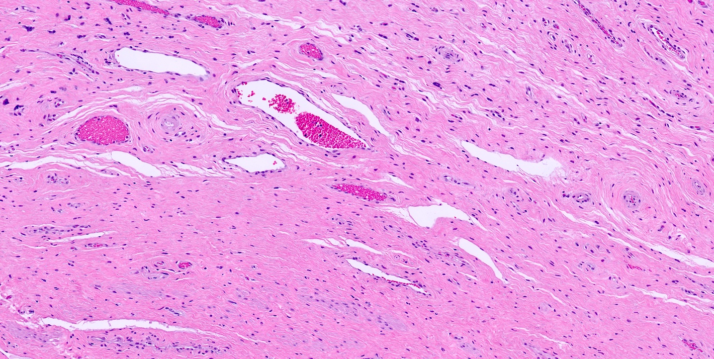
- Lamina propria (subepithelial stroma) is composed of loose connective tissue with elastic fibers, rich venous and lymphatic networks, spindle to stellate and some multinucleated stromal cells
- Muscle
- Outer longitudinal and thin inner circular layer of smooth muscle
- Adventitia is composed of inner dense connective tissue layer and outer loose connective tissue layer containing peripheral nerves, blood vessels and lymphatics
- Wolffian (mesonephric) duct, also known as Gartner duct, runs deeply along lateral vaginal walls; single small duct surrounded by a cluster of small glands lined by cuboidal epithelium with eosinophilic secretion in lumen
- Maturation index
- Ratio of parabasal to intermediate to superficial cells of vaginal epithelium (sampled at middle third of lateral vaginal wall)
- Sample is often obtained simultaneous with Pap smear to detect hormonal effects in menopausal and postmenopausal women
- Increased maturation in vaginal epithelium may be due to estrogenic effect of tamoxifen (Clin Exp Obstet Gynecol 1998;25:121)
- Lined by nonkeratinized stratified squamous epithelium and is composed of basal, parabasal, intermediate and superficial cell layers
- Female urethra
- Lined by urothelium (proximal two - thirds), stratified and pseudostratified columnar and squamous epithelium (distal third)
- Basal layers composed of either low columnar or cuboidal cells, followed by several layers of polyhedral cells
- Most superficial layer is composed of round, dome shaped umbrella cells that are occasionally multinucleated and flattened according to amount of distention
- Nonkeratinized squamous epithelium: cuboidal (deepest), polymorphous (middle), squamous / flattened (superficial) (J Urol 1987;138:775)
- Periurethral (Skene) glands (homologous to the prostate gland) open into the distal portion
- Lined by columnar or cuboidal epithelium with surrounding connective tissue and smooth muscle
- Minor vestibular glands (homologous to glands of Littre in males) open along the entire length
- Tubuloacinar mucinous glands with uniform, pale eosinophilic to clear cytoplasm and basally flattened nuclei
- Lined by urothelium (proximal two - thirds), stratified and pseudostratified columnar and squamous epithelium (distal third)
Microscopic (histologic) images
Contributed by Pooja Srivastava, M.D.
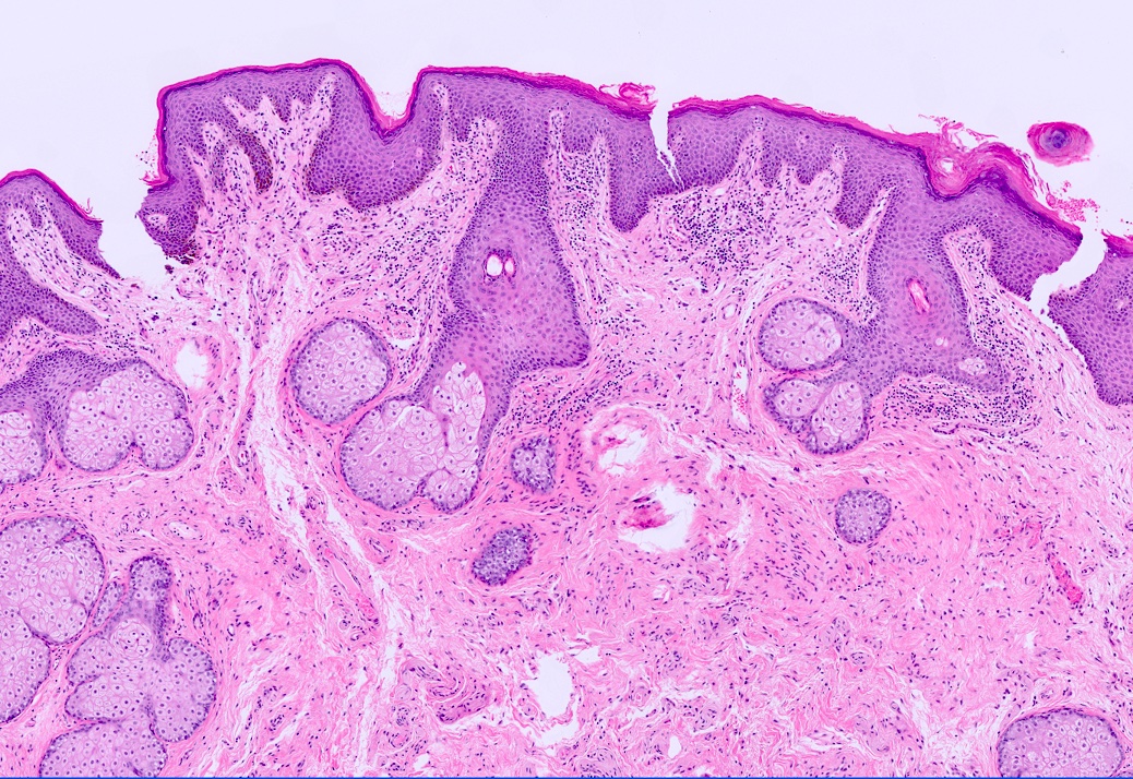
Labia majora overview
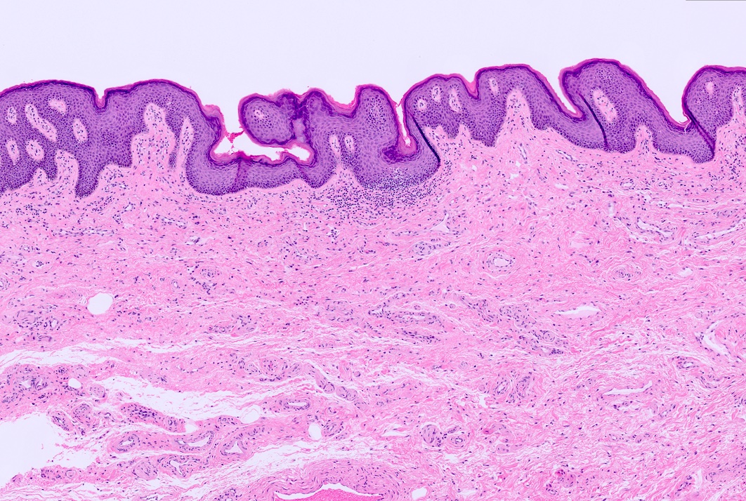
Labia minora overview
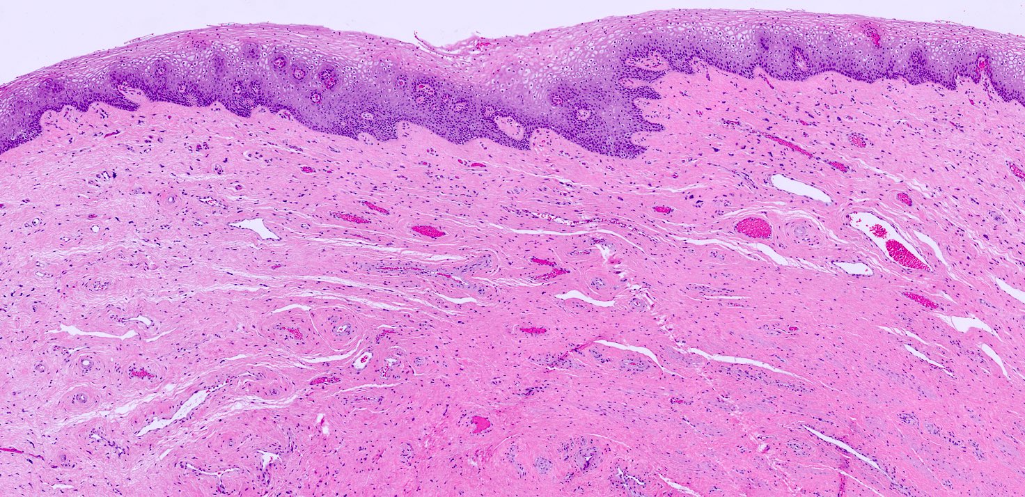
Vagina overview
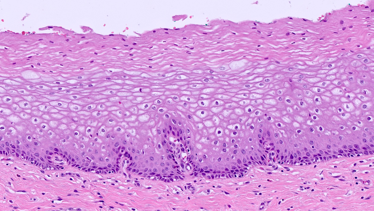
Vagina epithelium
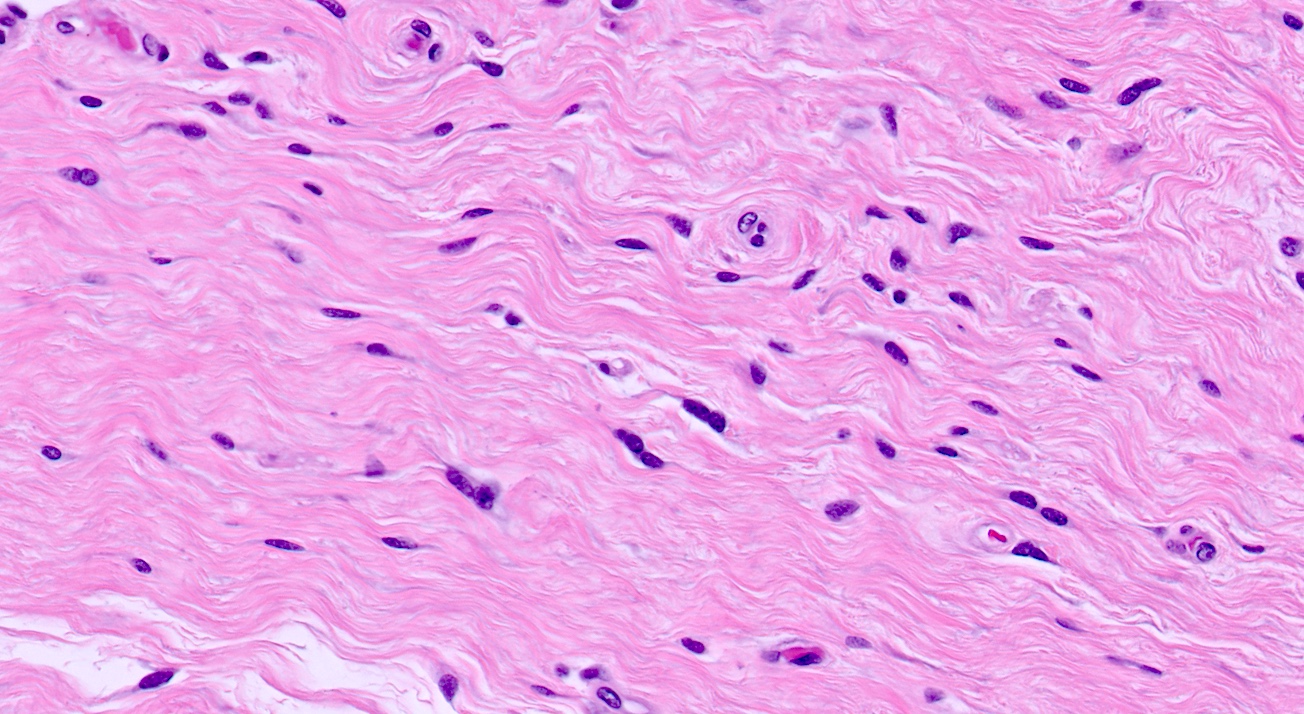
Vagina lamina propria
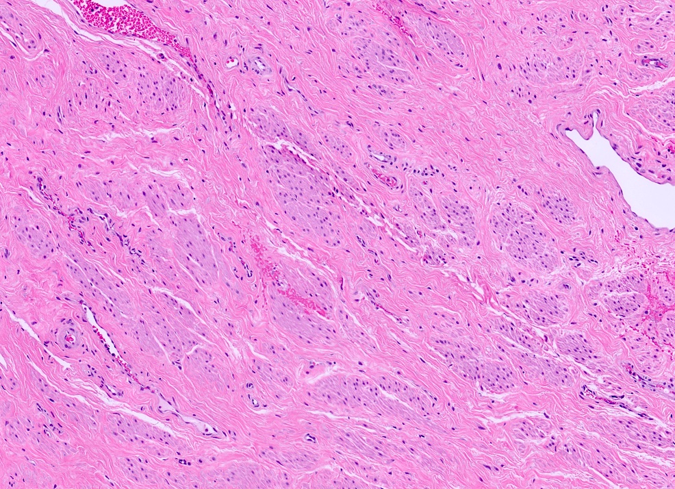
Vagina muscular layer
Virtual slides
Images hosted on other servers

Vulva normal histology
Videos
Normal histology of vagina
Anatomy of female urethra
Histology of female urethra
Board review style question #1
What is the site of biopsy shown in this image?
- Cervix
- Labia majora
- Labia minora
- Uterus
- Vagina
Board review style answer #1
B. Labia majora. Labia majora is composed of keratinized stratified squamous epithelium with hair follicles and eccrine, apocrine and sebaceous glands. Answer A is incorrect because the ectocervix is lined by stratified squamous epithelium overlying fibrous stroma and endocervix is lined by simple, columnar mucinous epithelial cells. Answer C is incorrect because labia minora is lined by keratinized stratified squamous epithelium, without any adnexa. Answer D is incorrect because uterus is lined by endometrial glands, stroma, myometrium and serosa. Answer E is incorrect because vagina has nonkeratinized stratified squamous epithelium and is composed of basal, parabasal, intermediate and superficial cell layers.
Comment Here
Reference: Anatomy & histology - vulva, vagina & female urethra
Comment Here
Reference: Anatomy & histology - vulva, vagina & female urethra
Board review style question #2
What is the epithelium of the labia minora classified as?
- Keratinized stratified squamous epithelium with adnexa
- Keratinized stratified squamous epithelium without adnexa
- Nonkeratinized stratified squamous epithelium with adnexa
- Simple, columnar mucinous epithelial cells
Board review style answer #2
B. Keratinized stratified squamous epithelium without adnexa. This describes the lining of labia minora. Answer A is incorrect because it describes the lining of the labia majora. Answer C is incorrect because the epithelium of labia minora is keratinized and without adnexa. Answer D is incorrect because this describes the lining of endocervix.
Comment Here
Reference: Anatomy & histology - vulva, vagina & female urethra
Comment Here
Reference: Anatomy & histology - vulva, vagina & female urethra
