

Bladder & urothelial tract
Urethral carcinoma
Female urethral carcinoma
Last author update: 1 April 2016
Last staff update: 19 April 2024 (update in progress)
Copyright: 2003-2024, PathologyOutlines.com, Inc.
PubMed Search: Urethral carcinoma[title] female


Table of Contents
Definition / general | Essential features | Epidemiology | Sites | Pathophysiology | Clinical features | Diagnosis | Radiology description | Prognostic factors | Case reports | Treatment | Gross images | Microscopic (histologic) description | Microscopic (histologic) images | Positive stains | Negative stains | Differential diagnosisCite this page: Chavez J. Female urethral carcinoma. PathologyOutlines.com website. https://www.pathologyoutlines.com/topic/vulvafemaleurethralcarcinoma.html. Accessed April 19th, 2024.
Definition / general
- Rare primary neoplasm of epithelial origin
- Secondary involvement by urothelial carcinoma of the bladder is much more common than a primary (Eur Urol 2013;64:823)
Essential features
- Urethral carcinoma is usually due to secondary involvement
- Primary urethral carcinoma is rare and the most frequent histologic types are urothelial carcinoma, squamous cell carcinoma and adenocarcinoma (not otherwise specified, clear cell)
Epidemiology
- Primary tumor accounts for < 1% of all genitourinary malignancies (Eur Urol 2013;64:823, BJU Int 2014;114:25)
- More common in women than men (Hematol Oncol Clin North Am 2012;26:1291)
- Urothelial carcinoma is the predominant histologic type (54 - 65%) followed by squamous cell carcinoma (16 - 22%) and adenocarcinoma (10 - 16%) (Urology 2006;68:1164)
- Secondary involvement of urethra post-cystectomy is seen in 3 - 4% of patients (Eur Urol 2013;64:823, Int J Surg 2015;13:148)
Sites
- Type depends on sex and location:
- Female urethra divided in proximal 2/3 and distal 1/3
- Proximal 2/3 usually urothelial carcinoma
- Distal 1/3 usually squamous cell carcinoma (BJU Int 2014;114:25)
- Frequently initially misdiagnosed as caruncle
- Adenocarcinoma present in both sexes; may originate anywhere along the urethra
- May arise from urothelial metaplastic mucosa or from periurethral glands in both sexes
- Clear cell adenocarcinoma is found predominantly in women and has a particular association with urethral diverticulum (J Urol 2008;180:2463, Int J Surg Oncol 2015;2015:790235)
- Indistinguishable from clear cell adenocarcinoma of the genital tract
- Female urethra divided in proximal 2/3 and distal 1/3
Pathophysiology
- Predisposing factors include:
- Iatrogenic chronic irritation (chronic catheterization / urethroplasty) (Eur Urol 2013;64:823)
- Urethral strictures (BJU Int 2014;114:25)
- Radiation therapy (BJU Int 2008;101:964)
- Chronic urethritis secondary to sexually transmitted diseases
- Recurrent urinary tract infections
Clinical features
- Most patients present with symptoms associated with locally advanced disease (Eur Urol 2013;64:823)
- Gross hematuria or bloody urethral discharge, dysuria, extraurethral mass
- Bladder outlet obstruction, pelvic pain, urethrocutaneous fistula
- Abscess formation, dyspareunia
- Approximately 1/3 of men and women present with involved regional lymph nodes
Diagnosis
- Clinical examination with palpation of external genitalia for suspicious indurations and pelvic exam in women (Eur Urol 2013;64:823)
- Urinary cytology
- Diagnostic urethroscopy and biopsy
Radiology description
- Aims to assess local extent and detect lymphatic and distant metastatic spread
- Magnetic resonance imaging for evaluating extent of tumor and monitoring response to neoadjuvant chemotherapy (Eur Urol 2013;64:823)
Prognostic factors
- Both sexes (Hematol Oncol Clin North Am 2012;26:1291, Eur Urol 2013;64:823):
- Advanced age (≥65 years) and race (African American)
- Stage, grade, nodal involvement and metastasis
- Size and proximal tumor location
- Presence of concomitant bladder cancer
- Type and modality of treatment
Case reports
- 49 year old woman with primary urethral urothelial cell carcinoma with distant lung and bone metastasis (BMC Cancer 2011;11:23)
- 56 year old woman with urothelial cell carcinoma in situ within a urethral diverticulum (Int Urogynecol J 2012;23:1801)
- 61 year old woman with urethral adenocarcinoma associated with intestinal type metaplasia (Int J Clin Exp Pathol 2013;6:1665)
- 65 year old woman with clear cell carcinoma of urethral diverticulum (J Formos Med Assoc 2013;112:489)
Treatment
- Localized disease in women:
- Primary radical urethrectomy; urethra sparing surgery is an alternative if negative margins are possible
- Local radiotherapy is an alternative, local toxicity needs to be taken into consideration (Eur Urol 2013;64:823)
- Advanced disease both sexes:
- Chemotherapy with cisplatinum based agents
- Uncertain value of lymph node dissection (Curr Opin Urol 2015;25:129
- References: Br J Urol 1998;82:835, Urology 1999;53:1126, Holland-Frei: Cancer Medicine, 6th Edition, 2003
Gross images
Contributed by Dr. Jesus Chavez and Dr. Debra Zynger
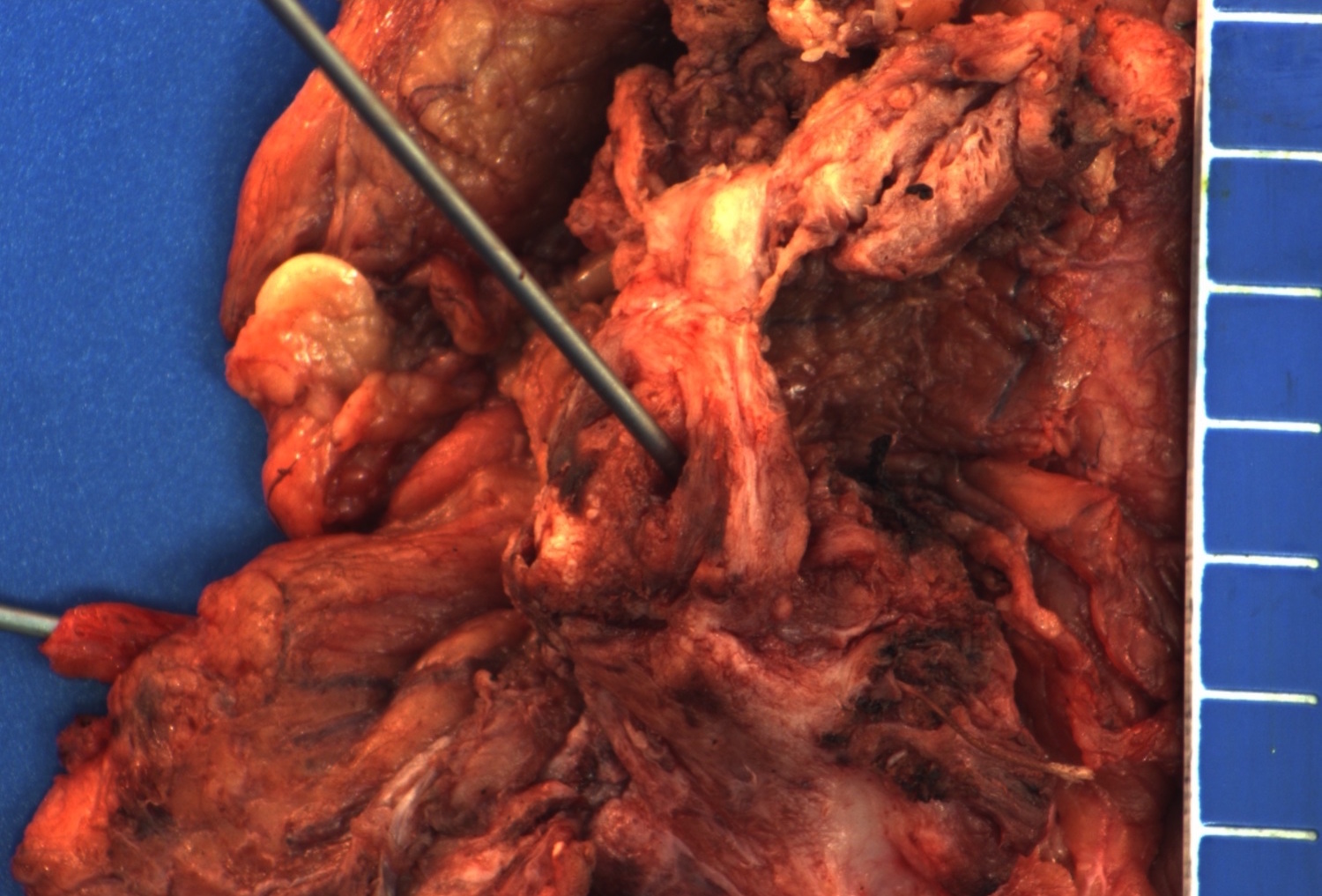
Urethra and periurethral tissue
Microscopic (histologic) description
- Urothelial carcinoma
- Cytologically malignant urothelial cells with visible cell membranes, marked nucleomegaly, irregular nuclei, prominent nucleoli, dark chromatin and abundant mitosis (Bostwick: Urologic Surgical Pathology, 3rd Edition, 2014)
- Squamous cell carcinoma
- Sheets of large, pleomorphic tumor cells with focal or abundant keratinization (depending of grade of differentiation), ample cytoplasm, intercellular bridges, high mitotic activity, prominent nuclear atypia
- Adenocarcinoma
- Composed of simple or pseudostratified columnar epithelium, apical cytoplasm and basally located hyperchromatic nuclei (Bostwick: Urologic Surgical Pathology, 3rd Edition, 2014)
- Occasional vacuolated cytoplasm with mucin or can be a true mucinous tumor with mucin pools
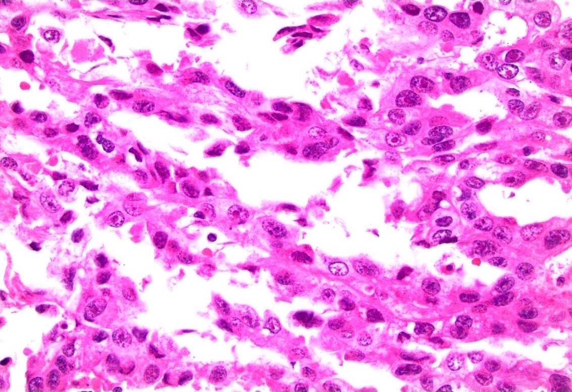
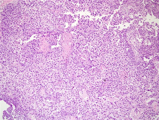
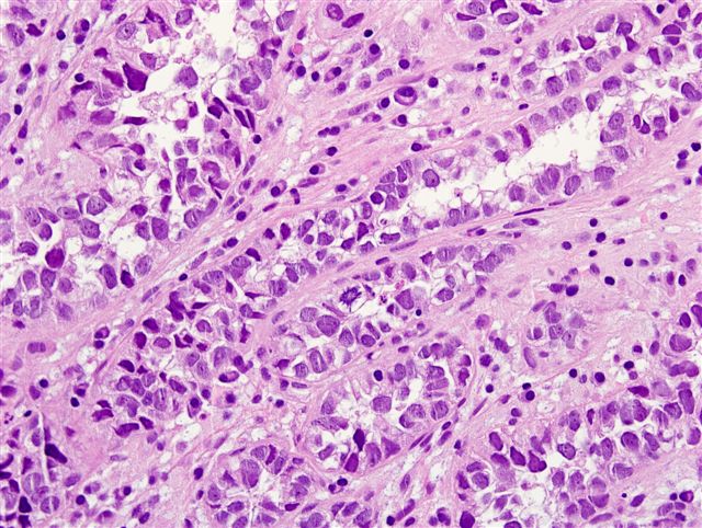
- Clear cell adenocarcinoma
- May have glandular, tubulocystic, solid / diffuse, papillary or micropapillary growth patterns
- Cuboidal, variably sized cells with abundant clear or eosinophilic cytoplasm and cytoplasmic vacuoles
- Nuclei that are hyperchromatic, pleomorphic and have prominent nucleoli
- Hobnail changes and extracellular mucoid material may be present
- Mitoses and necrosis are often seen
Microscopic (histologic) images
Contributed by Dr. Jesus Chavez and Dr. Debra Zynger
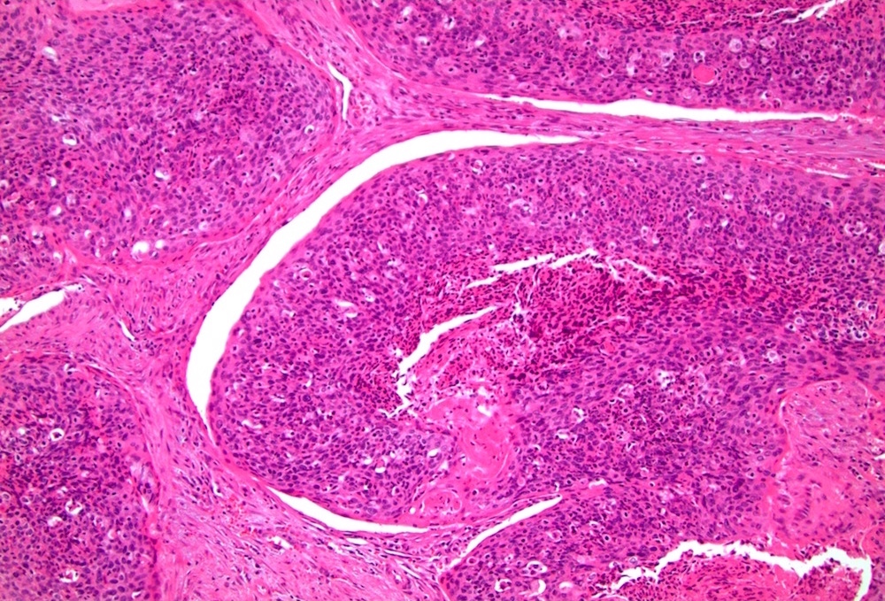
High power, low grade
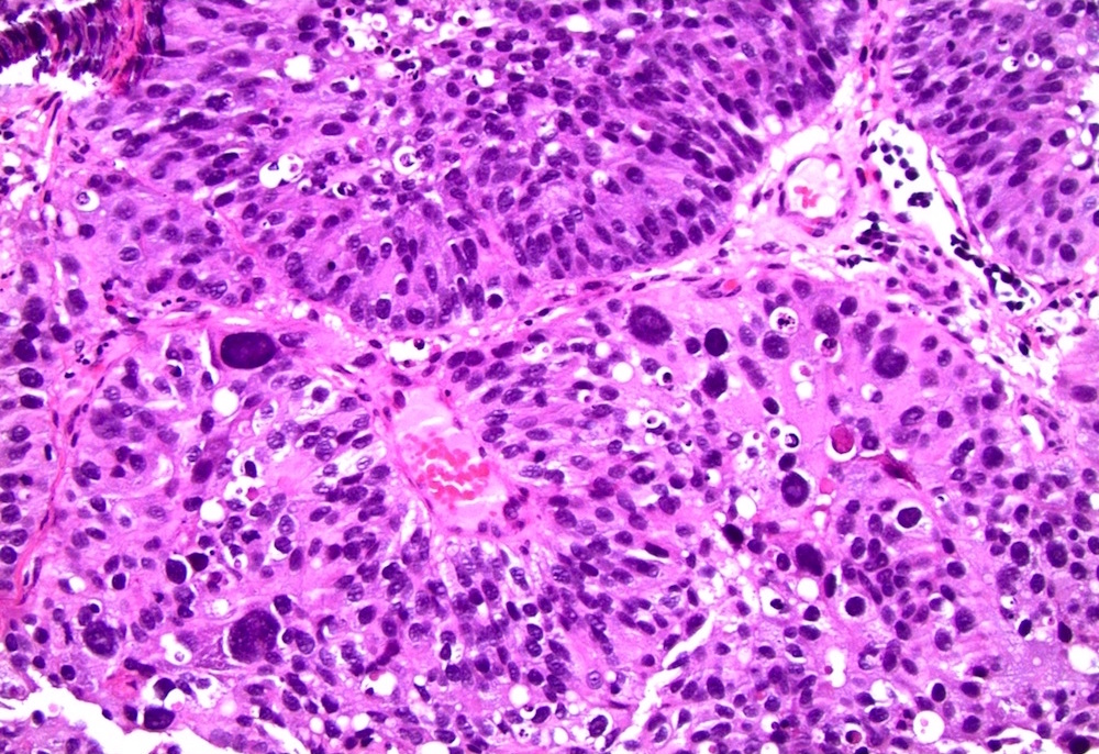
Noninvasive high grade
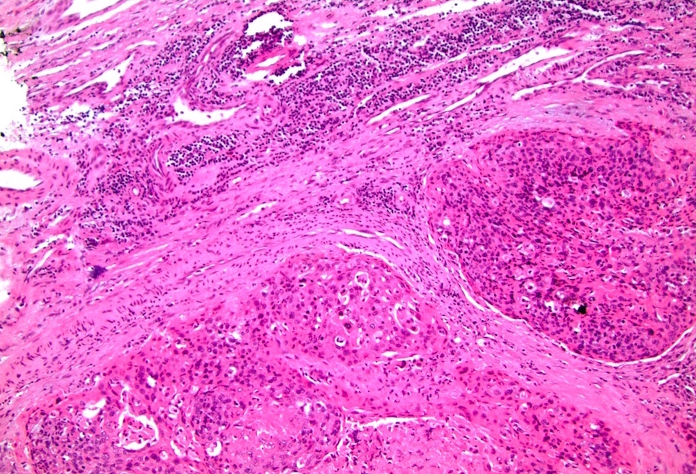
Invasive high grade
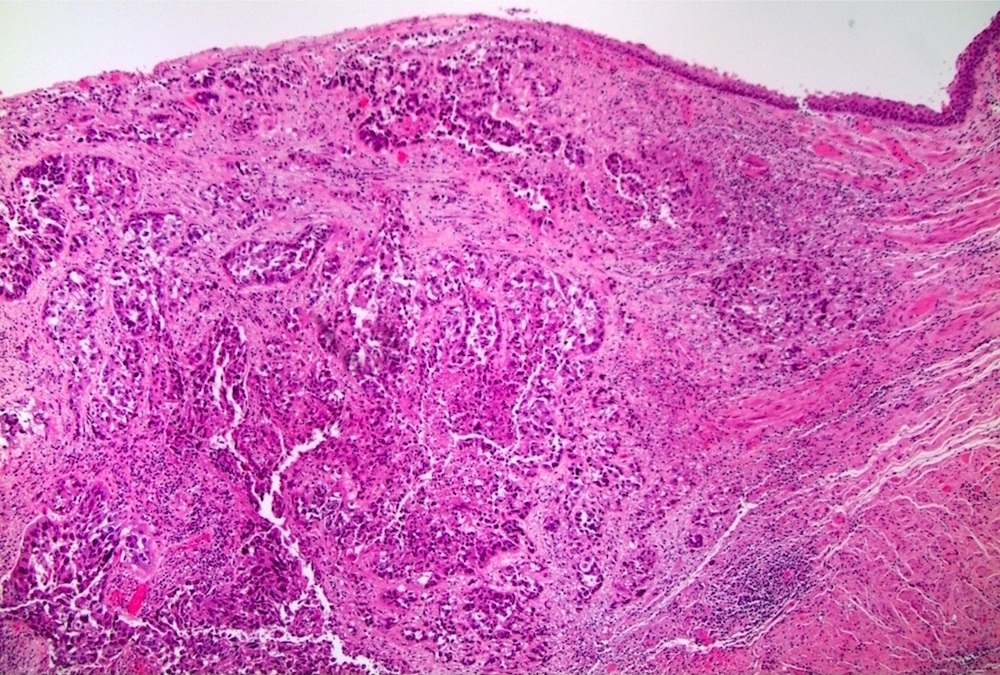
Necrosis and keratinization
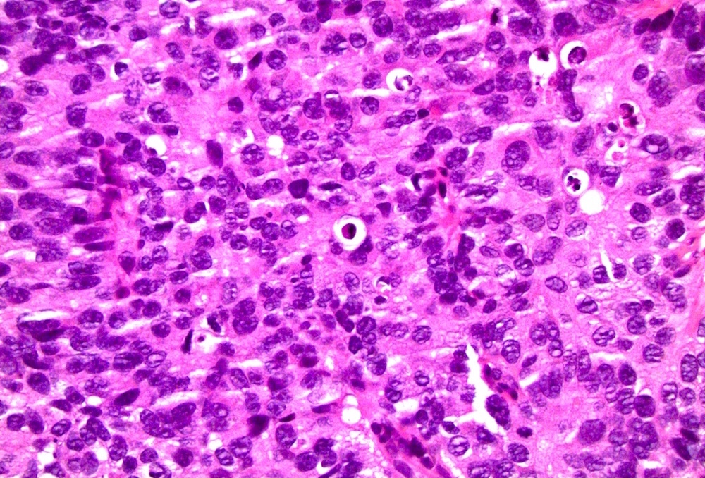
Elderly woman: metastatic
Clear cell adenocarcinoma
Low power, prominent necrosis
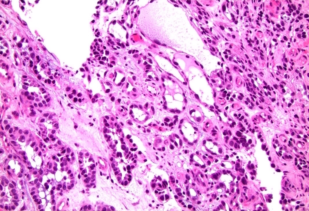
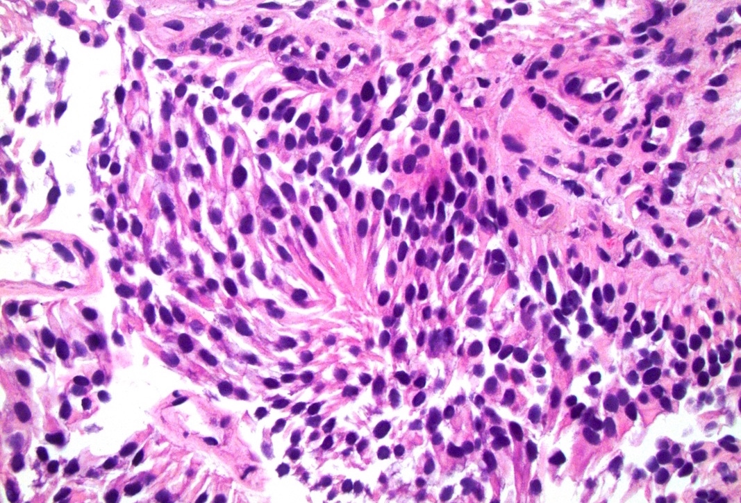
Can mimic nephrogenic metaplasia
With hobnailing
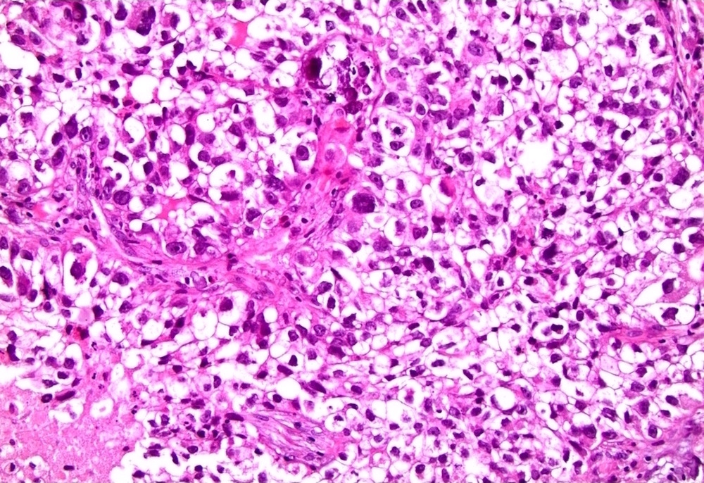
With prominent clear cells and diffuse, sheet-like growth
Case #194

Various images
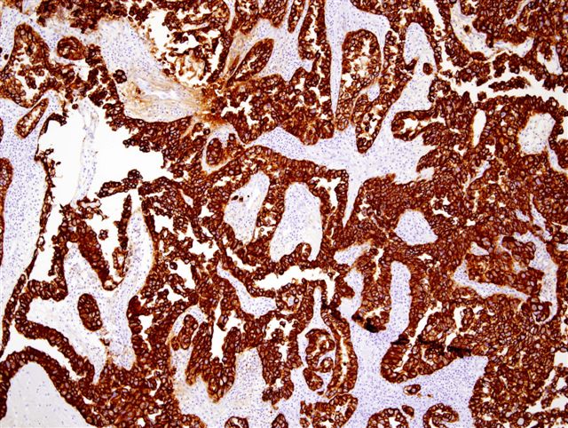
CK7
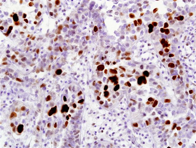
p53
Positive stains
- Adenocarcinoma
- CK20 (variable), CDX2 (variable), cytoplasmic beta-catenin
- Clear cell adenocarcinoma
- AMACR (racemase) (75%), vimentin (75%), p53 (100%) (Virchows Arch 2013;462:193)
- PAX8 (50%), CK7 (50%) (Virchows Arch 2013;462:193)
- Squamous cell carcinoma
- High molecular weight cytokeratin (CK903, CK5/6), p63, p16 (HPV related)
- Urothelial carcinoma
- p53 (80%), CK20 (transurothelial in carcinoma in situ, variable in invasive urothelial carcinoma), CK7 , GATA3 (Hum Pathol 2013;44:2760), High molecular weight cytokeratin (CK903, CK5/6), p63
Negative stains
- Urothelial carcinoma
- E-cadherin, CD44s, PAX8, PSA, PSAP
- Squamous cell carcinoma
- Adenocarcinoma
- Nuclear beta-catenin (colon usually has nuclear staining)
- PAX2, PAX8
- Clear cell adenocarcinoma
Differential diagnosis
- Adenocarcinoma of adjacent anatomic sites (prostate and colon)
- Nephrogenic metaplasia (versus clear cell adenocarcinoma)
- Squamous cell carcinoma of vulva or cervix with extension
- Urothelial carcinoma of bladder with extension (more frequent than primary)
