
7 August 2008 – Case of the Week #126
To view the images or references, you must click on the links in blue. Links in green are to journals with free full text-no registration. You can also access these cases by visiting our Home Page, then click on the Case of the Week button on the left hand side.
This email is sent only to subscribers. To subscribe or unsubscribe, email NatPernick@Hotmail.com, indicating subscribe or unsubscribe to Pathology Case of the Week. There is no charge. We do not sell, share or use your email address for any other purpose. We also have free email subscriptions for Pathologist/PhD jobs (biweekly), Other laboratory jobs (biweekly), Pathology website news (monthly) and Pathology new books (monthly). Email us to subscribe.
We don’t make any demands from the users of our free website, but we are requesting that you pledge support for the charity we established, The Detroit College Promise. As discussed on its home page at www.DetroitCollegePromise.org, this charity will pay for college scholarships for almost all Detroit Public School students, and motivate children to go to college, including many who never thought it was possible. It will also demonstrate to the public, including other physicians, the importance of pathologists, as our pathology website has founded this charity. This is particularly important at a time when other physicians are stealing our work and treating us as lackeys, and hospital administrators routinely violate federal law by denying us Part A payments. We are asking for you to fill in our pledge form (click here) for whatever amount you believe appropriate, whether $10, $25, $100 or more, or sending us an email with this information. Thanks for your support.
We thank Dr. Renuka Agrawal, Loma Linda University Medical Center, California (USA) for contributing this case and much of the discussion. To contribute a Case of the Week, email NatPernick@Hotmail.com with the clinical history, your diagnosis and diagnostic microscopic images in JPG, GIF or TIFF format (send as attachments, we will shrink if necessary). Please include any other images (gross, immunostains, etc.) that may be helpful or interesting. We will write the discussion (unless you want to), list you as the contributor, and send you $35 (US dollars) by check or PayPal for your time after we send out the case. Please only send cases with high quality images and a diagnosis that is somewhat unusual (or a case with unusual features).
Case of the Week #126
Clinical History
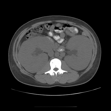
A 19 year-old Hispanic man presented with hypertension and renal insufficiency (serum creatinine of 7.1 mg/dl). The peripheral blood smear showed pancytopenia but no circulating blasts. A CT scan (image) demonstrated bilateral renal enlargement with lymphadenopathy in the retroperitoneum and neck.
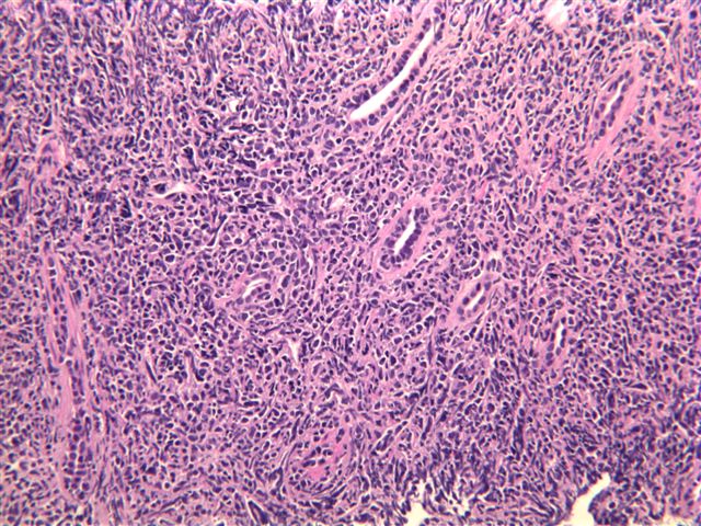
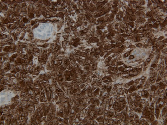
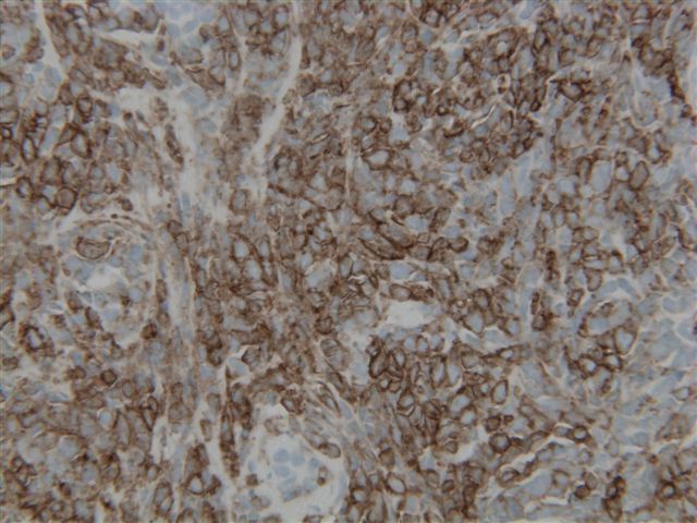
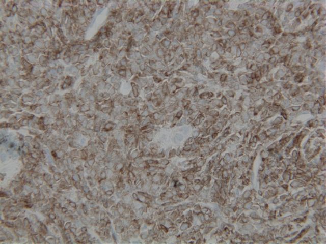
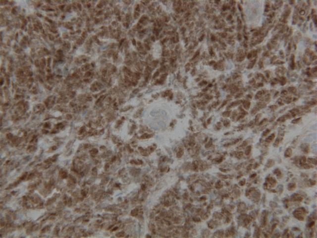
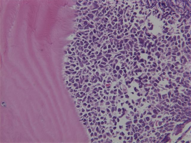
A core biopsy was obtained (image #1, #2), and immunostains were performed (CD10, CD34, CD79a, TdT)
What is your diagnosis?
Diagnosis:
Pre-B acute lymphoblastic lymphoma involving the kidneys
Discussion:
A bone marrow biopsy was also obtained (image), which showed a diffuse infiltration by tumor cells with a similar morphology as the renal biopsy.
The renal biopsy showed a diffuse infiltrate of monotonous blastoid cells obliterating the normal renal architecture. The bone marrow biopsy showed a similar population of cells replacing most of the marrow. Immunophenotyping by flow cytometry and immunohistochemistry showed that the tumor cells co-express CD19, CD20, CD10, CD34, CD38, CD79a, HLA-DR and TdT.
Precursor B-cell acute lymphoblastic leukemia/lymphoma (ALL) is a common pediatric hematologic malignancy. Although renal failure due to tumor lysis is a recognized complication of treatment, initial presentation with renal failure is distinctly uncommon. ALL must be considered among the causes of acute renal failure when the kidneys are enlarged. Careful morphologic study and immunophenotyping by flow cytometry or immunohistochemistry is helpful to arrive at the correct diagnosis, and to avoid confusion with other small blue cell tumors which may involve the kidney, such as Wilm’s tumor (J Pediatr Hematol Oncol 2008;30:471), small cell carcinoma or Ewing’s sarcoma/primitive neuroectodermal tumor.
Additional references:
1. Weinstein Howard J, Tarbell Nancy J. Leukemias and lymphomas of childhood. Cancer Principles and Practice of Oncology. 5th edition; Ch.44: Section 2, 2145-65
2. Boueva A, Bouvier R. Precursor B-cell lymphoblastic leukemia as a cause of a bilateral nephromegaly. Pediatr Nephrol 2005;20:679
3. Mehta A, Gulati K, Jain M, Gulati S. Non-Hodgkin lymphoma in a child presenting as nephromegaly and acute renal failure. Indian Pediatr 2001;38:407
4. Gilboa N, Lum GM, Urizar RE. Early renal involvement in acute lymphoblastic leukemia and non-Hodgkin lymphoma in children. J Urol 1983;129:364
5. Nizze H et al. Primary renal manifestations in malignant lymphomas and leukemia. Pathologe 2003;24:460
6. John William J, Foon Kenneth A, Patchell Roy A. Paraneoplastic syndromes Cancer Principles and Practice of Oncology. Vol.2; Ch.46: 2397-422.
7. PathologyOutlines.com - Leukemia, Acute chapter
Nat Pernick, M.D., President
PathologyOutlines.com, Inc.
30100 Telegraph Road, Suite 404
Bingham Farms, Michigan (USA) 48025
Telephone: 248/646-0325
Email: NatPernick@Hotmail.com
Alternate email: NatPernick@gmail.com