
4 March 2010 - Case #171
All cases are archived on our website. To view them sorted by case number, diagnosis or category, visit our main Case of the Month page. To subscribe or unsubscribe to Case of the Month or our other email lists, click here.
Thanks to Dr. Vladimir Osipov, Medical College of Wisconsin, Milwaukee, WI (USA), for contributing this case. This case was reviewed in May 2020 by Dr. Jennifer Bennett, University of Chicago and Dr. Carlos Parra-Herran, University of Toronto.

Orchard Pathology
Orchard Pathology is a complete diagnostic information system designed to accommodate the comprehensive pathology laboratory. Orchard Pathology is used as a stand-alone system for anatomic testing or combined with Orchard Harvest LIS to integrate clinical. The shared database provides access to the patient's history and enables the consolidation of clinical results to the pathology report.
Unlike older legacy systems that do not utilize pathology-specific word processing, Orchard Pathology stores report information in discrete data fields that enhance EMR integration and simplify the process to mine data for evaluations; correlation studies; quality assurance; and regulatory, statistical, and other management reports.
Advertisement
Case #171
Clinical history:
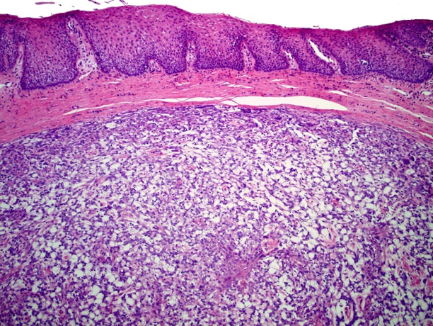
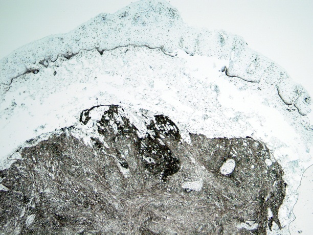
A middle aged woman presented with a vaginal nodule, which had been growing slowly over a period of several months. It was submitted as a vaginal cyst and measured 1 cm in size.
Microscopic images:


What is your diagnosis?
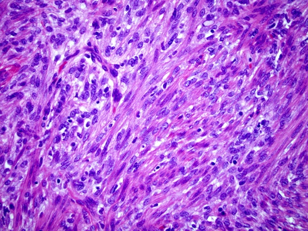
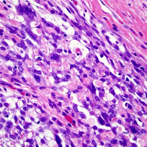
Diagnosis: Spindle cell epithelioma of the vagina
Discussion:
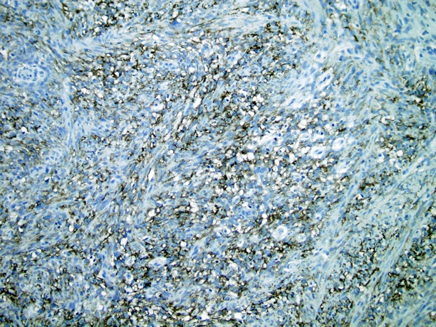
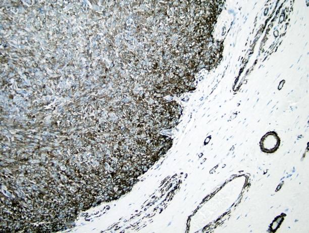
The tumor is well circumscribed but unencapsulated. There are myoid spindle cells (figure 4), epithelioid cells (figure 3) and cells forming cribriform patterns with mucin production (figure 2). There are occasional spherules present (figure 5). The tumor cells are immunoreactive with SMA, BCL2, CD99 and cytokeratin (not shown).
Spindle cell epithelioma, also called mixed tumor of the vagina, is a rare tumor characterized by a mixture of well differentiated epithelial cells and stromal cells, with no / rare atypia or mitotic figures. The tumors are usually not connected with the overlying epithelium. Unlike mixed tumors of other sites, these tumors do not have myoepithelial cells (Am J Surg Pathol 1993;17:509).
Spindle cell epithelioma typically is immunoreactive for cytokeratin, muscle actin, CD10, CD34, vimentin, BCL2 and desmin, with variable staining for h-caldesmon and EMA (Mod Pathol 2004;17:1243, Arch Pathol Lab Med 2001;125:547). They are usually negative for S100 and lack CD99 membranous staining (but often have cytoplasmic staining).
Based on the staining patterns, they are thought to arise from a pluripotential cell with the ability to differentiate in both epithelial and mesenchymal directions. Diagnosis is based on morphology and immunostains are usually not definitive.
These tumors have benign behavior. Simple excision is generally curative (Am J Surg Pathol 1981;5:413).
All cases are archived on our website. To view them sorted by case number, diagnosis or category, visit our main Case of the Month page. To subscribe or unsubscribe to Case of the Month or our other email lists, click here.
Thanks to Dr. Vladimir Osipov, Medical College of Wisconsin, Milwaukee, WI (USA), for contributing this case. This case was reviewed in May 2020 by Dr. Jennifer Bennett, University of Chicago and Dr. Carlos Parra-Herran, University of Toronto.
Orchard Pathology is a complete diagnostic information system designed to accommodate the comprehensive pathology laboratory. Orchard Pathology is used as a stand-alone system for anatomic testing or combined with Orchard Harvest LIS to integrate clinical. The shared database provides access to the patient's history and enables the consolidation of clinical results to the pathology report.
Unlike older legacy systems that do not utilize pathology-specific word processing, Orchard Pathology stores report information in discrete data fields that enhance EMR integration and simplify the process to mine data for evaluations; correlation studies; quality assurance; and regulatory, statistical, and other management reports.
Website news:
(1) Thanks for your support! On 2 March 2010, we had a record 11,017 visits, and last month, we had a record average of 8,425 visits per day (includes weekends).
Visit and follow our Blog to see recent updates to the website.
(1) Thanks for your support! On 2 March 2010, we had a record 11,017 visits, and last month, we had a record average of 8,425 visits per day (includes weekends).
Visit and follow our Blog to see recent updates to the website.
Case #171
Clinical history:
A middle aged woman presented with a vaginal nodule, which had been growing slowly over a period of several months. It was submitted as a vaginal cyst and measured 1 cm in size.
Microscopic images:
CD99
BCL2
Smooth muscle actin
What is your diagnosis?
Click here for diagnosis and discussion:
Diagnosis: Spindle cell epithelioma of the vagina
Discussion:
The tumor is well circumscribed but unencapsulated. There are myoid spindle cells (figure 4), epithelioid cells (figure 3) and cells forming cribriform patterns with mucin production (figure 2). There are occasional spherules present (figure 5). The tumor cells are immunoreactive with SMA, BCL2, CD99 and cytokeratin (not shown).
Spindle cell epithelioma, also called mixed tumor of the vagina, is a rare tumor characterized by a mixture of well differentiated epithelial cells and stromal cells, with no / rare atypia or mitotic figures. The tumors are usually not connected with the overlying epithelium. Unlike mixed tumors of other sites, these tumors do not have myoepithelial cells (Am J Surg Pathol 1993;17:509).
Spindle cell epithelioma typically is immunoreactive for cytokeratin, muscle actin, CD10, CD34, vimentin, BCL2 and desmin, with variable staining for h-caldesmon and EMA (Mod Pathol 2004;17:1243, Arch Pathol Lab Med 2001;125:547). They are usually negative for S100 and lack CD99 membranous staining (but often have cytoplasmic staining).
Based on the staining patterns, they are thought to arise from a pluripotential cell with the ability to differentiate in both epithelial and mesenchymal directions. Diagnosis is based on morphology and immunostains are usually not definitive.
These tumors have benign behavior. Simple excision is generally curative (Am J Surg Pathol 1981;5:413).
